StudentShare
Contact Us
Sign In / Sign Up for FREE
Search
Go to advanced search...
Free

The Role of Mitosis and Miosis In Cancer Tistes Antigen (CTA) - Literature review Example

Cite this document
Summary
This paper is about the role of mitosis and meiosis in cancer testis antigen. It's a well-known fact, that error in these two processes lead to the unstoppable division of cells and violation of differentiation. Together it may occur cancer in patients…
Download full paper File format: .doc, available for editing
GRAB THE BEST PAPER96.3% of users find it useful
The Role of Mitosis and Miosis In Cancer Tistes Antigen (CTA)
Read Text Preview

Extract of sample "The Role of Mitosis and Miosis In Cancer Tistes Antigen (CTA)"

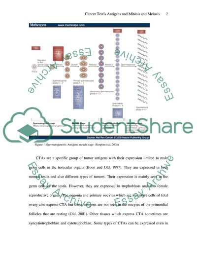
Mitosis and Meiosis and Cancer Testis Antigens It is a well known fact that the germ cell line derives from the primordial germ cells. These cells basically migrate through the dorsal mesentery of the embryo and then enter the fetal gonad that is developing in the genital ridge. In males, in this genital ridge, the primordial germ cells are enclosed by somatic Sertoli cells and are known as prospermatoonia (Simpson, 2005). These proliferate for a few days and then rest at G0 or G1 of cell cycle until puberty. When the individual attains puberty, proliferation of the prospermatogonia resumes, thus initiating spermatogenesis. Spermatogonia are the male germline stem cells and they proliferate from puberty all through the adult life. The spermatogonia either renew themselves or undergo differentiation and turn into spermatozoa. Two meoitic divisions are involved in this step and a tetraploid of primary spermatocytes are first formed which further undergo meiosis to form haploid sperms. Cancer Testis Antigens or CTAs like MAGE and NY-ESO-1 are basically expressed in these spermatogonia. Synaptonemal complex protein and other CTAs, those associated with meiosis are expressed in the spermatocytes (Simpson, 2005). Figure-1: Spermatogenesis: Antigens at each stage (Simpon et al, 2005) CTAs are a specific group of tumor antigens with their expression limited to male germ cells in the testicular organs (Boon and Old, 1997). They are expressed in both normal testis and also different types of tumors. Their expression is mainly seen in the germ cells of the testis. However, they are expressed in trophoblasts and also female reproductive organs. The oogonia and primary oocytes which are immature cells of fetal ovary also express CTA but these antigens are not seen in the oocytes of the primordial follicles that are resting (Old, 2001). Other tissues which express CTA sometimes are syncytiotrophoblast and cytotrophoblast. Some types of CTAs can be expressed even in nongametogenic tissues like liver, pancreas and spleen. Some recent reports have suggested that expression of certain CTAs like SSX, N-RAGE, MAGE, NY-ESO are expressed in both fetal and adult human mesenchymal stem cells of bone marrow, but subsequent to differentiation of adipocytes and oocytes, expression of these CTAs is down regulated. Thus researchers have suggested that expression of CTA can be a useful stem cell marker, in addition to characteristic of gametogenesis (Ghafouri-Fard, and Modarressi, 2009).. The restricted expression of CTAs in undifferentiated germ cells and soamatic cells is suggestive of their role in embryonic development. is it due to their restriction in expression that these antigens are targets for cancer vaccines. As of now, more than 140 members spread along 70 families of CTAs have been identified (Simpson, 2005). Expression of CTAs is almost restricted to male germ cells in the testis and also various malignancies. There is enough evidence that regulation of methylation has a significant role in the control of expression of CTA. or some CTAs like MAGE-A1, the primary silencing mechanism is methylation of DNA and simple demethylation is sufficient and necessary for production of expression. According to De Smet et al (1999), whenever there is heavy methylation, repression of gene expression in cells occurs, despite existence of various transcription factors necessary for expression and 5-aza-2’-deoxycytidine, the demethylation agent can induce MAGE-A1transcription in cell cultures. In yet another study by De Smet et al (2004), demethylation, along with persistent inhibition of remethylation locally in some tumor cells, influences the site specific hypomethylation of MAGE-A1. The functions of CTA in the biological functions of germ cell lines and also various tumors is actually poorly understood. The central question with regard to CTA is whether expression of CTAs contributes to tumorigenesis or is it a functionally irrelevant by-product of cellular transformation process, with possibility of global chromatin changes.However, several clues have merged with regard to the role of CT antigens like MAGE in human tumorigenesis (Simpson, 2005). The only CTA that has been extensively studied is MAGEA1. This antigen has been subjected to evaluation and analysis through yeast two-hybrid assay. The main binding partner for this CTA is transcriptional regulator SKI-interacting protein (SKIP). Binding of the CTA to SKIP is mainly dependent on the extreme carboxyl terminus of the MAGE protein, which is a domain also shared by MAGEA4 and even this binds to SKIP. SKIP mainly connects various DNA-binding proteins to other proteins that wither repress or activate transcription and participate in a wide range of signaling pathways including those involving other molecules like glucocorticoids, estrogens, retinoic acid, vitamin D, tansforming growth factor and Notch1 (Simpson, 2005). One interesting aspect in the Notch-1 pathway is that, MAGEA1 binds to SKIP and recruits histone deacetylase, thus disrupting SKIP-mediated Notch-1. Thus, in this signal pathway, MAGEA1 acts as transcriptional repressor. The function of MAGEA1 in the germ cell lineage is actually not elucidated. However, there is enough evidence to suggest that SKIP is involved. MAGEA1 represses the expression of various genes required for differentiation, in early stages of development as in spermatogonia, but not during later stages of development. It has been proposed that similar inhibition of differentiation of cells related to cancer probably contributes to tumorigenesis. Other CTAs which have been studied and incriminated in tumorigenesis are MAGEA11 and MAGEA4. While the former modulates the internal domain interactions and thus plays an important role in the androgen-receptor function, MAGEA11-stabilised ligand free androgen receptor in the cytoplasm leads to accumulation of the CTA and thus has dual amplifying effect on androgen signaling (Simpson, 2005). Thus, CTA being genes that control germ cell gene expression, alter the normal stem cells and contribute to gametogenesis and tumorigenesis. References Boon, T. & Old, L. (1997). J. Cancer tumour antigens. Curr. Opin. Immunol. 9, 681-683. Old, L. J. (2001). Cancer/testis (CT) antigens- a new link between gametogenesis and cancer. Cancer Immun. 1, 1-8. De Smet, C., Lurquin, C., Lethé, B., Martelange, V., Boon, T. (1999). DNA methylation is the primary silencing mechanism for a set of germ line- and tumor-specific genes with a CpGrich promoter. Mol Cell Biol., 19, 7327 – 7335. De Smet, C., Loriot, A., Boon, T. (2004). Promoter- dependent mechanism leading to selective hypomethylation within the 5 region of gene MAGE-A1 in tumor cell., Mol Cell Biol., 24, 4781 – 4790. Ghafouri-Fard, S., and Modarressi, M. (2009). Cancer-Testis Antigens: Potential Targets for Cancer Immunotherapy. Arch Iranian Med., 12 (4), 395 – 404. Simpson, A.J.G., Caballero, O.L., Jungbluth, A., et al. (2005). Cancer/Testis Antigens, Gametogenesis and Cancer: Sidebar: Schematic Summary of Spermatogenesis in Humans. Nat Rev Cancer, 5(8), 615-625. Read More
Tags
Cite this document
  • APA
  • MLA
  • CHICAGO
(“The Role of Mitosis and Miosis In Cancer Tistes Antigen (CTA) Literature review - 1”, n.d.)
Retrieved de https://studentshare.org/medical-science/1421274-the-role-of-mitosis-and-meiosis-in-cancer-testis-antigen-cta
(The Role of Mitosis and Miosis In Cancer Tistes Antigen (CTA) Literature Review - 1)
https://studentshare.org/medical-science/1421274-the-role-of-mitosis-and-meiosis-in-cancer-testis-antigen-cta.
“The Role of Mitosis and Miosis In Cancer Tistes Antigen (CTA) Literature Review - 1”, n.d. https://studentshare.org/medical-science/1421274-the-role-of-mitosis-and-meiosis-in-cancer-testis-antigen-cta.
  • Cited: 0 times

CHECK THESE SAMPLES OF The Role of Mitosis and Miosis In Cancer Tistes Antigen (CTA)

The Different Stages of the Mitosis

In a multi-cellular organism, two types of cell division occur: they are mitosis and meiosis.... Prophase is the first stage of mitosis which is visible.... Telophase is the last stage of mitosis in which the chromosomes begins to loosen itself from the thicker rigid structure into thin filaments.... The cells undergoing the four stages of mitosis such as prophase, metaphase , anaphase and telophase were identified in the slide using the 10x magnification....
5 Pages (1250 words) Lab Report

Compare and Contrast Meiosis vs Mitosis

Running Head: MITOSIS AND MEIOSIS Comparison and Contrast of mitosis and Meiosis [Name of the Writer] [Name of the Institution] Comparison and Contrast of mitosis and Meiosis The eukaryotic cells get divided by partitioning its chromosomes into two indistinguishable sets of chromosomes and this process occurs in the nucleus of the cell, this process of cell division is called mitosis1.... hellip; In simple words, the parent cell gets separated into two daughter cells that are similar to the parent cell in the process of mitosis....
6 Pages (1500 words) Essay

Similarities and Differences in Mitosis and Meiosis

These mechanisms are presented in the form of mitosis and meiosis.... This short essay compares and contrasts the two mechanisms of cell division viz namely mitosis and Meiosis.... DNA replication is skipped in interphase and the cells proceed to divide exactly by the mechanism of mitosis.... mitosis comprises of standard steps of Interphase, Prophase, Prometaphase, Anaphase and Telophase.... Lehninger, 2008) In the Prophase in mitosis, four chromatids combine to form a pair of chromosomes which are joined by a centromere....
3 Pages (750 words) Essay

The Life Cycles of Sexually-Reproducing Organisms

The slide was examined for identifying any stage of mitosis.... ResultsMitosisThe four phases of mitosis- Prophase, Metaphase, Anaphase and Telophase- were observed under the microscope.... Also called the direct cell division it occurs just by elongation and constriction of the nucleus and cytoplasm resulting in two daughter cells. mitosis is a division of a parent cell into two identical daughter cells having the same number and kind of chromosomes as in the parent cell....
5 Pages (1250 words) Lab Report

The Mitotic Phase of the Cell Cycle

There are six stages in mitosis and these include interphase, prophase, prometaphase, metaphase, anaphase and lastly the telophase.... Animals for instance are known to undergo a form of mitosis called open mitosis.... mitosis refers to a form of cell division where the eukaryotic cells separate the chromosomes existing in its cell nucleus into two identical sets formed in two separate nuclei.... mitosis is generally succeeded immediately by cytokinesis also called cytoplasmic division which… The two processes define the mitotic phase of the cell cycle that divides the mother cell into two daughter cells that are identical (Snedden, 1-48). mitosis is a complex process that takes place only in mitosis mitosis refers to a form of cell division where the eukaryotic cells separate the chromosomes existingin its cell nucleus into two identical sets formed in two separate nuclei....
1 Pages (250 words) Essay

Cancer Testis Antigens and Meiosis

2010). CTA has tumor-restricted pattern of expression in male germ cells in the testis in the early phases of spermatogenesis along with their The antigen HOM-TES-14 which is repeatedly encountered during CDNA expression library is encoded by the SCP-1 gene, which is mainly expressed during prophase of mitosis in spermatocytes and involved in homologous chromosomes pairing therefore is less restricted in its expression.... Sertoli and Leydig cells are tubular cells which do not express cta....
9 Pages (2250 words) Literature review

Mitosis, Meiosis, and Mutation

For each virtual tissue sample, count the number of cells in each stage of mitosis and record your observations below.... There are four simulation tissue samples; each of which is “stained” so that you can observe the stages of mitosis in the sample cells.... issue SampleInterphaseProphaseMetaphaseAnaphaseTelophaseSample #1: Normal Tissue91000Sample #2: Normal Tissue90001Sample #3: Cancer Tissue72010Sample #4: Cancer Tissue71101Analysis (20 points)It was observed that 90 percent (nine cells) of the normal cells were in the anaphase stage of mitosis in the first sample while only 10 percent was in prophase....
2 Pages (500 words) Assignment

Cell Division and the Musculoskeletal System

… The paper “Cell Division and the Musсulоskеlеtаl System, Importance of mitosis and Meiosis” is a well-turned example of coursework on biology.... The paper “Cell Division and the Musсulоskеlеtаl System, Importance of mitosis and Meiosis” is a well-turned example of coursework on biology.... mportance of mitosis and MeiosisMitosis is important in sexual reproduction as the process allows the development of a cell to a grown individual....
6 Pages (1500 words) Coursework
sponsored ads
We use cookies to create the best experience for you. Keep on browsing if you are OK with that, or find out how to manage cookies.
Contact Us