StudentShare
Contact Us
Sign In / Sign Up for FREE
Search
Go to advanced search...
Free

Application of Microscopy in Biomedical Sciences - Lab Report Example

Cite this document
Summary
The author of the paper "Application of Microscopy in Biomedical Sciences" will begin with the statement that in the past Biologist were confined to the traditional transmission electron microscope and correlated the morphology with molecular and biochemical data…
Download full paper File format: .doc, available for editing
GRAB THE BEST PAPER91.6% of users find it useful
Application of Microscopy in Biomedical Sciences
Read Text Preview

Extract of sample "Application of Microscopy in Biomedical Sciences"

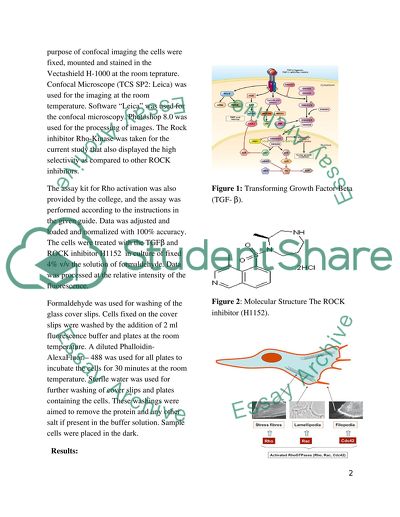
Application of Microscopy in Biomedical Sciences Introduction: In the past Biologist were confined to the traditional transmission electron microscope and correlated the morphology with molecular and biochemical data. Use of the electron microscopy provides details in changes of cells due to ecological effects on living cells. Fluorescence emission in the fluorescence microscopy is not only characterized by the position and intensity but also the wavelength, lifetime and polarization. Use of fluorescence imaging has made it easier the observation of photo physical events. Rotation and mobility of the fluorescence is measured through the TR-FAIM as “Time-resolved fluorescence anisotropy imaging”. New applications are used to find the path of the unexpected discoveries (Suhling, French and Pillips 2004). Atomic Force Microscopy is the most important technique used in the biomedical applications, but it cannot compete with the traditional electron microscopy and confocal microscopy because of speeds at which later techniques capture the images (Haupt, Pelling and Horton, 2006). Some other scholars also emphasized on the importance of confocal microscopy used for the observation of living cells. Use of the confocal microscopy is now common as non-ionizing radiations are employed, which are also used for the tissue preparation and study of the living cells. TGFβ stands for the transforming growth factor β, a family of the secreted factors, which are involved in the growth regulation, migration, differentiation, apoptosis, adhesion in the multistep processes for the wound healing, and angiogenesis. For the epithelial cells, the most important factor of family TGFβ is the TGFβ 1. It also acts as the growth inhibitor and expresses the early gene JunB. TGFβ 1 also plays an important role for the morphology and transcriptional programme of cells. Endothelial cells differ from the epithelial cells, and show the additional features in order to achieve the specific functions. In these cells, the TGFβ 1 also controls the process of angiogenesis (Varon et al., 2008). TGFβ performs the dual role as metastasis promoter and tumour suppressor and keeps the balance between Smad3 and Smad2. Smad2 is found to be deleted or mutated in human cancers. ROCK Inhibitor such as Y27632 is used for inhibition of the GFP expression (Stuelten et al., 2007). Materials and Methods: Anti-Endo180 and B3/25 used as the anti-transferring receptor were taken from the Medical College in New York. American Diagnostic Inc. provided the Mouse anti-human LDLR and Mouse anti Human Upar. The wild type known as the Endo 180 was also cultured. Confocal Imaging and Immunostaining: Cell Surface labeling and immunistaining of the Endo 180 were carried out. For the purpose of confocal imaging the cells were fixed, mounted and stained in the Vectashield H-1000 at the room teprature. Confocal Microscope (TCS SP2: Leica) was used for the imaging at the room temperature. Software “Leica” was used for the confocal microscopy. Photoshop 8.0 was used for the processing of images. The Rock inhibitor Rho-Kinase was taken for the current study that also displayed the high selectivity as compared to other ROCK inhibitors. The assay kit for Rho activation was also provided by the college, and the assay was performed according to the instructions in the given guide. Data was adjusted and loaded and normalized with 100% accuracy. The cells were treated with the TGFβ and ROCK inhibitor H1152 in culture of fixed 4% v/v the solution of formaldehyde. Data was processed at the relative intensity of the fluorescence. Formaldehyde was used for washing of the glass cover slips. Cells fixed on the cover slips were washed by the addition of 2 ml fluorescence buffer and plates at the room temperature. A diluted Phalloidin-AlexaFluor – 488 was used for all plates to incubate the cells for 30 minutes at the room temperature. Sterile water was used for further washing of cover slips and plates containing the cells. These washings were aimed to remove the protein and any other salt if present in the buffer solution. Sample cells were placed in the dark. Results: Figure 1: Transforming Growth Factor-Beta (TGF- β). Figure 2: Molecular Structure The ROCK inhibitor (H1152). Figure 3: Actin Cytoskeleton Figure 4: Mushroom with toxin Phalloidin in the cap. Figure 5: Visualization of Actin Cytoskeleton Discussion: The stimulation of the cultured cells was carried out with the transforming factors known as the TGF-β in the current work. This TGF-β was soluble and produced by the cells and stored in the same cells of the extracellular matrix. The TGF-β was bind to the cells of the surface receptor and multiple signalling paths were activated. Activation of signalling paths resulted into a directed and a wide range of the cellular functions as shown in figure 1. It has been seen that production of extracellular matrix (ECM), cells proliferation, and cells migration was controlled by the TGF-β. An increased in the Migration or motility of cells is an important function of the metastatic cancer. This condition is only possible when disease spreads to other tissues of the body parts. This emerged concept is known as the change in behaviour from “epithelial to the mesenchymal transition” (EMT). This EMT involved the breaking away of the epithelial cells from other groups of cells, and formed the epithelial sheets, which were seen as lined ducts in the glandular tissues as shown in figure 1. In the current work, Rho-kinase also known as the ROCK was used as the inhibitor of the cellular enzyme as shown in figure 2. This is the key effecter of the TGF-β during the period of the promotion of the EMT. ROCK regulated the cells motility by itself activation or the small GTPase as Rho. Activation of the RHOCK resulted into the regulatory of myosin light chain that acted as motor part and provided a contractile response for the “”actin cytosleleton” at the rear (back end) of the moving cells forwardly. A contractile response supported in propelling of the cells forward as through a disassembly produced from adhesion of cell-matrix. The disassembly mechanism for the cell adhesion and cell-matrix was also seen. Adhesion disassembly and cells contractility was promoted. This finding also confirmed the report of (Ezratty et al., 2005) as they identified the downstream target for the microtubule adhesion assembly in the fibroblasts. It was significant when determined the activation of Rho-Rock during the tail retraction or the de-adhesion of the rear cells. Confocal microscopy and immunoflourescent of the cells treated with the ROCK inhibitor showed dramatic accumulation of the Endo 180 cells in a large number of untreated tails. This confirmed that Endo 180 with endosomes were more accumulated in the motility process or in the migrating cells. It also showed that a higher trafficking of endosomes occurred during the cell migration. In this work, the cell nucleus was visualized with the same time of the actin cytoskeleton. By using the 4,6-diamidino-2-phenylindole, the cell nucleus was visualized that reflected the fluorescent stain. The stain was bind to the A-T rich region of DNA. The wavelength of the 4,6-diamidino-2-phenylindole absorption ranged between 358 nm (ultraviolet) and 461 nm (blue). Fluorescent Microscopy resulted into 4, 6-diamidino-2-phenylindole visualization through the blue/cyan filter. Study of Claxton, Fellers and Davodson (n.d) also characterised the fluorescence based on the absorption and other fluorescent properties. The fluorescence was expressed as the ratio between the number of emitted photons and number of absorbed photons. The fluorescent Microscope visualised the actin cytoskeleton and properties of Phalloidin. This Phalloidin as a potent toxin that was taken from the death cap of the mushrooms as shown in the figure 4. Chromtain condensation was also visulaized by the use of the conformal microscopy. This is evident from the study of Tatton and Rideout (1999) who found that Parkinson disease resulted into death of the neuron cells via the apoptosis. Structural detail of nucleus was provided by the identification of the apoptotic nuclei through confocal microscopy. Cells treated with the apoptosis inducers showed the condensation of the chromatin material by using the fluorescence microscope. Nuclei of the cells also demonstrated the staining. Those cells, which had undergone apoptosis, loosed the nucleolar staining. Conclusions: The outlook of the fluorescence microscopy and its applications in the biomedical sciences is bright. Development of confocal microscopy demonstrated the greater precision in manipulation of cells and molecules. In conjunction with the demands of immunoflourescent dyes, the imaging of the living cells has shown more progress in bio-medical sciences. Study of the cell structure such as nucleus and other cell organelles is now easier with the help of microscopic instruments. Use of microscopic applications revealed the useful results. Motility or migration of cells is observed under the confocal microscope. Properties of the materials also observed under the microscope equipments used in the biomedical sciences. Visualization of 4,6-diamidino-2-phenylindole was seen observed by using the absorption of wavelength ranged from 358 nm to 461 nm. Visualisation of the Actin Cytoskeleton was also observed with several related results under the use of Phalloidin. Cellular integrity of live, dead as well as apoptosis is monitored through the fluorescent dyes. It is also suggested that development of the Hybrid Confocal microscopy will yield more advantages than the current development in the biomedical sciences. References: Abcam (2012). Nuclear Condensation Assay Kit – Green Fluorescence, available fromhttp://www.abcam.com/ps/products/139/ab139479/documents/ab139479%20Nuclear%20Condensation%20Assay%20Kit%20-%20Green%20Fluorescence%20(website).pdf Accessed on 28/02/2013. Claxton, N., Fellers, T., and Davidson, M. (n.d). Laser Scanning Confocal Microscopy, The Florida State University, Tallahassee, Florida 32310. Ezratty, E.J., M.A. Partridge, and G.G. Gundersen. 2005. Microtubule-induced focal adhesion disassembly is mediated by dynamin and focal adhesion kinase. Nat. Cell Biol. 7:581–590. Haupt, B., Pelling, A., and Horton, M. (2006). “Integrated Confocal and Scanning Probe Microscopy for Biomedical Research”, The Scientific World JOURNAL 6, P.P.1609–1618. Stuelten, C., Kamarju, A., Wakefield, L., Roberts, A. (2007). “Lentiviral reporter constructs for fluorescence tracking of the temporospatial pattern of Smad3 signaling”, BioTechniques 43. P.P. 289-294. Suhling, K., French, P., and Phillips, D. (2004). Time-resolved fluorescence microscopy, The Royal Society of Chemistry and Owner Societies 2005. Tatton, N.A., and Rideout, H.J. (1999). “Confocal microscopy as a tool to examine DNA fragmentation, chromatin condensation and other apoptotic changes in Parkinsons disease.”, Parkinsonism & Related Disorders. Volume 5, Issue 4 , p.p.179-186 Varon, C., Rottiers, P., Enzan, J., Reuzeau, E., Basoni, C., Krame, Genot, E. (2008). “TGFβ1 regulates endothelial cell spreading and hypertrophy through a Rac–p38-mediated pathway”, Biol. Cell. 100, p.p. 537–550. Read More
Cite this document
  • APA
  • MLA
  • CHICAGO
(“Application of Microscopy in Biomedical Sciences Lab Report - 1”, n.d.)
Retrieved from https://studentshare.org/health-sciences-medicine/1616250-application-of-microscopy-in-biomedical-sciences
(Application of Microscopy in Biomedical Sciences Lab Report - 1)
https://studentshare.org/health-sciences-medicine/1616250-application-of-microscopy-in-biomedical-sciences.
“Application of Microscopy in Biomedical Sciences Lab Report - 1”, n.d. https://studentshare.org/health-sciences-medicine/1616250-application-of-microscopy-in-biomedical-sciences.
  • Cited: 0 times

CHECK THESE SAMPLES OF Application of Microscopy in Biomedical Sciences

Motive to Get a Bachelor of Science Degree in Healthcare Radiation Technology

nbsp;In conclusion, I am requesting the Admissions Committee to consider two main ideas when reviewing my application: 1) I am a caring and compassionate individual with a burning desire to help people through difficult and even painful life situations, and 2) My secondary goal to fulfill my aspiration of working in the field of human science....
1 Pages (250 words) Admission/Application Essay

Why I Want to Be a Doctor

The paper "Why I Want to Be a Doctor" states that the author has realized that the individual patient's care, creativity as well as comfort, are the fundamental parts which are utmost important for medical education.... nbsp;He would able to fulfill the desire to contribute positively towards society....
2 Pages (500 words) Admission/Application Essay

The Biological Sciences Borders on the Obsessive

I have tremendous interest in the biological sciences.... It is my avowed objective to excel in biological sciences.... The paper describes places of learning that incorporated coeducation.... To its credit, such bigotry was conspicuous by its absence, at Stanford University....
5 Pages (1250 words) Admission/Application Essay

Considering The Ohio State University

am interested in applying my love of mathematics in the field of biomedical Engineering.... The possibilities in this field are astounding and knowing that mathematics is at the very core of biomedical research makes the major an attractive one.... Ohio State University has a stellar reputation as a biomedical research and engineering center.... I believe that Ohio State University is a place where I can learn how to meld my love of pure mathematics with biomedical science....
1 Pages (250 words) Admission/Application Essay

Science

Moreover, reserves of hydrocarbons are depleting fast all over the world.... Most studies suggest that they cannot last more than… Further, the biggest disadvantage of fossil fuels is that its increasing consumption leads to global warming and environment degradation.... In this perspective, it is crucial to find alternative sources of energy to replace fossil fuels for our needs....
4 Pages (1000 words) Admission/Application Essay

The Field of Medical Technology

That is why I wish a career that will develop my skills and provide experience in science or applied sciences.... application Essay Medical technology and clinical laboratory science are generally very important parts of medicine and science.... The present high level of medicine currently strongly depends on the features and efficiency of patients' diagnostics....
1 Pages (250 words) Admission/Application Essay

Master of Science in Business Analytics

 This essay focuses on supplying business decision-makers with the right data is one of the biggest challenges faced by contemporary organizations.... During product development and market research, businesses rely on big data to make important decisions....  …   For example, consumer electronics goods companies rely on performance data collected over time to decide whether to launch products, shelve them, or enhance them to meet market demand....
2 Pages (500 words) Admission/Application Essay

Neuroscience Brain Science

This background has made me be able to attain highly in my education, in sports and other extracurricular activities I engage in.... … I am highly interested in medicine and particularly neuroscience or the science of the brain.... In order to get into medical school, I will need to complete a pre-health track course specifically in neuroscience....
2 Pages (500 words) Admission/Application Essay
sponsored ads
We use cookies to create the best experience for you. Keep on browsing if you are OK with that, or find out how to manage cookies.
Contact Us