StudentShare
Contact Us
Sign In / Sign Up for FREE
Search
Go to advanced search...
Free

Mechanisms of Protein Folding In Vitro - Essay Example

Cite this document
Summary
This work called "Mechanisms of Protein Folding In Vitro" describes the incorrect folding or misfolding of proteins that can result in serious health consequences. The author outlines confrontational changes taking place in vitro, advance, and subsequent paradigm-shifting breakthroughs and impacts on the field of protein folding…
Download full paper File format: .doc, available for editing
GRAB THE BEST PAPER92.7% of users find it useful
Mechanisms of Protein Folding In Vitro
Read Text Preview

Extract of sample "Mechanisms of Protein Folding In Vitro"

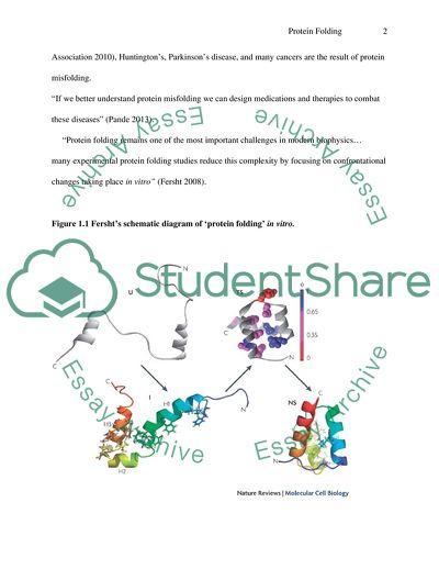
s Mechanisms of Protein Folding In Vitro 2 March Introduction The inquiry approach continues in a vigorous quest to problem solve, brainstorm, troubleshoot. Indeed, inquiry captures the mind’s critical thinking and enduring persistence to answer questions that trouble our modern and postmodern societies. A question posed to the collaborative bioscientists, biochemists, biotechnologists and many others of the biophysical disciplines… What is in vitro protein folding? According to Pande (2013) and collaborative bioscientists, “Defining protein folding… Proteins are biology’s workhorses, its ‘nanomachines’. Proteins help you body break down food into energy, regulate your moods, and fight disease. Before proteins can carry out these important functions, they assemble themselves, or ‘fold’. While protein folding is critical and fundamental to virtually all biology much of the process remains a mystery” (Pande 2013). Further research has indicated that the incorrect folding or misfolding of proteins can result in serious health consequences. Well known diseases such as Alzheimer’s, Mad Cow (BSE), Creutzfeldt-Jacob Disease (CJD – a rare degenerative, invariably fatal brain disorder. NINDS 2012), Amyotrophic lateral sclerosis (ALS – also referred to as Lou Gehrig’s disease, a progressive neurodegenerative disease that affects nerve cells in the brain and spinal cord. ALS Association 2010), Huntington’s, Parkinson’s disease, and many cancers are the result of protein misfolding. “If we better understand protein misfolding we can design medications and therapies to combat these diseases” (Pande 2013). “Protein folding remains one of the most important challenges in modern biophysics… many experimental protein folding studies reduce this complexity by focusing on confrontational changes taking place in vitro” (Fersht 2008). Figure 1.1 Fersht’s schematic diagram of ‘protein folding’ in vitro. (Source of data: Fersht, Readcube 2008) Fersht (2008) gives voice to further insights and dynamics on biophysics “… every breakthrough that opens new vistas also removes the ground from under the feet of other scientists. The scientific joy of those who have seen the new light is accompanied by the dismay of those whose way of life has been changed forever. The publication of the first structures of proteins at atomic resolution fifty years ago astounded and inspired scientists in every field, but caused others to flee or scoff. Advance and subsequent paradigm-shifting breakthroughs and impacts on the field of protein folding have met with some resistance before universal acceptance” (Fersht 2008). Protein Folding Mechanisms In Vitro Fig. 1.2 Anfinsen’s schematic diagram of Hydrophobic collapse, Framework model and Nucleation growth model (Source of information and data: Radford, S.; School of Biochemistry and Molecular Biology University of Leeds, UK). 2013, 2000. The past twenty-five years has revealed substantial breakthroughs in understanding and effective application of ‘protein folding mechanisms’. Radford (2000) of the University of Leeds (Leeds, UK), School of Biochemistry and Molecular Biology, also meaningfully shares on the motivating scientific dynamics of biophysics and protein folding. “… We have reached an exciting stage with consensus beginning to emerge that combines both theoretical and experimental approaches. Furthermore, new fields and disciplines have emerged, including in vitro protein folding and the study of protein misfolding diseases. In today’s post-genomic world, understanding protein folding has never been more important. The topic has wide-ranging impacts in fields from structural biology to materials science” (Radford 2000). Key concepts and terminology used during the biophysics of in vitro analysis of protein folding – hydrophobic collapse, nucleation growth, Framework model, molten globules – are illustrated in Figure 1.2. The Anifinsen Experiments (Fig. 1.2) demonstrate the ‘Framework model’ of protein folding. Experimental observations of rapid formations of secondary protein structures or molten protein globules are analyzed as well as individual secondary structure elements. Subsequently, there is the ‘coalescence and rearrangement’ of secondary structure elements to form the ‘nucleation condensation model’. “This model is supported by protein engineering studies and various theoretical calculations. There is the formation of ‘hydrophobic’ residues along with expansions of the nucleus” (Robinson, 2013). Figure 1.3 offers another schematic diagram of ‘Hydrophobic Collapse; Framework model; Nucleation model’. Figure 1.3: Diagram of Hydrophobic collapse; Framework Model; Nucleation Condensation: Theories of Protein Folding mechanisms (Source of information: Andrzej Kolinski Research Group: The Laboratory of Theory of Biopolymers. Warsaw University. (2011). During biomolecular and biophysical experimentation in the Matysiak Lab (University of Maryland), the Biomolecular Modeling Group conducts modern theoretical and computational studies that construct effective scientific support for experiments toward a thorough description or characterization of protein folding and dynamics mechanisms. Course-grained in vitro protein models for the characterization of folding landscapes are used as descriptions of protein folding processes and frameworks for the interpretation of research study results. (Fig. 2.1). Figure 2.1 Illustration of In Vitro Protein Folding Model Mechanisms (Source of information: The Biomolecular Modeling Group: Matysiak Lab University of Maryland. (2013). The Biomolecular Modeling Group explains the purpose for in vivo modeling. “Recent advances in protein modeling and development of methodologies are allowing theoretical and experimental synergistic characterization of folding landscape. The protein folding landscape can provide us with a theoretical framework that can also be used to characterize the physical and chemical factors regulating protein misfolding mechanisms” (Biomolecular Modeling Group 2013). The Biomolecular Modeling Group further clarifies their recent experimental goals. “Our group develops models and methods to quantitatively map the route from the unfolded protein to the correctly folded state on a protein free energy landscape. The main goal is to investigate how certain perturbations such as mutations or changes in environmental conditions may have dramatic disease-triggering changes on the landscape” (Biomolecular Modeling Group 2013). Lessons Learned from Studying Protein Folding Mechanisms The purposive research studies of in vitro protein folding landscapes are very important tools for researching processes of protein folding. Vital insights and literature reviews have been gained from such studies. Current research methodologies of protein biophysics demonstrate how experiments should be designed. Nickson and Clarke (2010) emphasised “the importance of synergy between experiment and theory… We also stress the importance of choosing the right system carefully; analyzing the essential structural and sequence databases available” (Nickson & Clarke 2010). Important research and insights on the protein folding fields and studies of homologous proteins has originated from investigating the differences between related proteins – proteins within the same families, but with different folding mechanisms; and proteins with different kinetic properties and different folding pathways. “The first importance of these studies is that they have allowed insight into some of the fundamental questions about protein folding mechanisms. These experimental studies have been particularly powerful when they have gone hand-in-hand with computational and theoretical work on folding. Studies of protein folding of families of proteins have been vital in developing our understanding of relative importance of topology, sequence, entropy (space)/enthalpy (energy) balance and secondary structure properties in determining folding mechanisms” (Nickson & Clarke 2010). Most of the experimental analysis about protein folding comes in the form of a ‘snapshot’ or schematic diagram – an experimentally designed structure of a distinct state on the folding pathway. (Figure 2.2). Figure 2.2: An In Vitro ‘Snapshot’ of the Protein Folding Pathway (Source of information: Nickson & Clarke 2010; ScienceDirect 2013) Figure 2.2 is a schematic diagram or ‘snapshot’ of the ‘four classical folding mechanisms’. (1) The ‘Framework model’ demonstrates that the elements of the secondary structure forms first. These then diffuse together, collide and produce the correct tertiary structure; (2) The ‘Hydrophobic Collapse’ model suggests that a protein collapses rapidly around its hydrophobic side-chains and proceeds to restructure or rearrange from the restricted confirmation of the ‘molten globule’ state; (3) The ‘Nucleation Propagation’ model demonstrates that interactions form a small amount of secondary structures which act as a nucleus; (4) The ‘Nucleation Condensation’ model demonstrates the metastable nucleus that is unable to trigger protein folding until a sufficient number of stabilizing interactions have occurred. Once these protein folding processes or mechanisms have occurred, the native protein structure condenses so rapidly that the nucleus is not fully formed and is within a transition state. Protein Folding In Vitro: A Kinetic and Thermodynamic Study According to biophysical research, proteins are the major functional elements in living cells. Amino acids are the primary structures of proteins. For these primary structures to be functional within the body, they will need to fold into tertiary structures which are the optimal form of protein packaging. According to biophysicists Hongxing Lei and Yong Duan of Beijung Institute of Genomics (Chinese Academy of Sciences, Beijing; UC Davis Genome Centre and Department of Applied Science, Davis; College of Physics, Huazhong University of Science and Technology, Wuhan, China), “… the ‘protein folding’ problem is concerning the detailed processes and transition from primary structure to tertiary structure” (Lei & Duan 2011). Designs of in vitro experimentation have focused on ‘protein folding’ as a mechanism concerning two major biophysical issues – kinetics and thermodynamics. During in vitro experimental analysis, thermodynamically, the native state is the most stable, dominant state for proteins. Kinetically, the emergent proteins take different pathways in reaching the native state. Both the thermodynamic and kinetic issues have been extensively and intensely investigated during experimental research as well as theoretical studies. As documented by Lei and Duan (2011), … “The pioneering work by Christian Anfinsen (1957, 1973) led to the creation of the ‘thermodynamic hypothesis’ – the Anfinsen’s Dogma – which states that the native state is unique, stable and kinetically accessible free energy”. Via the Anfinsen Dogma, many research studies have been done to explain kinetically accessible folding pathways. “Towards this end, several well-known theories have been presented. (1) The ‘Framework Theory’ or ‘Diffusion-Collision Theory’ supports that the formation of secondary structure is the initial step and foundation of protein folding; (2) The ‘Nucleation Condensation Theory emphasizes the specific contact as initiation points of both secondary structure formation and protein folding” (Fersht 1995). In the kinetic and thermodynamic theories, the ‘Hydrophobic Hydration Theory’ supports that the repulsion between hydrophobic residues and water environments is what catalyzes the spacial distributions of polar/non-polar residues and, eventually, global protein folding. Recently, the theory known as the ‘Funnel Theory’ is dynamically illustrated as a ‘funnel shape’ in which conformational space – entropy – and conformation energy – enthalpy – gradually decrease during kinetic ‘traps’ in the pathways of global protein folding. The catalytic force behind protein folding is not yet known in the Funnel Theory. Design and Management of Protein Health Via In Vitro Protein Folding Studies In the research Laboratories of William E. Balch, major biophysical goals are being achieved through in vitro protein studies. The key goals of the Balch Laboratory are: (1) to define how membrane trafficking pathways work and are integrated with the ‘proteostasis’ program to generate protein function… Proteostasis is essential to maintain the stem cell environment, to dynamically drive development, to protect us from the environment and pathological challenges that occur daily and during ageing; (2) determine the biochemical and structural roots of protein folding disorders that dramatically impact the human healthspan (ex. Cystic fibrosis) ; and (3) learn how therapeutic management of proteostasis biology can be used to restore the abilities of the misfolded protein to normal function to benefit human lifespan” (Scripps Research Institute 2013). The Balch Laboratory offers an in-depth explanation and exploration into the insights and laboratory experimentation of what is to be considered one of the most dynamic research studies of biophysics. “Through a multidisciplinary approach and application of diverse state-of-the-art cell biological, biochemical, molecular systems and structure approaches, we hope to gain critical insight into the fundamental principles of integrated proteostasis cellular programs… one that manages both protein folding and trafficking and a new understanding of the role of proteostasis in controlling human health and ageing” (Balch & Scripps Research Laboratory Institutes 2013). The studies and analysis are on-going. The Balch/Scripps Laboratory acknowledges that these pathways will enable or further the development of small-molecular PRs that adjust the protein folding environment in order to restore cellular functions and benefit human lifespan. Protein folding in the context of proteostasis and multidimensional approaches are used to address the critical role of proteostasis in human health and disease. Figure 3.1: Protein Folding In Vitro Simulation (Source of diagram: Lei & Duan. 2011). Historical Research Study and Analysis: Principles that Govern Folding of Protein Chains by Christian B. Anfinsen During research analysis on protein folding, biophysicist Christian Anfinsen meaningfully shares in vitro observations on the ribonuclease molecule. Excerpt from the Anfinsen Experiments, 1973… “The telegram that I received from the Swedish Royal Academy of Sciences specifically cites… ‘Studies on ribonuclease, in particular, the relationship between the amino acid sequences and the biologically active conformation…’. The work that my colleagues and I have carried out on the nature of the process that controls the folding of polypeptide chains into the unique three-dimensional structures of proteins was, indeed, strongly influenced by observations on the ribonuclease molecule. Many others, including Anson and Mirsky in the 1930’s and Lumry and Eyring in the 1950’s had observed and discussed the reversibility of denaturation of proteins… The original observa- tions that led to this conclusion were made together with my colleagues Michael Sela and Fred White 1956-1957. These were, in actuality, the beginnings of a long series of studies that rather vaguely aimed at the eventual total synthesis of the protein. As we all know, Gutte and Merrifield at the Rockefeller Institute and Ralph Hirschman and his colleagues at the Merck Research Institute have now accomplished this monumental task. (from the desk of C. Anfinsen, 1957, 1973). Further Analysis on Mechanisms of Protein Folding: In Search of Answers (Pande Lab 2012) Inquiry into the research studies experimental analysis, and purpose of research (answering the inquiry… why?) on the mechanisms of protein folding is a dynamically growing method in the search for cures and mechanisms for ‘protein folding diseases’ of this postmodern era. Defining the purpose of proteins is an ongoing study. According to research descriptions and definitions of Vijay Pande (2012) of Stanford University, and interesting comparison is constructed… “proteins are necklaces of amino acids – long chain molecules that come in a variety of shapes and sizes” (Pande 2012). Proteins are a foundational basic for biological studies used in saving lives and getting things done. As enzymes, proteins are a vital catalyst in all of the biochemical reactions which enable biology to function and work. As the basic structures within the body, proteins are the main constituents of bone, muscles, hair, skin and blood vessels. As the structural elements of antibodies, proteins enable the immune system to attack viral or other invasive antigens. Proteins enable muscle movement and processes signals from the sensory system. It is for the study of proteins that biophysicist V. Pande and collaborative scientists have conducted biochemical and biophysical studies on sequencing of the human genome…the genome, the blueprint for all of the proteins in the biological discussion. The purposive research studies of V. Pande and colleagues is on how and why to understand the function of proteins. Amino acid sequence is not enough to determine and calculate the function of specific proteins. In order for antibodies and enzymes to carry out bodily functions, they must take on the particular shape necessary to function; this is known as ‘folding’ In vitro experimental studies demonstrate how protein, nucleotides go through folding processes while functioning as the enzymatic amino acids and antibodies. According to V. Pande, “… out of an astronomical number of possible ways to fold, a protein can pick one with microseconds… and intriguing biological mystery. One of our project goals is to simulate folding in order to understand how proteins fold so quickly and reliably. Our goal is to learn about what happens when ‘misfolding’ occurs” (Pande 2012). Protein Folding and Disease Misfolding of the protein, nucleotide structures result in disease in the living body. Diseases such as BSE (Mad Cow disease), Alzheimer’s disease, cystic fibrosis, forms of inherited emphysema, and many forms of cancer are the outcomes of protein misfolding. Misfolding causes proteins to ‘clump or aggregate’. In turn, these aggregated protein globules can gather in the brain where the causes the symptoms of Mad Cow or Alzheimer’s disease. The processes of ‘protein folding’ are complex and challenging during laboratory study. The processes of protein folding occur so quickly that observation of ‘self-assembly’, as Pande (2012) refers to the folding, is the challenge of experimental analysis. The process occurs in approximately a millionth of a second. Quite an analysis. Protein folding and misfolding of protein in vitro: The Horizon Symposium 2003 An international panel of experts was brought together to collaborate and discuss current thinking on protein folding research, from basic cell biology to therapeutic strategies for treating protein folding diseases. Among the panel of experts were researchers, biophysicists and scientific writers from the University of Cambridge (UK), Brigham and Women’s Hospital (Boston), Harvard Medical School, The Nature Publishing Group, University of San Francisco, and Brescia, Italy. Christopher M. Dobson (Department of Chemistry, University of Cambridge, United Kingdom) brings together discussion threads of the 2005 Meetings or Horizon Symposium in order to give a personal ‘snapshot’ of the state of work and play in this dynamically exciting biophysical interdisciplinary field. As enlightened during the Horizon Symposium, the topic of ‘protein folding and disease’ was discussed under the topic of what is broadly accepted as ‘folding and misfolding of proteins in vitro. As researchers have studied, “… It is generally accepted that the native states of protein is the thermodynamically most stable of the polypeptide chain under physiological conditions. Furthermore, it is generally accepted that the three-dimensional structure of the protein structure and the fundamental mechanism of folding is encoded in the amino-acid sequence” (Dobson 2003). During in vitro studies, ‘new views’ or ‘snapshots’ of the protein folding mechanisms to be revealed or uncovered without the complications of the biological environment, according to Dobson (2003). Chris Dobson further clarifies that these experiments involve the unfolding of the protein in a denaturant such as urea and probing of the unfolding process using biophysical methods. Such experimental methods were revolutionized by the introduction of the protein engineering approach. This method or approach involved in vitro observations in which the effects of mutations of the kinetics and thermodynamics of folding can provide considerable insights in how amino acid sequence encodes the fold. The studies and experiments of the protein folding field have been intensely influenced by the development of theoretical approaches including Monte Carlo and molecular dynamics simulation in vitro techniques. “Given that we are beginning to get to grips with the physical basis of the folding process, the key question is how biology has regulated or controlled the intrinsic ability of some polypeptide sequences to fold to specific structures. The key difference between in vitro and in vivo folding, in the broadest sense of the word, is that biology has evolved mechanisms to control and regulate the whole process” (Dobson 2003). Yet, as answer, the understanding of chemistry is enabling the rational design of drugs to combat the expanding family of ‘confrontational’ diseases. Works Cited Andrzej Kolinski Research Group. The Laboratory of Theory of Biopolymers (Warsaw University). Theories of Protein Folding Mechanisms: The three classical mechanisms of protein folding. (2011). Retrieved from http://biocompchem.uw.edu/pl/ news.php. [WEB]. ALS Association. Create a world without ALS. What is ALS? (2010). Retrieved from http://www.alsa.org/about-als/what-is-als.html. [WEB]. Anifinsen, C. B. Principles that govern the folding of protein chains. Science. (1973). Retrieved from http://cdn.intechopen.com/pdfs/20173/InTech-Kinetics_and_ thermodynamics_of_protein_folding.pdf. [WEB]. Biomolecular Modeling Group. Matysiak Lab (University of Maryland). Course-grained protein models for the characterization of folding landscapes. (2013). Retrieved from http://biocomp.chem.uw.edu.pl/news.php. [WEB]. Dagget, V. & Fersht, A. Protein folding and binding: Moving into unchartered territory. (2009). Retrieved from http://dx.doi.org/10.1016/j.sbi.2009.01.006 [WEB]. Dobson, C. Protein folding diseases. Horizon Symposium: Connecting Science to Life. Retrieved from http://www.nature.com/horizon/proteinfolding/background/disease.html Fersht, A. R. ReadCube: From the first protein structures to current knowledge of protein folding: delights and skepticisms. (2008). Nature Reviews; Molecular Cell Biology. Retrieved from http://www.readcube.com/articles/10.1038/nrm2446.[WEB]. Jackson, S.E. (University of Cambridge, UK). Protein Folding, Engineering of. Encyclopedia of Condensed Matter Physics. (2005). Retrieved from http://dx.doi.org/10.1016/BO-12-369401-9/00382-X. [WEB]. Lei, H. & Duan, Y. Kinetics and Thermodynamics of Protein Folding. (2011). Intech: open science/ open minds. Retrieved from http://www.intechopen.com/ Books/thermodynamics-kinetics-of-dynamic-systems/kinetics-and-thermodynamics.[WEB]. Moore, R.; Taubner, L. & Priola, S. Prion protein misfolding and disease. (2009). Retrieved from http://dx.doi.org/10.1016/j.sbi.2008.12.007. [WEB]. National Institute of Neurological Disorders and Stroke (NINDS). Creutzfeldt-Jakob Disease Fact Sheet: National Institute of Health. (2012). Retrieved from http://www.ninds.nih.gov/disorders/cjd/detail_sjd.htm. [WEB]. Nickson, A. & Clark, J. (University of Cambridge: Department of Chemistry: MCR Centre for Protein Engineering). What lessons can be learned from studying the folding of homologous proteins? (2010). Methods. Retrieved from http://dx.doi. org/10.1016/BO-12-369401-9/00382-X. [WEB]. Pande, V. What if you could help find a cure? (2013). Retrieved from http://folding. stanford.edu/[WEB]. Pande, V. (Stanford University). The sequence behind Folding ‘at’ home (2000-2012). Retrieved from http://folding.stanford.edu/English/Science. [WEB]. Radford, S.E. & Elsevier, B. N. (School of Biochemistry and Molecular Biology: University of Leeds, UK). Trends in Biomedical Sciences – Protein folding: Progress made and promises ahead. (2013, 2000). ScienceDirect; SciVerse. Retrieved from http://www.sciencedirect.com/science/article/pii/SO9680004000 17072 [WEB]. Robinson, D. Protein Folding and Biospectroscopy (Lecture 4) 2013. Retrieved from http://robinson.chem.nottingham.ac.uk/teaching/F14PFB/lecture4.pdf. [WEB]. Scripps Research Institute. Design and Management of Protein Health: The Laboratory of William E. Balch. (2013). Retrieved from http://www.scripps.edu/balch/ [WEB]. Travaglini-Allocatelli, C.; Ivarsson, Y.; Jeinth, P.; Gianni, S. Folding and stability of globular proteins and implications for function. (2009). Current Opinion in Structural Biology. Retrieved from http://www.sciencedirect.com/science/article/pii/ SO959440X08001759. [WEB]. Read More
Cite this document
  • APA
  • MLA
  • CHICAGO
(Mechanisms of Protein Folding In Vitro Essay Example | Topics and Well Written Essays - 3000 words, n.d.)
Mechanisms of Protein Folding In Vitro Essay Example | Topics and Well Written Essays - 3000 words. https://studentshare.org/biology/1795771-review-the-mechanisms-of-protein-folding
(Mechanisms of Protein Folding In Vitro Essay Example | Topics and Well Written Essays - 3000 Words)
Mechanisms of Protein Folding In Vitro Essay Example | Topics and Well Written Essays - 3000 Words. https://studentshare.org/biology/1795771-review-the-mechanisms-of-protein-folding.
“Mechanisms of Protein Folding In Vitro Essay Example | Topics and Well Written Essays - 3000 Words”. https://studentshare.org/biology/1795771-review-the-mechanisms-of-protein-folding.
  • Cited: 0 times

CHECK THESE SAMPLES OF Mechanisms of Protein Folding In Vitro

The function and mechanism of AlkB

The mechanism also verified through in vitro assays on purified AlkB involves the binding of Fe2 and 2-oxoglutarate to the enzyme core followed by binding of methylated middle base to the ‘lid'.... Investigations on crystal structure and mechanism of AlkB have involved the use of both native and recombinant protein.... Recombinant enzyme, entire as well as truncated form is produced through homologous expression; the former cloned in pET21b vector and expressed in BL21 (DES) competent cells; … Investigations on crystal structure and mechanism of AlkB have involved the use of both native and recombinant protein....
4 Pages (1000 words) Research Paper

Adipose Tissue and Resveratrol

Dasgupta and Milbrandt (2007) show that both in vivo and in vitro, Resveratrol increases the activity of neuronal Adenosine Monophosphate-activated Kinase (AMPK).... This decrease in the decrease in protein synthesis, specifically during the S phase of mitosis, may be attributed to Resveratrol's antioxidant activity....
2 Pages (500 words) Essay

Main Types of End Point Used in Toxicity Testing in Cellular System

As a result, other non-animal solutions were developed, which use mostly cells and cultures in vitro, and in turn can expect faster results that better resemble human cellular processes (Andersen and Krewski, 2009).... Still, the use of cell lines in cell-based toxicity screening assays seem promising through the use of in vitro tests with increased loads in assay while using viable cells such as primary cells, immortalised cell lines, human cells and stem cell-derived systems, and these can improve both outcome paces and predictive value of the tests in relation to human cell system toxicity (Basketter, et al....
4 Pages (1000 words) Essay

The Amyloid Fibrils Formation

In pH 5.... and 7.... , the fluorescence of tyrosine remained unchanged while at pH 9.... intensity decreased significantly after 40h indicating conformational change and/or quenching of tyrosine fluorescence.... Overall fluorescence of tyrosine at this pH remained lower than at other two pH values tested (Fig 1....
7 Pages (1750 words) Lab Report

Necrotizing Ulcerative Gingivitis

Melnick and coworkers investigates the susceptibility of necrotizing ulcerative gingivitis and in doing so they build up their study on the knowledge that impaired immune defense mechanisms and genetic factors play a role in susceptibility of this disease.... Introduction: Necrotizing ulcerative gingivitis or acute necrotizing ulcerative gingivitis occurs most frequently in young adults of 16 to 30 years of age....
5 Pages (1250 words) Essay

New Wine in a New Bottle

The essay "New Wine in a New Bottle" takes us further in our journey towards an understanding of the mechanisms involved in alcohol dependence and tolerance of drugs like diazepam.... Lead investigator Jing Ling and her colleagues at the University of California provide another step forward towards a better understanding of the mechanisms involved in ethanol abuse.... In this study, the investigators set out to find out whether the function and the subunit composition of GABAARs could be changed by just one intoxicating of ethanol and study the mechanisms involved in these changes with regard to the hippocampal CAI and dentate gyrus regions....
7 Pages (1750 words) Essay

Disrubtion of the tight junction ZO-1 by cytokine in intestinal epithelial and probiotic treatment

(2011) found in an in-vitro study that pre-treatment of intestinal cells with the probiotic Lactobacillus casei resulted decreased consequences of inflammation.... It is present in various types of epithelial Disruption of Tight Junction ZO by Cytokines in Intestinal Epithelial Cells and Probiotic Treatment Zonula occludens -1 (ZO-1) was discovered in 1986 as the first intracellular protein component of the tight junction (Bazzoni and Dejana, 2004).... The junction-associated protein, zonula occludens-1, localizes to the nucleus before the maturation and during the remodeling of cell- cell contacts....
2 Pages (500 words) Essay

Caveolin as the Principal Marker of the Caveolae

Since the caveolin serve as the caveolae coat protein, the caveolin is considered as a major element of the caveolae which represents as… Commonly found in the caveolae, caveolins play a significant role in the process of receptor-independent endocytosis (Williams and Lisanti; Tang, Scherer and Okamoto; Jasmin, Frank and Lisanti 6). Caveolin has a ID Number & Caveolin protein As a good example of scaffolding protein, the caveolin is considered as the principal marker of the caveolae (Gussak and Antezelevitch 234)....
1 Pages (250 words) Essay
sponsored ads
We use cookies to create the best experience for you. Keep on browsing if you are OK with that, or find out how to manage cookies.
Contact Us