StudentShare
Contact Us
Sign In / Sign Up for FREE
Search
Go to advanced search...
Free

Muscular Tissue Types and Nervous Tissue Types - Essay Example

Cite this document
Summary
The "Muscular Tissue Types and Nervous Tissue Types" paper summarizes how the bright field light microscope works, the steps in preparing a tissue section, ready for mounting on a microscope slide and ready for staining, and outlines the Haematoxylin and Eosin staining method and the results…
Download full paper File format: .doc, available for editing
GRAB THE BEST PAPER95.7% of users find it useful
Muscular Tissue Types and Nervous Tissue Types
Read Text Preview

Extract of sample "Muscular Tissue Types and Nervous Tissue Types"

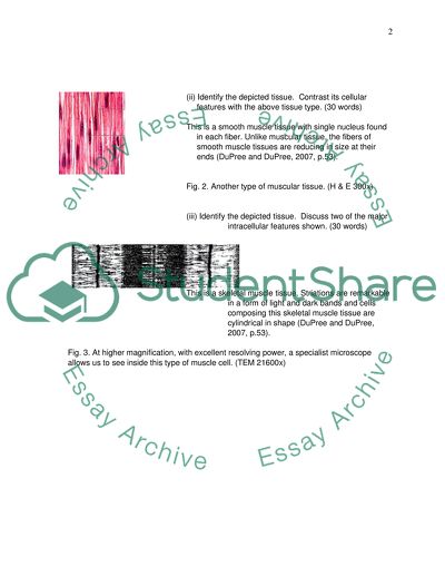
1 a. Summarize how the bright field light microscope works. Bright field microscope is used in simple illumination technique to observe samples like cells and tissues that are no longer alive. The principle behind this optical illumination is to allow light to illuminate dense areas of samples, providing contrasting effect making them visible as image ready to be magnified (Karp, 2009, p.718). In most cases, illumination technique by bright field light is made possible by applying stains to improve contrast by absorption by specimens. b. Summarize the steps in preparing a tissue section, ready for mounting on a microscope slide and ready for staining. Tissue sample removed from its immediate source should be fixated with fixing fluids like acetic acid, formaldehyde or ethanol in order to prevent it from being digested by enzymes present in it. Fixation therefore preserves protoplasm, prevents autolytic changes and bacterial growth (Pawley, 2006, p. 370). After this, tissue sample should undergo dehydration, clearing and embedding. These processes ensure tissue sample is dehydrated or cleared from fixated agents and solidified to provide strong support to tissue blocks to make cutting in thin sections possible (Pawley, 2006, p.370). c. Outline the Haematoxylin and Eosin staining method, its purpose and the results. In bright field light microscopy staining for routine histological examination of tissue sections enhances natural contrast making tissues or cells more visible. Haematoxylin reacts with basophilic components of cells producing bluish tint to dark blue stain, Eosin stains pinkish colour to pink of acidophilic components of cells (Bancroft and Gamble, 2008, p.126). 2.) a. Muscular Tissue Types (i) Identify the depicted tissue. Discuss at least two (2) major cellular features. (30 words) This is a cardiac muscle tissue. Its fibers are joined by intercalated discs, thick plasma membranes, shown as horizontal lines in the picture (DuPree and DuPree, 2007, p.53). The single central nucleus is also visible. Fig. 1. These cells can communicate electrically with each other. (H&E 600x) (ii) Identify the depicted tissue. Contrast its cellular features with the above tissue type. (30 words) This is a smooth muscle tissue with single nucleus found in each fiber. Unlike muscular tissue, the fibers of smooth muscle tissues are reducing in size at their ends (DuPree and DuPree, 2007, p.53). Fig. 2. Another type of muscular tissue. (H & E 300x) (iii) Identify the depicted tissue. Discuss two of the major intracellular features shown. (30 words) This is a skeletal muscle tissue. Striations are remarkable in a form of light and dark bands and cells composing this skeletal muscle tissue are cylindrical in shape (DuPree and DuPree, 2007, p.53). Fig. 3. At higher magnification, with excellent resolving power, a specialist microscope allows us to see inside this type of muscle cell. (TEM 21600x) (Question 2 Continued) b. Nervous Tissue Types (i) State the system, organ and two types of tissues seen. (30 words) This is a central nervous system, particularly a spinal cord formed by nervous tissue. Neurons and neuroglia are remarkable in this image showing the interconnecting cells and their supports (Chiras, 2011, p.78). Fig. 4. An organ, sectioned, stained and shown using a wide view objective. (Luxol fast blue/cresyl violet 20x TM) (ii) Discuss the role of at least three cellular features depicted. (30 words) This is a neuron which contains nucleus in its cell body that is responsible for synthesizing proteins and membranes while the axon conducts electrical impulse while the dendrites collect information (Hollenbeck and Bamburg, 2003). Fig. 5. The digital camera allows us to enlarge certain cellular features, while still resolving the fine detail of the cell. (H&E 400x TM, enlarged) (iii) This is a human spinal ganglion. Discuss satellite cells, their role and arrangement in a ganglion. (30 words) Satellite cells are situated in dorsal root ganglia somewhere in either side of the spinal cord and they enclosed cell bodies containing sensory neurons that are found inside the ganglia (Tiidus, 2008, p.77). Fig. 6. Tissue sections allow us to infer details about the function of individual cell types. (H&E Methylene blue 400x TM, enlarged) (Question 2 Continued) c. Connective Tissues Types Ordinary staining technique could not enhance visibility of the collagen fibres of hyaline cartilage (Amitrano and Tortora, 2012, p.65). Protoplasm and nuclei under intranuclear network are found in this specimen. Figure 7. Hyaline cartilage. (Picrosirius-hematoxylin stain. Medium magnification) Silver and gold methods are commonly applied to enhance visibility of reticular connective tissue (Young, 2006). Interlacing reticular fibers and reticular cells form network (Schwartz, 2012). Figure 8. Reticular connective tissue (Bielschowsky’s Silver Method. Higher Magnification) Giemsa method of staining can be effective when applied in bone connective tissues (Young, 2006). This image shows collagenous connective tissue matrix saturated with salts, and osteoblasts forming osseous tissue. Figure 9. Membrane bone mandible (Bergman et al., 2012) (Cat, Müllers* fluid, H. & E., A. 162 x; B. & C. 1416 x.) (Question 2 Continued) d. Epithelial Tissues H&E could readily give better result for absorption and magnification (Young, 2006). This image shows tubule edge and basement membrane stains with darker pink (Bernd, 2012). Figure 10. Kidney medulla with simple cuboidal epithelium lining the lumina of tubules (H.E. Medium Magnification) H&E gives better optical illumination for this tissue (Young, 2006). The nuclei are found in the cell’s base. The cells are then connected by tight connections (Bernd, 2012) Figure 11. Simple columnar epithelium (H.E. Medium Magnification) (Blue Histology, 2012) H&E staining gives better optical illumination for this tissue (Young, 2006). This image depicts glomerulus and Bowman’s capsule line with simple squamous epithelial tissue (Bernd, 2012). Figure 12. Simple squamous epithelium (H.E. Medium Magnification) (Blue Histology, 2012) 3.) Blood vessel histology from the cardiovascular system - Artery Artery is one of the blood vessels carrying blood from the heart to all body tissues. With this very critical function, it is not surprising this blood vessel’s structure is highly complex. Artery has four layers and in the middle and outer part of it are connective tissues (Singh, 2011, p.178). In the middle part of artery are connective tissues that are composed of elastic and collagenous fibres. The same components of connective tissues can be found outside of the artery. However, in the external part, irregular connective tissues are remarkable which also contains elastic and collagenous fibres. The connective tissues that are situated somewhere at the external part are connected with smooth muscle tissues. In the same way, the internal connective tissues are also connected with smooth muscle tissues (Singh, 2008, p.150). Artery is also composed of epithelial tissues known as endothelium (Singh, 2011, p.178). This tissue is found somewhere in the internal part as lining, connecting next to connective tissues. In other words, the connective tissues found somewhere in the middle part of artery are situated between endothelium and smooth muscle tissues. With this framework or structure of the artery, it is therefore built with flexibility to adjust with heart’s activity and constant flow of blood. As stated, artery is also composed of smooth muscle tissues aside from connective tissues and epithelial tissues. This tissue is indeed relevant to the artery most especially in ensuring to maximize its function to carry blood from the heart to all body tissues. Smooth muscle tissues are directly linked with both and between elastic and collagenous fibres of regular and irregular connective tissues. As the artery is always sets in motion as it constantly channels blood for distribution in the circulatory system, the combined characteristics of smooth muscles and connective tissues are the right designed to ensure both elasticity and strength. In the blood vessels and in particular in the case of arteries, blood has to travel long distances in it, and it should also ensure vasoconstriction or vasodilation as its important capability in order to alter blood flow. This capability of artery to alter blood flow is tantamount to the actual design or structure of tissues composing it. With the combined elasticity and strength, found in the integrated characteristics of smooth muscle tissues and connective tissues, altering blood flow and blood travel for long distances are at their optimum level ensuring that the cardiovascular activity of the heart would continue to distribute blood to each tissue of the body. The artery with its intricate structure and design therefore has a special function in the cardiovascular system and even in all related body systems involving important body organs. Respiratory histology – Lungs The lungs have elastic connective tissue. This can be visibly and individually found around the alveoli or trachea as it holds blood vessels. In its actual image, alveoli are nothing but just like empty spaces surrounded by tissues. These are connective tissues that are also connecting to arteries and veins (Singh, 2011, p.226). Connective tissues play a significant role in keeping the structure of the lungs, while it helps to ensure the organ’s framework in order to guarantee optimum function of the flow of air, blood, and nutrients. Connective tissues are therefore not only creating a framework for the lungs, they also stand as to give individual structure for a specific part of the organ, such as the alveoli, in order to help optimize its function. The epithelial tissue lines the trachea and bronchi. Its two main cells include ciliated cells and goblet cells (Singh, 2011, p.226). They sit on basement membrane. This contains fibres made from proteins. Beneath that membrane the cells secret protein. As proteins are secreted in here epithelia are therefore responsible for forming the lung structure, particularly of the alveoli or air sacs where the actual gas exchange takes place. In other words, epithelial tissues are important linings of the trachea and bronchi in order to ensure strong framework of the lungs where the actual exchange of gas occurs (Singh, 2011). In the lungs, there are also veins and arteries where blood continually flows. These arteries are made of connective tissues inside and outside while they cover the smooth muscle tissue. This clearly shows that in order for the lungs to function well, connective tissues and smooth muscle tissues at some point have to work in a coordinated manner. As the lungs cannot work without oxygen, arteries therefore are essential in their general function as they carry blood with full oxygen. The supply of fresh oxygen and blood would ensure the lung could optimize its function while it tries to perform gas exchange. As stated earlier, the lungs have elastic connective tissues that are all over the said organ. The lungs would shrink and expand respectively as an organism inhales and exhales. If connective tissues are not elastic enough for this activity, then the lungs could not perform successful gas exchange. It therefore makes sense that an elastic connective tissues are around the lungs especially all over air sacs or alveoli where the actual exchange of gas occurs. Air sacs can be more flexible in the actual exchange of gas because of the surrounding elastic connective tissues and prolific epithelial tissues that line the trachea and bronchi. Arteries also perform certain flexibility as they are made up of connective tissues and smooth muscle tissues. References Amitrano, R. J. and Tortora, G. J. (2012) Anatomy and Physiology Laboratory Manual: Update. 7th ed. Belmont: Cengage Learning. Bancroft, J. D., and Gamble, M. (2008) Theory and Practice of Histological Techniques. 6th ed. Philadelphia: Elsevier Health Sciences. Bergaman, R. A., Afifi, A. K., and Heidger, P. M. (2012). Connective Tissue. [online]. Retrieved from http://www.anatomyatlases.org/MicroscopicAnatomy/Section03/Section03.shtml [Accessed 19th October 2012]. Bernd, K. (2012). Epithelial Cells. [online]. Retrieved from http://www.bio.davidson.edu/people/kabernd/BerndCV/Lab/EpithelialInfoWeb/Simple%20Cuboidal%20Epithelium.html [Accessed 19th October 2012]. Blue Histology (2012) Tissue Study. [online]. Retrieve from http://www.carlalbert.edu/index.php?id=496 [Accessed 19th 2012]. Chiras, D. (2011) Human Biology. 7th ed. Sudbury: Jones & Bartlett Learning. DuPree, J. R., and DuPree, P. (2007) Anatomy & Physiology Workbook For Dummies. Hoboken: John Wiley & Sons. Hollenbeck, P. J., and Bamburg, J. R. (2003) Neurons: Methods and Applications for the Cell Biologist. Danvers: Academic Press. Karp, G. (2009) Cell and Molecular Biology: Concepts and Experiments. 6th ed. West Sussex: John Wiley & Sons. Pawley, J. (2006) Handbook of Biological Confocal Microscopy: Language of Science. 3rd ed. Madison: Springer. Schwartz, S. L. (2012) Anatomy and Physiology. [online]. Retrieved from: http://faculty.irsc.edu/faculty/jschwartz/Lab%20Notes%20edited%202.htm [Accessed 19th October 2012]. Singh, I. (2011) Textbook of Human Histology: With Colour Atlas & Practical Guide. 6th ed. Panama: JP Medical Ltd. Singh, I. (2008) General Anatomy. Noida: JP Medical Ltd. Tiidus, P. M. (2008) Skeletal Muscle Damage and Repair. Auckland: Human Kinetics. Young, B. (2006) Wheater’s Functional Histology: A Text and Colour Atlas. 5th ed. Philadelphia: Elsevier Health Sciences. Read More
Cite this document
  • APA
  • MLA
  • CHICAGO
(“Human Biology Essay Example | Topics and Well Written Essays - 1500 words”, n.d.)
Human Biology Essay Example | Topics and Well Written Essays - 1500 words. Retrieved from https://studentshare.org/biology/1605814-human-biology
(Human Biology Essay Example | Topics and Well Written Essays - 1500 Words)
Human Biology Essay Example | Topics and Well Written Essays - 1500 Words. https://studentshare.org/biology/1605814-human-biology.
“Human Biology Essay Example | Topics and Well Written Essays - 1500 Words”, n.d. https://studentshare.org/biology/1605814-human-biology.
  • Cited: 0 times

CHECK THESE SAMPLES OF Muscular Tissue Types and Nervous Tissue Types

Clinical and Metabolic Consequences of Type 1 and Type 2 Diabetes

Type 2 Diabetes Mellitus on the other hand is a progressive disease that results from a set of complex metabolic disorders originating from coexisting defects in multiple organ sites that include insulin resistance in muscle and adipose tissue, gradually reducing pancreatic insulin secretion, lack of regulation in hepatic glucose production, inappropriate glucagons secretion and reduced production of gastrointestinal incretins (Barr, Myslinski & Scarborough, 2008)....
8 Pages (2000 words) Term Paper

The Co-Ordination of the Human Body by the Endocrine and Nervous System

This paper "The Co-Ordination of the Human Body by the Endocrine and nervous System" is being carried out to overview the nervous system in humans, discuss the integration of endocrine and nervous system (overlapping of functions) and the role of the hypothalamus and pituitary gland.... The endocrine system is instrumental in regulating metabolism, growth, development, and puberty, and tissue function and also plays a part in determining mood....
10 Pages (2500 words) Research Paper

Diabetes, complications and solutions in Chiropody/Podiatry care

There are two types of diabetes called Type 1 and Type 2 diabetes.... Diabetes mellitus is a condition whereby the amount of glucose or sugar in the blood is more than what the hormonal insulin could convert into energy or fuel needed by the body.... Type 1 diabetes is developed when....
20 Pages (5000 words) Essay

The Function and Responsibility of the Nervous Tissue

This essay concerns the functions of nervous tissue.... Hence, it is stated that the function and responsibility of the nervous tissue is to sense stimuli and transmit signals to and from different parts.... It is also highlighted that the basic unit of nervous tissue is neuron.... The function and responsibility of the nervous tissue is to sense stimuli and transmit signals to and from different parts.... The basic unit of nervous tissue is neuron....
2 Pages (500 words) Essay

The Different Characteristics of the Specie

Translocations are of two types that are reciprocal translocations and Robertsonian translocations.... Inversions are of two types paracentric and pericentric.... The paper 'The Different Characteristics of the Specie' focuses on many species that have been recognized and each of these species has a distinct set of characteristics....
8 Pages (2000 words) Case Study

The Debate about Stem Cells

The use of stem cells is a path-breaking discovery in medical science, and embryonic stem cells have the versatility to transform into almost any type of tissue in the body.... tem cells are extremely effective in curing certain terminal diseases, and diseases resulting from damage to the nervous system and the brain (Medical uses of stem cells).... ardiac problems, muscular dystrophy, kidney ailments, cancer, and other such diseases, have proved to be extremely detrimental to the physical and psychological well being of humans....
5 Pages (1250 words) Research Proposal

Effect of Aggrandizing Working of the CVS

The paper "Effect of Aggrandizing Working of the CVS" presents that the nervous system has 4 main parts the central nervous system (CNS) and the peripheral nervous system (PNS), somatic and autonomic nervous systems.... The CNS consists of two parts: the brain enclosed in the cranium....
11 Pages (2750 words) Case Study

Muscular Dystrophy - Clinical Monitoring Methods

ypes of Genetic Lesion for Muscular DystrophySome of the notable genetic lesions that cause the different types of muscular dystrophy include; the cystic lesion, which is the main cause of the congenital muscular dystrophy.... The paper 'muscular Dystrophy - Clinical Monitoring Methods, the Genetic and Non-Genetic Analysis Methods' is a convincing version of the essay on health sciences & medicine.... Duchenne and Becker MD are the most common muscular dystrophies that are recognized by clinical methods....
6 Pages (1500 words) Essay
sponsored ads
We use cookies to create the best experience for you. Keep on browsing if you are OK with that, or find out how to manage cookies.
Contact Us