StudentShare
Contact Us
Sign In / Sign Up for FREE
Search
Go to advanced search...
Free

Tissue Culture and PCR - Essay Example

Cite this document
Summary
This work called "Tissue Culture and PCR" describes PCR techniques that use heat to change temperature through the different cycles. The author outlines the requirements for any basic PCR technique are thus, two primers, DNA template, Taq polymerase, divalent cations, a buffer, deoxynucleotide triphosphates, and a monovalent cation…
Download full paper File format: .doc, available for editing
GRAB THE BEST PAPER95.4% of users find it useful
Tissue Culture and PCR
Read Text Preview

Extract of sample "Tissue Culture and PCR"

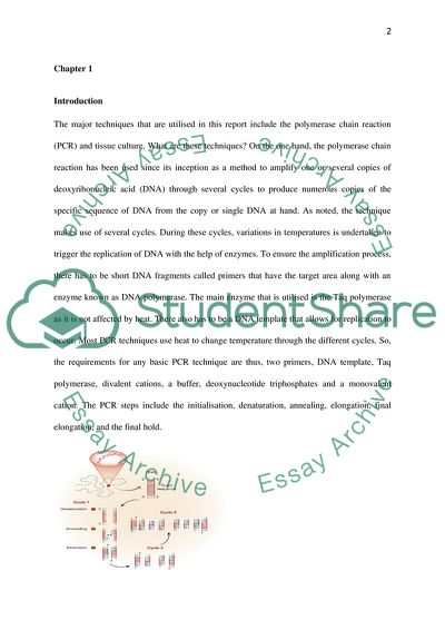
Tissue Culture and PCR Contents Chapter 2 Introduction 2 Chapter 2 5 Materials and Methods 5 Chapter 3 7 Section 7 Tissue culture 7 Section 2 10 Polymerase Chain Reaction 10 Chapter 4 16 Discussion and Conclusion 16 References 18 Chapter 1 Introduction The major techniques that are utilised in this report include the polymerase chain reaction (PCR) and tissue culture. What are these techniques? On the one hand, the polymerase chain reaction has been used since its inception as a method to amplify one or several copies of deoxyribonucleic acid (DNA) through several cycles to produce numerous copies of the specific sequence of DNA from the copy or single DNA at hand. As noted, the technique makes use of several cycles. During these cycles, variations in temperatures is undertaken to trigger the replication of DNA with the help of enzymes. To ensure the amplification process, there has to be short DNA fragments called primers that have the target area along with an enzyme known as DNA polymerase. The main enzyme that is utilised is the Taq polymerase as it is not affected by heat. There also has to be a DNA template that allows for replication to occur. Most PCR techniques use heat to change temperature through the different cycles. So, the requirements for any basic PCR technique are thus, two primers, DNA template, Taq polymerase, divalent cations, a buffer, deoxynucleotide triphosphates and a monovalent cation. The PCR steps include the initialisation, denaturation, annealing, elongation, final elongation, and the final hold. Figure 1: Diagrammatic representation of the PCR principle (Garibyan & Avashia, 2013) On the other hand, tissue culture is a technique whose aim is to evaluate the environments in which the tissue life can be extended outside that in the ideal organism. There are two tissue culture types. They include the primary and the culture of established cell lines. Primary cultures are made up of cells that are directly obtained from organisms or complex tissue slices. While established cell lines cultures are made up of cells obtained from transformed cells in vitro or from tumours (Mather & Roberts, 1998). The counting of cells is done using a haemocytometer, which is made up of an exceptional glass slide that has a precisely ruled grid with accurate measurements. It is used to count Chlorella cells and even blood cells but it is not suitable for counting of bacteria cells. It is used to count both dead and living cells or viable and non-viable cells that can be distinguished using distinctive staining procedures. The number of cells is counted per mm 3 is counted using the formula (N/80) × 4,000. N is the cell number counted in 80 small squares (Adds & Larkcom, 1999) Further, Ribonucleic acid (RNA) is extracted to give nuclear or cytoplasmic RNA. The process of extraction involves the lysis of cells with the help of buffers. The buffers that are mainly used include those that have tough agents like sodium dodecyl sulphate (SDS) and those that are gentle like hyptonic Nonidet P-401 (Farell, 2010). The gentle buffers ensure that the integrity of the nucleus is maintained. After cell lysis, the cell and protein debris is then removed by centrifugation. Enzyme inhibitors that inhibit nucleases are included in the buffer while ensuring proper handling of extracted RNA. The outward exhibition of genes through genetic transcription and translation is termed as gene expression. Gene expression analysis does exist to evaluate the gene pattern expressed at the gene transcription level under certain conditions in a cell (NCBI, 2015). According to Mahmood and Yang (2012), the western blot method makes use of three essentials to locate particular proteins from an intricate protein mixture isolated from cells. The three elements are separation through size, relocation to a membrane, and marking of the target protein utilising an appropriate primary and secondary antibodu for observation (Mahmood & Yang, 2012). Tunimayacin is a nucleoside antibiotic that has uracil. It also has a long fatty-chain acid, tunicamine that is a carbon amino-deoxydialdose, and N-acetylglucosamine (Montreuli, et al., 1995). The antibiotic is derived from Streptomyces lysosuperficus (CIBA Foundation Symposium, 2008). It is formed as a mixture of compounds that are very much related but differ by structure and size of the chain of fatty acids. The antibiotic has been shown to hinder asparagine-linked precursor oligosaccharide formation. It is from this oligosaccharide that every N-linked oligosaccharide is obtained through hindrance of the initial step in the dolichol pyrophosphate oligosaccharide pathway. Hence, all proteins synthesised in its presence lack N-linked oligosaccharide (Rosenborg, 2006). B104 neuroblastoma cells keep various differentiated neural functions in culture and are used to study the effect of tunimayacin (Bittar, 1996). Figure 2: 2D structure of tunimayacin (NCBI PUBCHEM, 2015). Cytochalasin D is a cytokinesis inhibitor that is cell permeable. It binds to the fast-growing actin end and hinders the linking and de-linking of the sub-units. G-actin filaments interact directly with it to cause the hindrance. Thus, it depolymerises actin and upsets the interaction of actin with other cytoskeleton compounds (Dunham-Ems, 2006). Chapter 2 Materials and Methods 1. Tissue Culture The materials that were used include fluorescent microscopy, chamber slides, haemocytometer, metabolic poisons, fixatives, and staining solutions. The tissue culture experiment was undertaken in several stages. The cells were seeded onto chamber slides. A sterile buffer was used to rinse the cells, 2 ml’s of 1% solution of trypsin in Hepes buffer was added to the cells. Trypsinization was stopped by adding FBS containing media. They solution was centrifuged, supernatant removed, and pellet re-suspended. Counting was done; the cells diluted to 0.2 × 10⁵ cell/ml, and seeded onto cover slips. They were then incubated. The counting of cells using a haemocytometer was done by disinfecting its surface with 70 % ethanol. After harvesting the cells, they were re-suspended in a pre-warmed growth medium. The cell suspension was mixed and 20 µl was transferred to each of the haemocytometer chamber. They were then observed under the microscope and counted. Afterwards the haemocytometer was cleaned. Treatment of cells with metabolic poisons was done by making 5 ml’s of media with 1µg/ml Cytocholasin D and another with 200 µg/ml Tunicamycin. Both tubes were then warmed at 37 0 C. They were then removed from the incubator after asserting their adherence to the disks. The media was then removed and 1 ml of Tunicamycin containing media to the first two empty wells and another of Cytocholasin D containing media to the first two remaining empty wells. They were then placed in an incubator overnight for incubation. The cells were fixed and labelled after the overnight incubation. PBS was used to clean them. 500 µl of 4 % paraformaldehyde was added to them. Fixation was allowed for 15 minutes and they were rinsed off PBS. 200 µl OF 0.2 % Triton-X was added for five minutes and then removed. PBS wsas used to clean them again and rhodamine phalloidin was added and left for one hour in the solution. They were washed again and the chamber wells were removed from the slide. They were then viewed using fluorescent microscopy. 2. RNA Isolation, cDNA Synthesis, and Real Time PCR The cells were lysed and 350 µl of 70 % ethanol added. Centrifugation was done for 15 s. 10 µl of DNases was added to 70 µl RDD DNase buffer. Buffers were used to wash coupled with centrifugation. 30 µl of RNas-free water was added, centrifuged, and discarded. The Nanodrop ND-1000 software was activated and used to measure the RNA. cDNA synthesis by reverse transcription was conducted by adding 1000 ng RNA on ice, 1 µl random primers and enough water to top up the volume to 10.6 µl. Denaturation was undertaken by incubating at 70 0 C for 5 minutes, then 4 0 C for 2 minutes in a PCR. 4 µl 5X buffer, 1 µl dNTP, 1 µl RNasin, and 1 µl reverse transcriptase was added in another tube, vortexed and centrifuged. On denaturation, 10.6 µl was added to the prepared reagent mixture and incubated at 25 0 C for 5 minutes, 42 0 C for 60 minutes, 70 0 C for 15 minutes, and stored overnight at 4 0 C. In the real time PCR procedure, a 1 in 10 dilution of the cDNA samples was prepared. A master mix was then prepared using 3.5X CHOP/GADD153. So was the case for other genes, BiP and 18S rRNA. 10 µl of the master mix was pipetted into the first three wells of the PCR plate. So was the case for the 18S master mix. The thermal cycling conditions for 40 cycles were 95 0 C for 20 s, 95 0 C for 3 s, 60 0 C for 30 s, 95 0 C for 15 s, 60 0 C for 60 s, and 95 0 C for 15 s. Chapter 3 Section 1 Tissue culture The tissue culture experiment was done in several stages. As Bancroft (2008) indicates, cells can be cultured on chambers, culture plates, coated slides, or chambers. The important factor is the processing technique that is used to grow them and if the cells are needed alive if they have to be fixed. The processing method determines the type of culture technique to be used. After the cells are removed from the membrane from which they are fixed onto, they have to be innoculated. It has been stated that innoculation is determined by the ideal conditions for the cell types at hand. That is, if the cells have to have their concentration raised before seeding can be done to obtain the right density for seeding, then centrifugation can be done prior to seeding. However, if the cell concentration is not an issue, then dlution can be done and direct seeding conducted. Centrifuging was necessary in this experiment. It has been noted that care should be taken during centrifugation to limit the amount of stress that the cells are exposed to. Failure to do so will lead to their destruction. Thus, this was done as evidenced by the required centrifugation time of 3 minutes at 1000 rotations per minute. Doing so ensured that the relative gravititional force was increased to enhance the formation of the pellet at the bottom of the tube. The pellet should be easy to resuspend in the new medium; however, if it is difficult, it shows that the cells were damaged from excess gravititional force (Martin, 1994). Aggregate formation can be avoided by resuspending in steps. It makes sure that dissolving takes place and there is uniformity in the cells obtained for counting. Nevertheless, the experiment did not need any stepwise resuspension. Seeding at a suitable density facilitates optimal growth of the cells that can be easily counted using the haemocytometer. The haemocytometer was ideal for use in the experiment as it was simple to undertake. As per its usage in the experiment, it was a thick glass slide that had minute grids curved out of the glass. The chambers were of fixed volume and as it is designed, the total volume if each square is 0.1 mm 3 (Wilson & Walker, 2010). With this knowledge, it was possible to count the number of cells per cubic centimetre. Only cells touching the top or left borders were counted and not those touching the bottom and right edges or cells outside the large squares. Accuracy was maintained by ensuring that for one the surface of the haemocytometer was clean and that the coverslip was placed firmly. Clumps that were present were counted as one cell. Poisoning of the cells with cytochalasin D resulted in the collapse of cells with two nuclei and the presence of numerous retraction fibres. The changes in cell morphology are distinctive and mechanism dependent as noted by Inglese (2006). While poisoning of the cells with tunicamycin led to the death of cells. It is vital to remember that tunicamycin is a reagent that induces stress on the endoplasmic reticulum (Acton, 2013). The results of the poisoning obtained from the experiment is depicted by the figures 3, 4, and 5 below. Figure 3: Image of the Control. Figure 4: Image of the tunicamycin treated cells. Figure 5: Image of cytochalasin D treated cells. The figure 3 with the control showed the cells in their original number and morphology. The cells. All the cells show their normal cell morphology and the nuclei are present in each of them. They are well spread out. Figure 4 shows the cells after been treated with tunicamycin. The cells are have distorted morphology with rounding been evident. Also, adhesion was hindered to a small extent as three quarters of the cells are visible. When compared with the control, they are brighter. In figure 5, cytochalasin D was used to treat the cells. When compared with the control, they show a slight alteration in the morphology and are more elongated than in the control. They also appear darker than the control. Their number has decreased tremendously in comparison with the control. Section 2 Polymerase Chain Reaction Before the polymerase reaction could be undertaken, isolation and quantification of RNA was done. The cells were vortexed along with the lysis buffer to ensure maximum binding of the RNA and the shearing of any DNA that was present. It is noted that the main impediment to isolation of RNA is RNase, endogenous, and exogenous (Grody, et al., 2009). The vortexing undertaken in this experiment was the mechanical form of cell disruption. The mechanical disruption allowed the RNA to come out of the solution and bind to the column. To ensure the integrity of the RNA, all the DNA present must be removed. Hence, DNase buffer is used. It contains DNase that eliminates any DNA that could have been left after vortexing. It is an important step that was done prior to the production of the single stranded cDNA (Baker, et al., 2007). Purity of RNA is vital, hence washing is done to remove digested DNA and the DNase I enzyme. Before cDNA synthesis could be undertaken, quantification of RNA was done to check on the yield and concentration. The Nanodrop ND-1000 software came in handy as it can measure minute volumes up to 0.5 µl (Stein, et al., 2011). Thus, it was an efficient method to utilise. After the quantification of the RNA, the utilisation of Real Time-PCR requires that cDNA be synthesised. When undertaking cDNA production, the RNA is used as a template and it is reverse transcribed into what is initialised as cDNA, which is complementary DNA. The process through which it was done used reverse transcription. There had to be a negative and positive control to ensure the authenticity of the experiment. That is, the positive reverse transcription, which has both the RNA and the reverse transcriptease enzyme and the negative reverse transcription that has RNA but lacks the reverse transcriptease enzyme. There was also a control without any no RNA but the enzyme is present. What is present is water (Xiong & Gendelman, 2014). After annealing with heat at varying temperatures for different durations, the enzyme extended the cDNA strand along the RNA from the RNA primer. The experiment yielded cDNA that was stored at 4 0 C. Now that the cDNAs was successfully synthesised. RT-PCR was used for amplification and quantification. So what happens? After dilution, mixing the cDNA with the master mix for the three replicate samples, preparation of master mixes for the other genes, BiP nad 18S rRNA, and setting up of the mixtures in the PCR appropriately. The cycles were initiated. In the first cycle, the single DNA strand is bound to the complementary primer to form a double strand with the aid of the heat stable Taq polymerase. After each cycle, it was possible to quantify the target sequence, which is the essence of RT-PCR. As explained by Bates (2012), in the simplest form, dyes that fluorescence are utilised. Every time the quanity of the double stranded product rises, the fluorescence increases. On completion of the RT-PCR, the relative quanitfication of the PCR was calculated. The relative quanitfication gives the size of the reference signal produced. Hence, it permits the evaluation of the variation in the expression of genes in a certain sample in comparison with another (Mackay, 2007). The computation for the two genes BiP and CHOP was done and the average Ct for each gene was calculated. The results are as shown in table 1 below. Table 1: Average Ct values for BiP and CHOP Sample Name Target Name Task Reporter Quencher Cт Mean Richard 18S Untreated SYBR None 10.60644 Ivor 18S Treated SYBR None 10.29971 Richard BiP Untreated SYBR None 22.57969 Ivor BiP Treated SYBR None 19.05764 Richard CHOP Untreated SYBR None 28.10001 Ivor CHOP Treated SYBR None 22.5997 Abeer 18S Untreated SYBR None 10.78669 Dalal 18S Treated SYBR None 10.4785 Abeer BiP Untreated SYBR None 22.73863 Dalal BiP Treated SYBR None 20.23221 Abeer CHOP Untreated SYBR None 28.09063 Dalal CHOP Treated SYBR None 23.22707 In step two, the ΔCt for CHOP and BiP is computed using the formula: ΔCt CHOP = Average Ct CHOP – Average Ct 18S rRNA Thus, 23.22707 – 10.4785 = 12.74857 For BiP: 20.23221 – 10.4785 = 9.75371 The RT-PCR gave an amplification plot from a pair of samples. The amplification curves of the three genes were colour coded. In the case of CHOP and BiP, the leftmost amplification curve is the 24 Hr tunicamycin-treated sample, while the rightmost curve is that of the untreated sample. This is shown in graph 1 below. Graph 1: Amplification curve of the three genes 18S, CHOP, and BiP The melt curves used to check for the specificity of the two genes. It depicts an overlay of three individual wells melt curves. The curves have been colour coded for each of the three genes. The melting temperature for CHOP is displayed as it was simply the first one selected for this overlay. They are as depicted in graph 2 below. Graph 2: Melt curves for the three genes Bip, CHOP, and 18S Chapter 4 Discussion and Conclusion Cytochalasin D falls in the category of cytochalasisn. Cytochalasins are a group of molecules that bind to the actin monomers rising the nucleoside triphosphatase functioning of actin. Research studies have shown that cytochalasin D disrupts the microfilaments that contain actin. The reason being that it has a greater affinity for actin (Fulton, 1984). It has also been demonstrated that cytochalasin D reversibly hinders the synthesis of proteins. It has also been shown to cause the release of mRNAs from the cytoskeletal structures. As per the experiment only a few number of cells were evident in the figure depicting the cytochalasin D. This is in tandem with research studies that showed apoptosis in mouse 3C6 T-cells. The metabolic poison also caused apoptosis in renal epithelial cells and endothelial cells of the aorta (Camenzind & De Scheerder, 2005). Tunicamycin is known to be a severe inhibitor of the synthesis of dolichosacchride. Thus, it has been found to hinder the main N-glycosylation. N-glycosylation is important for the folding and structure of proteins as it enhances synthesis of N-glycans. It also helps in the sustaining of the stability of cellular proteins (Hopsu-Havu, et al., 1997). Thus, the tunicamycin poisoned cells had a distorted cell morphology in tandem with research findings. They indicate that it destroys transformed cells in the culture and changes their morphology and adhesion hence the decrease in number (Keeler, 1991). The amplification curves of the tunicamycin treated gene samples showed abnormal curves that were edged. However, for the untreated samples, the curves showed a normal outline. An indication of the hindrance of N-glycosylation. Hence the formation of proteins is destroyed leading to the edged curves (Kellems, 1992). The melt curves were very distinct and had high peaks. It was an indication of the high purity of the produced samples. The melt curves also showed high specificity of the three genes. These results are an indication of the efficiency of RT-PCR in amplification of genes. Hence, it is the common method of choice for gene expression (Logan, et al., 2009). It can be concluded that the two metabolic poisons tunicamycin and cytochalasin D do have effect on the growth of cells. Their effect on the cell morphology and causation of cell death is quite evident. RT-PCR is an effective technique for the quantification and amplification of genes. In essence, the purity of the genes produced was very high as shown by the peaks. References Acton, Q. A., 2013. Hydroxylamines—Advances in Research and Application. Atlanta: ScholarlyEditions. Adds, J. & Larkcom, E., 1999. Tools, Techniques and Assessment in Biology: A Course Guide for Students and Teachers. Cheltenham: Nelson Thornes. Baker, G., Lajtha, A., Dunn, S. & Holt, A. eds., 2007. Handbook of Neurochemistry and Molecular Neurobiology: Practical Neurochemistry Methods. New York: Springer Science & Business Media. Bancroft, J. D., 2008. Theory and Pracice of HistologicAL Techniques. 6 ed. Philadelphia: Elsevier Health Sciences. Bates, A., 2012. Molecular Biology. s.l.:Garland Science. Bittar, E., 1996. Cell Chemistry and Physiology: Part IV, Part 4. Middlesex: Elsevier. Camenzind, E. & De Scheerder, I. eds., 2005. Local Drug Delivery for Coronary Artery Disease: Established and Emerging Applications. Abingdon: CRC Press. CIBA Foundation Symposium, 2008. Myopia and the Control of Eye Growth. West Sussex: John Wiley & Sons. Dunham-Ems, S., 2006. Fibroblast Growth Factor Receptor-1 Trafficking and Nuclear Function: Lessons from Protein Movement in Live Cells. Ann Arbor: ProQuest. Farell, R. E., 2010. RNA Methodologies: A Laboratory Guide for Isolation and Characterization. 3 ed. New York: Academic Press. Fulton, A. B., 1984. The Cytoskeleton: Cellular Architecture and Choreography. London: Alice Fulton,. Garibyan, L. & Avashia, N., 2013. Polymerase Chain Reaction. Journal of Investigative Dermatology, Volume 133, pp. 1-4. Grody, W. W., Nakamura, R. M., Kiechle, F. L. & Strom, C. eds., 2009. Molecular Diagnostics: Techniques and Applications for the Clinical Laboratory. London: Academic Press,. Hopsu-Havu, V. K., Jarvinen, M. & Kirschke, H. eds., 1997. Proteolysis in Cell Functions. Amsterdam: IOS Press. Inglese, J., 2006. Measuring Biological Responses with Automated Microscopy. London: Academic Press. Keeler, R. F., 1991. Handbook of Natural Toxins: Toxicology of Plant and Fungal Compounds. New York: CRC Press. Kellems, R. E., 1992. Gene Amplification in Mammalian Cells: A Comprehensive Guide. New York: CRC Press. Logan, J. M. J., Edwards, K. J. & Saunders, N. A. eds., 2009. Real-time PCR: Current Technology and Applications. Norfolk: Horizon Scientific Press. Mackay, I. M. ed., 2007. Real-time PCR in Microbiology: From Diagnosis to Characterization. Norfolk: Horizon Scientific Press. Mahmood, T. & Yang, P.-C., 2012. Western Blot: Technique, Theory, and Trouble Shooting. North American Journal of Medical Sciences, 4(9), pp. 429-434. Martin, B. M., 1994. Tissue Culture Techniques: An Introduction. Boston: Springer Science & Business Media. Mather, J. P. & Roberts, P. E., 1998. Introduction to Cell and Tissue Culture: theory and technique. New York: Springer Science & Business Media. Montreuli, J., Vliegenthart, J. F. G. & Schachter, H., 1995. Glycoproteins I. Amsterdam: Elsevier. NCBI PUBCHEM, 2015. Tunicamycin from Streptomyces sp.. [Online] Available at: http://pubchem.ncbi.nlm.nih.gov/compound/71312646 [Accessed 05 March 2015]. NCBI, 2015. Gene Expression. [Online] Available at: http://www.ncbi.nlm.nih.gov/genome/probe/doc/ApplExpression.shtml [Accessed 04 March 2015]. Rosenborg, I. M., 2006. Protein Analysis and Purification: Benchtop Techniques. 2 ed. Boston: Springer Science & Business Media. Stein, G. S. et al. eds., 2011. Human Stem Cell Technology and Biology: A Research Guide and Laboratory Manual. Hoboken: John Wiley & Son. Wilson, K. & Walker, J., 2010. Principles and Techniques of Biochemistry and Molecular Biology. 7 ed. Cmbridge: Cambridge University Press. Xiong, H. & Gendelman, H. E. eds., 2014. Current Laboratory Methods in Neuroscience Research. London: Springer. Read More
Cite this document
  • APA
  • MLA
  • CHICAGO
(Tissue Culture and PCR Essay Example | Topics and Well Written Essays - 3250 words, n.d.)
Tissue Culture and PCR Essay Example | Topics and Well Written Essays - 3250 words. https://studentshare.org/biology/1862335-tissue-culture-and-pcr
(Tissue Culture and PCR Essay Example | Topics and Well Written Essays - 3250 Words)
Tissue Culture and PCR Essay Example | Topics and Well Written Essays - 3250 Words. https://studentshare.org/biology/1862335-tissue-culture-and-pcr.
“Tissue Culture and PCR Essay Example | Topics and Well Written Essays - 3250 Words”. https://studentshare.org/biology/1862335-tissue-culture-and-pcr.
  • Cited: 0 times

CHECK THESE SAMPLES OF Tissue Culture and PCR

Itabari Njeri: Whats in a Name

According to The Name Site (2010), “your name is your identity and a window on your culture and self.... According to The Name Site (2010), “your name is your identity and a window on your culture and self.... The author defined culture based on the perceptions, views, and collective information of reactions towards her distinct and unique name.... Using this as a framework, the author defined culture based on the perceptions, views, and collective information of reactions towards her distinct and unique name....
2 Pages (500 words) Essay

What Is Term Cool Means

People also view cool in the form of aesthetic appeal; it is taken as an attitude adopted by intellectuals and artists, who have aided in its infiltration into the culture.... Cool can be in the positive or negative sense, although it is mainly used in the former.... Cool, as a principle, for example, means remaining… Yet again, it may mean lack of excitement in the negative sense....
5 Pages (1250 words) Essay

Resisting Disability Epithets

Its communication culture manifests an abundance of expressing disability slur or disability epithet, defined as “an abusive or contemptuous word or phrase…commonly used as a simple synonym for a term of abuse or slur”, with frequent references to words such as “spazzos” and “retards” within their communication patterns.... The main party involved in Stacey, who is disturbed by the communication culture, particularly due to her being “the mother of a child with an intellectual disability”....
4 Pages (1000 words) Assignment

The Zoot Suit Riot and the UFW Strikes

Concurrently, a group of Mexican Americans who became enthusiastic in jazz began to don zoot suits, originally defined as “an African American youth fashion, closely connected to jazz culture, the zoot suit was co-opted by a generation of Mexican American kids, who made it their own” (American Experience: Zoot Suit culture par.... "Zoot Suit culture....
2 Pages (500 words) Assignment

Summary of Cultural, Tradition and Ritual Practices in African Society

Ndebele culture, traditions and rituals.... Many people in the African society have values and beliefs that are attached to their cultural practices, traditions and rituals that shape their lives in the society (Nel par.... ).... These… Tradition is an inherited pattern of behaviors or beliefs that are passed from one generation to another....
2 Pages (500 words) Essay

How Schools Mirrior the Dominate Culture

Schools have adopted an “English only” policy in which How Schools Mirror the Dominate culture For a culture to be identified as dominant in a society, its members must either be the majority or possess more power than other groups.... The dominant culture of the white people is mirrored in many ways in schools; these are just a few examples....
1 Pages (250 words) Assignment

Geertz and Pacanowsky Cultural Approach to Organization

These are great scholars who did a lot of research on the area of organizational culture and came up with a theory how to understand the organizational culture.... He agrees that culture is not organization, itself, is a culture and does not necessarily need to have one.... From the film 'Developing Organizational culture,' Tom examines how the theory can be applied in promoting an organizational culture within a workplace.... He says that culture needs to be inclusive....
4 Pages (1000 words) Essay

Balancing the Benefits and Drawbacks of Studying Abroad

Studying abroad provides better exposure to the diversified culture and environment as well as imparts a sense of self-reliance in the student.... An effort towards building their social network together with culture and food is also necessary.... Again it also erodes the concerned person of his own culture....
5 Pages (1250 words) Essay
sponsored ads
We use cookies to create the best experience for you. Keep on browsing if you are OK with that, or find out how to manage cookies.
Contact Us