StudentShare
Contact Us
Sign In / Sign Up for FREE
Search
Go to advanced search...
Free

Analysis and Review of Data on the Impact of the Training Process on the Pulse Rate - Essay Example

Cite this document
Summary
The paper "Analysis and Review of Data on the Impact of the Training Process on the Pulse Rate" tells that the way the pulse rate recovers after an exercise provides inkling as to the heart condition of a person. This study illustrates that exercise increases heartbeat and therefore, pulse rate…
Download full paper File format: .doc, available for editing
GRAB THE BEST PAPER94.1% of users find it useful
Analysis and Review of Data on the Impact of the Training Process on the Pulse Rate
Read Text Preview

Extract of sample "Analysis and Review of Data on the Impact of the Training Process on the Pulse Rate"

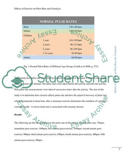
?EFFECTS OF EXERCISE ON PULSE RATE AND URINALYSIS A. EFFECTS OF EXERCISE ON PULSE RATE Introduction When the heart beats, it produces a vibration dueto the pulsating wave of blood in the arteries. This is manifested as pulse that can be felt most in the following areas: temporal artery above the ear; mandibular artery on the lower bone; carotid artery in the neck; femoral artery in the groin, and; the radial artery in the wrist (Rosdahl & Kowalski 2008, p. 518). A pulse rate is equal to the number of heart vibrations or beats in a minute, which may vary depending on age, activities, general health, sex, emotions, pain and modifications. For example, pulse rate is higher when a person is engaged in some kind of strenuous activity and is lower when sleeping; babies and children have higher pulse rates than adults, and; athletes have generally lower pulse rates than the average because of well-developed cardiovascular system (Lindh et al 2009, p. 573). The way the pulse rate recovers after an exercise provides inkling as to the heart condition of a person. Figure 1 shows the pulse rates considered normal for various age groups. Babies and children are shown as having the highest pulse rates. Several factors exist, however, that can increase the normal heart rate and among them are the following: anxiety; some medication; trauma; pain, infection; cardiac abnormalities; circulation problems; increased body temperature, and; exercise (Ingram & Lavery 2009, p. 55). Fig. 1 Normal Pulse Rates of Different Age Groups (Lindh et al 2009, p. 573) To illustrate how pulse rate is affected by exercise, the subject in this case is made to run up and down the stairs in 2 minutes. His pulse rates were taken before the activity and after the activity. Post pulse rate measurements were taken 6 successive times after the activity. The aim of this study is to determine how exercise affects pulse rate and how the speed of recovery of pulse rate, which corresponds to heart beat, after a strenuous exercise determines the condition of a person’s coronary health. A slower heart rate is associated with coronary disease. Results The following are the data gathered on the pulse rate of the subject: resting pulse rate, 75bpm; immediate post-exercise, 109bpm; first minute post-exercise, 105bpm; second minute post-exercise, 90bpm; third minute post-exercise, 89bpm; fourth minute post-exercise, 80bpm; fifth minute post-exercise, 80bpm. Discussion This study illustrates that exercise increases heart beat and therefore, pulse rate. The data gathered before the activity and immediately after the activity show that the person’s initial pulse rate corresponds to the normal pulse rate for an adult as shown in Fig. 1. After a two-minute strenuous activity, however, his pulse rate accelerated to more than 34 bpm at 109 bpm. This increased pulse rate gradually delerated, and returned to almost normal but only after 5 minutes. In the first minute post-exercise, the pulse rate decreased by 4 bpm, in the second by 15 bpm, in the third by 1 bpm, in the fourth by 9 bpm and in the fifth, the pulse rate has stabilized. In five minutes, the pulse rate decelerated by 29 bpm or an average of 5.8 bpm per minute (See Fig. 2). Fig. 2 Heart Rate Recovery of Subject The gradual return of the pulse rate to its normal rate is called heart rate recovery. Heart rate recovery has been used as a diagnostic/prognostic tool for heart conditions. The rationale for this is that the heart’s response to exercise is a reflection of “the balance between the central nervous system withdrawal of vagal tone and an increase in sympathetic tone, an abnormal heart rate response to exercise is also likely related to abnormal autonomic balance” (Froelicher & Myers 2006, pp. 113-114). Various studies have been made regarding heart rate recovery as a diagnostic tool for possible heart ailments. Cole et al studied over 2000 individuals for more than 6 years and established that a recovery rate of 12bpm or less at 1 minute after post exercise is abnormal and has a 4.0 mortality risk. Eventually, abnormal rate recovery was defined as 42bpm decrease or less 2 minutes after exercise with a mortality risk twice that of the group with normal rates. According to Basson and Lerman (2009), the 12bpm benchmark is to be used only when an exercise uses a cooling down protocol, but when absent an 18bpm or less for the first minute after exercise should be used as benchmark for abnormal heart rate recovery. On the other hand, Froelicher et al conducted their own study for 13 years and concluded that abnormal heart rate recovery is best defined as recovery rate of 22bpm or less at two minutes corresponding to a hazard ratio of 2.6 (Froelicher & Myers 2006, p.114). Conclusion Applying the various findings of researchers that conducted heart rate recovery, the subject herein seems to be at high risk. Using the Cole et al model, the subject has failed with a mere 4bpm decrease after 1 minute as it is both within the 12bpm or less at 1 minute frame and the 42bpm or less after 2 minutes ofexercise because he generated only a 19bpm decrease at the end of the first two minutes, but since no cooling down protocol is mentioned in their case, the 18bpm should be utilized in this case. Nonetheless, even under this model, the subject has still abnormal heart rate recovery. Similarly, the subject is also at high risk in accordance to the more stringent model of Froelicher et al, which sets 22bpm or less at two minutes post-exercise as the benchmark for individuals with abnormal heart condition. B. EFFECTS OF EXERCISE ON URINALYSIS Introduction The body’s urinary system, consisting of two kidneys, two ureters, one bladder and one urethra, works to rid the body of its waste product. Aside from that primary function, it also regulates the volume and composition of blood by producing urine and excreting it. A urinalysis is a test taken on a urine sample to determine the condition not only of the kidneys, but also of related organs. The test is used to detect kidney diseases, urinary tract infection and diabetes, among others. For example, the presence of abnormal particles, protein, blood cells or crystals may indicate kidney malfunction. The presence of glucose and/or ketones may indicate diabetes and bacteria or a high level of white blood cells of urinary tract infection (University of California 1995, p. 512). Exercise causes changes to occur in the mechanism of kidneys, the organs that regulate fluid and electrolytes excretion. Strenuous activities decrease renal blood flow and increase glomerular filtration pressure because of the constricting effect of increased epinephrine and nor-epinephrine to the afferent and efferent arterioles. Glomerular filtration is the process by which the kidneys remove waste product of the body from the bloodstream. Exercise also increases water reabsorption (Mellion et al, 2003, p. 245). In this particular exercise, urinalysis is conducted on 20 individuals who were made to run up and down the stairs for two minutes before the test. The results of the urinalysis are indicated below and discussed for their implications thereafter. The objective of this exercise is to establish the effect of exercise on body functioning that may reflect on the results of a urinalysis test subsequently taken thereafter. These changes are evidenced by the increase or decrease of some of the substances that usually show up in the test or the presence or absence of substances that are usually detectable or not detectable during urinalysis. Results The data collected after urinalysis test was conducted showed the following results: glucose was negative in all subjects; bilirubin was negative in 14 subjects and positive in 6 although the amounts were small; ketone was negative in 18 and positive, in trace amounts, in two; 2 subjects had specific gravity of 1.015, 3 had 1.010, 4 with 1.025, 5 with 1.020 and 6 with 1.030; red blood cells were negative in 13 subjects, trace amounts in 5, and large amounts in 2; 4 subjects had pH of 5, 3 with 6, 8 with 6.5, 2 with 7, and 3 with 7.5; protein was negative except for 4 subjects who had trace amounts; urobilinogen was negative in all; nitrite was negative in all except for 1 subject with trace amount, and; leukocytes was negative in 11 subjects, 5 with trace amounts, 1 with large amount, 2 with small amount, 1 with moderate amount. Discussion At issue here is the presence, or absence, and the implication of the amounts of glucose, pH, specific gravity and protein detected in some of the subjects after the urinalysis was undertaken. PH, which is the abbreviated form of potential hydrogen ion concentration, is the measurement of urine’s acidity or alkalinity and its values ranges from 4.5 to 8.0, the average being 6.0, which is slightly acidic. Higher values point to alkalinity while lower values to acidity. Death is possible with a too acidic blood, known as acidosis, or a too alkaline blood system, known as alkalosis. To maintain blood pH homeostasis, the kidneys and the lungs assist the body to maintain the perfect ph of 7.35 to 7.45 by consistently adjusting body secretions. Medications, diets and pathological conditions affect the ph balance of urine. Acidic urine may be caused by a high-protein diet, certain medications, renal tuberculosis, high fever and uncontrolled diabetes. On the other hand, alkaline urine may be caused by a high-protein diet, citrus fruits, some medications, urinary tract infection and even dairy products (Lindh et al 2009, p. 1224). In the present case, the urinalysis indicates that 13 of the subjects have pH values higher than 6, which is considered the average in urine pH, signifying alkalinity of the subjects’ urine and only 7 have acidic value readings. A pH of 7, which indicates is a high vegetarian diet, may be indicative of Fanconi’s syndrome, urinary tract infection or metabolic or respiratory alkalosis. On the other hand, a pH below 7, a high-protein diet, may indicate renal tuberculosis, phenylketonuria, alkaptonuria, pyrexia and acidosis (Lippincott Williams 2007, p. 240). Urine specific gravity indicates its density, which is the proportion of dissolved solid constituents to the total volume of urine. The reference point is the density of water at 1.000. Urine is expected to have a higher specific density because it contains dissolved solid wastes, such as phosphates, urea, proteins and sugars, of the body. Specific gravity measures urine’s capacity to concentrate. Nonetheless, an extremely specific gravity may indicate the presence of such substances as glucose, a very dense substance. The normal specific gravity of urine is 1.005 to 1.035, with average range at 1.010-1.025. The results herein show that all subjects are within the normal range, most of which lean towards the upper level, viz. 15 subjects have 1.025 and higher, which is not surprising considering that the subjects underwent physical exercises that may have caused some level of dehydration, which may increase specific gravity reading. This is because when the body is dehydrated, it gives off less fluid in proportion to the denser dissolved solids in the body (Lindh 2009, p. 1222). Glucose in urine may be attributed to the diabetes mellitus condition, and in this group none was positive for it. For normal people, glucose should be absent from urine because of the high renal threshold of glucose (Pollak 2009, 251). This should be more so in the case during exercise because glucose is being used up by the body for energy. Nonetheless, the absence of glucose in urine and the use of urinalysis to test for diabetes mellitus are not reliable to conclude the absence of the disease. This is because glucose has a high renal threshold, which means that it will take a high amount of glucose in the blood for the kidneys to intervene and take out excess glucose as compared to other substances in the body. Protein or albumin is normally undetectable in urine and therefore, registers as negative in a urinalysis. This is because protein molecules are too big and is immediately filtered by the glomerulus. The abnormal presence of protein in urine signifies the inability of the kidneys to function and filter this substance. When a person exercises, however, the permeability of the glomerulus is increased, allowing the passage of protein and albumin to urine and consequently, become detectable in urinalysis. In the present test, four had trace amounts of it and the rest are negative. The four subjects with positive protein findings do not readily imply kidney conditions, but may have been engendered by the physical strenuous exercise. This warrants a closer examination, however, as the majority did not exhibit a similar finding (Pollak 2009, p. 251). Conclusion This study shows that exercise can affect the results of urinalysis with normally undetectable substances becoming detectable and previously normal levels of body elements suddenly either increased or decreased. Specific gravity, for example, goes up because exercise can cause some level of dehydration leaving urine denser than usual. Similarly, protein which is normally not detectable in urinalysis for normal individuals may show up in urinalysis conducted after exercise, without being indicative that the individual is suffering from some kind of kidney malfunction. References: Basson, C. & Lerman, B. (2009). Topics in Structural Heart Disease. New York: Demos Medical Publishing. Froelicher, V. & Myers, J. (2006). Exercise and the Heart. 5th Edition. Phildelphia: Elsevier Health Sciences. Ingram, P. & Lavery, I. (2009). Clinical Skills for Healthcare. UK: John Wiley and Sons, 2009 Lindh, W., Pooler, M., Tamparo, C., amd Dahl, B. (2009). Delmar's Comprehensive Medical Assisting: Administrative and Clinical Competencies. 4th Edition. NY: Cengage Learning. Lippincott Williams & Wilkins (2007). Deciphering Diagnostic Tests. PA, USA: Lippincott Williams & Wilkins Mellion, M., Putukian, M. and Madden, C. (2003). Sports Medicine Secrets. 3rd Edition. Philadelphia: Elsevier Health Sciences. Pollak, A. (ed). (2009). Critical Care Transport. UK: Jones & Bartlett Learning. Rosdahl, C.B. & Kowalski, M. (2008). Textbook of Basic Nursing. 9th Edition. Lippincott Williams & Wilkins. University of California (1995). The New Wellness Encyclopedia. New York: Rebus, Inc. Read More
Cite this document
  • APA
  • MLA
  • CHICAGO
(“Effects of exercise on pulse rate and urinalysis Essay”, n.d.)
Retrieved from https://studentshare.org/psychology/1424546-effects-of-exercise-on-pulse-rate-and-urinalysis
(Effects of Exercise on Pulse Rate and Urinalysis Essay)
https://studentshare.org/psychology/1424546-effects-of-exercise-on-pulse-rate-and-urinalysis.
“Effects of Exercise on Pulse Rate and Urinalysis Essay”, n.d. https://studentshare.org/psychology/1424546-effects-of-exercise-on-pulse-rate-and-urinalysis.
  • Cited: 0 times

CHECK THESE SAMPLES OF Analysis and Review of Data on the Impact of the Training Process on the Pulse Rate

Effect of Different Exercise Durations on Arterial Stiffness

Effect of Different exercise Durations on Arterial Stiffness Institution Effect of Different exercise Durations on Arterial Stiffness Arterial stiffness is an established cardiovascular risk factor.... hellip; On the other hand, the intense effects are not well established....
9 Pages (2250 words) Research Paper

Antenatal and Intrapartum Competencies

Initial assessment (observation, history taken, physical examination, social, cultural and emotional assessment) using a systematic and logical approach, using appropriate terminology: Entails reviewing the patient's health record, then, testing the blood pressure, temperature, pulse rate and undertaking urinalysis tests (NHS, 2013 n.... Finally, observe the fetal heart rate, while avoiding using cardiotocography, where the pregnancy is in the low-risk category (NHS, 2013 n....
12 Pages (3000 words) Essay

Clinical Examination

His pulse rate was 92, blood pressure 100/92, peripheral pulses were equal volume on both the sides with carotids being palpably normal.... On examination, he had harsh vesicular breath sounds throughout the lung fields, with features of laboured breathing at a rate of 22 per minute at rest.... The apical pulse was located in the seventh intercostal space 1....
13 Pages (3250 words) Case Study

CASE STUDY ABDOMINAL AORTIC ANEURYSM

The patient was very cold, tachycardia with a pulse rate 100-105beats/minute, respiration 24-30/min, in respiratory distress and cyanosed.... urinalysis showed protein presence.... On examination femoral pulse was absent.... PAST MEDICAL HISTORY:-He smoked for 20yrs 2 packets a day....
12 Pages (3000 words) Essay

Management of Long-Term Cardiorespiratory Symptoms

His personal history revealed little to no exercise, and he spent most of the day at home watching television.... David is a 71 year old male who lives with his wife.... He is a retired labourer.... Since his retirement he has put on a large amount of weight and now has a BMI of 27....
14 Pages (3500 words) Essay

Problem based SOAP notes for three patients

Instruction in side effects of Viagra.... This is a 72 year old black male, retired school teacher who is presenting with symptoms of erectile dysfunction.... He describes that he is able to have an erection but it does not stay long enough to participate in intercourse.... He was diagnosed three years ago with Type 2 diabetes and is somewhat hypertensive....
3 Pages (750 words) Essay

The Effect of Exercise on Pulse Rate

The Effect of exercise on pulse rate Background of the Study The rationale for carrying out pulse measurement is to both heart rate and rhythm, since the heart pumps blood throughout the body, the rhythm with which this is done may be detected through the wrist, neck and upper arm.... The current study was designed to determine the effect of exercise on pulse rate.... The rationale for carrying out pulse measurement is to both heart rate and rhythm, since the heart pumps blood throughout the body, the rhythm with which this is done may be detected through the wrist, neck and upper arm....
3 Pages (750 words) Essay

Effect of Exercise on Blood Pressure and Pulse Rate

This paper reports on the analysis, interpretation of the results obtained after conducting an experiment on the short-term and immediate effects of exercise on pulse rate and blood pressure.... To find out more about the effects of exercise on pulse rate and blood pressure, an experiment was conducted.... The differences in the results were used to determine the effects of exercise on pulse rate and blood pressure.... The influences of exercise on pulse rate and blood pressure can be scientifically ascertained....
4 Pages (1000 words) Essay
sponsored ads
We use cookies to create the best experience for you. Keep on browsing if you are OK with that, or find out how to manage cookies.
Contact Us