StudentShare
Contact Us
Sign In / Sign Up for FREE
Search
Go to advanced search...
Free

RF Burns - Causes and Prevention - Assignment Example

Cite this document
Summary
This assignment "RF Burns - Causes and Prevention." shows that Magnetic Resonance Imaging is a medical imaging technique used to visualize the body’s internal structures. MRIs have three types of magnetic fields. First, is a large superconducting magnet producing the main magnetic field…
Download full paper File format: .doc, available for editing
GRAB THE BEST PAPER96.4% of users find it useful
RF Burns - Causes and Prevention
Read Text Preview

Extract of sample "RF Burns - Causes and Prevention"

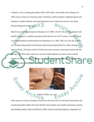
?Radiofrequency Burns – Causes and Prevention Introduction Magnetic Resonance imaging is a medical imaging technique used to visualize the body’s internal structures. MRIs have three types of magnetic field. First, is a large superconducting magnet producing the main magnetic field; second is the Radio Frequency field (RF); and third are the gradient magnets. In relation to the radio frequency field, risks of RF burns have been known to arise. Most RF burns are however mostly caused by local and persistent heating of the skin, including the underlying tissue. This paper shall now discuss the different causes of RF burns, specifically RF burns during Magnetic Resonance Imaging (MRI) procedures, including its prevention measures. This study is being carried out in order to establish guidelines in the use and application of radiofrequency, mostly in terms of its safety and prevention of injuries. Body Causes 1. Heating of implants An RF pulse would often be transmitted by the MRI to stimulate protons through energy exchange. The pulse is an electromagnetic wave coming from the RF coils (Lin, 2008). RF power during MR scans are changed into heat within the tissue of the patient due to resistive losses (Eising et.al., 2010). This absorption of energy is specifically considered the SAR (Specific Absorption Rate). The specific effects of heating during MR scanning and burns usually refer to conductive implants and normal tissue. Most studies discuss the impact of conductive wires, including pacemaker cables, ECG cables, and metallic stents (Eising et.al., 2010). Issues in the use of aneurysm clips in the brain, cochlear implants, implanted spinal cord stimulator, metallic implants, and metal fragments in one or both eyes can also cause burns (Premier Diagnostic Center, n.d). Based on an experimental study by Nakamura et al. (2001), 55-235 volts could be induced with the RF irradiation in a metallic loop placed inside the bore of a 0.5-T scanner. A resistor which was inserted sparked and then burned out (Nakamura et al., 2001). This was seen only with the axis of loops being parallel to the linearly polarized transmitting field. For cables, heatings at 20 degrees Celsius, 26 Celsius, and 63.5 Celsius have been assessed. Assessments indicate that the heating of the ECG cable is known to cause fire. Small implants can lead to issues with the heating of skin seen in tattooed patients caused by the heating of metallic elements of the ink used for the tattoo (Klitscher et al., 2010). Implants (Wills.com, n.d.) Some reports on excessive heating or burns have also been seen for iron-based oxide tattoos and transdermal patches added with metal elements; these patches may include testosterone, nicotine, and clonidine patches (Tope and Shellock, 2002). Tattoos using ferromagnetic compounds can cause react to the MRI machine (Ross and Metava, 2011). In general, the image would somehow appear distorted following the MRI; however, the injuries usually represent burns on the patient’s skin. As such, any redness or apparent injury on the skin has to be managed medically (Ross and Matava, 2011). As seen in the image below, the ferromagnetic compounds of the tattoo caused the RF burns, mostly manifesting as burns and redness on the patient’s skin on the site of the tattoo. Tattoo with ferromagnetic compounds following MRI (Melina, 2010) Currents may be triggered by two fields. The first is the pulsed magnetic-gradient field, and the second being the pulsed radiofrequency field (Jacob et al., 2010). These two fields increase their intensity over time and may eventually cause an electromotive force through a conductive loop. Heating will then come about from the current originating from the loop; with the amount of heating based on the resistance in the conductive processes (Dempsey and Condon, 2001). The pulsed magnetic field is triggered by a large coil which encompasses the coils and is often found near the patient to be scanned. The issue of excessive heating is based on the distance of the RF coil to the patient, including the frequency of the RF emitted. Prevention During the MR scanning, the patient must also be protected from contact point heating injury. Only the approved padding by the manufacturer must be applied to protect the patient from RF burns (Chapple, 2003). Non-conducive padding must be at least 0.25 inches in thickness in order to secure protection. Patients must also never be allowed to directly impact on the magnet bore or surface coil (Busch et al., 2005). Obese patients cannot be evaluated in the small magnetic bore because they would not fit into it and may suffer burns through possible contact with the magnet bore. RF injuries may arise immediately; it is therefore important to not depend on the patient’s warning of discomfort during the process. A patient alert bulb must therefore also be provided to all patients during the scanning (GE Healthcare, 2008). MRI Protection pad (protects patient by preventing contact with the bore of the MRI machine) (Newmatic Medical, n.d.) Patient teaching would also assist ensure safety of the patient, with the nurse or the technician explaining the procedure and preparing the patient for the procedure. Monitoring the patient during the MRI also forms part of the safety of the patient). Through monitoring, quick responses can be made and the extent of the burns can be reduced (Busch et al., 2005). MRI monitoring (allow quick responses to be made) (Soung Imaging, 2006) 2. Heating of tissue In the tissue heating of the human head, a numerical model has been applied (Eising et al., 2010). For their experiment, they sought to determine the effects of heating to the human head, and the point at which dangerous effects are seen. Their experiment included scanning of a human cadaver, with dangerous levels seen at 8.0 T and insignificant heating seen by 0.7 Celsius. For the cornea, an increase in temperature 1.8 Celsius was observed. Issues involving cataractogenesis was not reached by such heating level. Nevertheless, MRT has been known to lead to focal heating with second and third-degree burns for patients established in case report with 1.5-T MRT (Knopp et al., 1998). Authors supported the avoidance of small areas of skin contact during MR scans. The figure below demonstrates the case of a cancer patient who was burned because of contact with an aluminium blanket; the aluminium reflected heat back to itself and caused the burn. Tissue heating (small areas of skin contact during MR scans) (Pray for Noah, 2011) Numerous reports have been indicated on burn injuries acquired during MRI scanning procedures (Dempsey and Condon, 2001). Their study sought to assess the different elements which may have caused these burns. The authors carried out experiments in order to assess three possible applications leading to heating in the copper wire during MRI. The first is direct electromagnetic induction in a conductive loop, the second is induction in a resonant conducting loop, and the third is the electric field seen in coupling with a wire, otherwise known as the antenna effect (Dempsey and Condon, 2001, p. 627). Temperatures recorded increased to 0.6 degrees Celsius for the loop and 61.1 degrees for the resonant loop; the resonant antenna was at 63.5 degrees Celsius. These findings indicate that burns are more likely to occur as a result of the resonant conducting loops coming from extended wires which then create the resonant antenna (Dempsey and Condon, 2001, p. 627). As was mentioned above, the absorption of the RF power by the tissues is known as the Specific Absorption Rate (SAR) which is measured in watts/kg (Martinos Imaging Center at McGovern Institute, 2006). The SAR for MRIs is based on different variables which include pulse sequence as well as coil parameters and the weight of the patient. However, the temperature of the tissue is increased due to exposure to radiation from the machine, based on the thermoregulatory system of the subject-patient (Martinos Imaging Center at McGovern Institute, 2006). This would explain why the use of 3T magnet does not allow for the use of blankets or any other restrictive clothing. In other words, the subject needs to dissipate or distribute the heat well. Physiological responses to the MR procedure depends on various factors, including the duration of exposure, the rate at which energy is deposited, the patient’s thermoregulatory response, the underlying health issue, and the general conditions in the MR system. In instances of thermal challenge, the body experienced heat loss through convection, conduction, radiation, and evaporation. These mechanisms are responsible for heat dissipation with the body seeking to ensure thermal homeostasis. Where the thermoregulatory effectors cannot totally dissipate the heat load, there is storage of heat, and increase in local tissue temperatures (Shellock, 2012). Prevention In order to prevent RF burns, periodic maintenance of the MRI machine is very much significant. Various patient precautions must also be taken in order to prevent such burns. First, it is important to protect the patient from metallic heating (Eising et al., 2010). All patients, visitors, and other staff members must be screened before they are allowed entry into the scanning room. In this case, a patient or anyone else having a ferrous or electronic device must never be allowed to enter the scan room or be scanned (Eising et al., 2010). The same is true for those with implants or prosthesis contraindicated for MR scanning. Currents in some metallic objects may lead to excessive heating which can then lead to burns in the tissues (Dempsey et al., 2001). The image below indicates safe clothing for patients during MRI procedures. In general, the clothing must be free of any metallic items, including buttons and zippers. Patient in MRI-safe hospital gown (Premier Diagnostic Center, n.d.) All patients must therefore be assessed for surgically implanted objects including those similar objects implanted due to injuries or accidents (Busch et al., 2005). All metallic objects on the patient must also be removed before the scan, this would include gold jewellery which can often lead to burns. Certain clothes with metallic dyes can also heat during the scan; only hospital issue gowns must therefore be worn by the patient during the scans (Vaughan et al., 2006). Shoes must also be removed during the scan. Some tattoos and cosmetics may also have metallic elements (Ross and Matava, 2011). Makeup must therefore be removed as they may heat during the scan. Those with permanent tattoos cannot undergo MR scans as their skin would likely suffer burns from the metallic content of their tattoos (Ross and Matava, 2011). 3. Electromagnetic Induction Heating The magnetic fields and the RF electromagnetic fields of the MRI can trigger voltages in conductive media which can then lead to current flow. Such circulating flows lead to power loss owed to ohmic heating, referred to as induction heating. Electromagnetic induction heating for the monitoring cables is considered the main cause of thermal burns during MRI. Loops formed in the monitor cable can increase the circuit’s inductance, causing larger currents to be created by the higher heating of the cable. The most unfortunate scenario for electromagnetic heating will likely be seen when circuits are in a resonant state. (Thomas, 2009) Prevention Cables must go under the pads (Thomas, 2009) 4. Heating due to the antenna effect The monitoring cables can sometimes be seen as RF wire antennae which are sometimes sensitive to the electric components, not the magnetic elements of the RF radiation for the MRI. The antenna attracts the electromagnetic waves and gains power from them. The waves then enter the antenna and relay electrical charges with matching currents. Resonant antennae manifest current and volts standing wave patterns seen reflected from open ends of the wire. Prevention 1. The cables should not touch the skin (2 cm). This is the same protective safety measure as indicated above. (Thomas, 2009) The patient must also be protected from excessive exposure to RF. If the patient is exposed to the RF faster than the rate of skin absorption and dissipation, the surrounding tissues can experience burns (Deo, 2012). The rate of tissue heat is based mostly on the patient’s weight, pulse sequence, number of slices, as well as the saturation. An increase in body temperature can also be hazardous to the patient, especially those with a decreased regulatory power (Wheaton et al., 2004). Lower capacity for thermoregulation can be based on existing patient conditions, including decreased circulatory processes, cardiac affectations, hypertension, diabetes, old age, fever, and the decreased ability to perspire (Wheaton et al., 2004). Patients manifesting these issues must be constantly monitored. 5. Thermal effects Thermal effects can also be seen after being subjected to elevated levels of RF energy, including persistent exposure to lower levels or directly contacting with powerful antenna (Silver 2006). These burns generally lead to physical damage to the tissues and burn the tissues from inside the body, gravitating outwards (Overall et al., 2010). The burns must be healed from the inside out and in some instances, blisters seen on the skin may not be too apparent for days following the burn. Burns healing from inside (Pray for Noah, 2011) Blindness can also be seen by the elevated levels of energy which can lead to thermal damage to the eyes (Segami and Nishimura, 2004). Limited thermal damage can also lead to sterilization without causing any pain. Fourth-degree burn of wrist and right forearm immediately after the session of MRI (Haik et al., 2009) The patients must also be protected from heating hazards arising from accessory devices. MR equipment which is not functioning well or is damaged must never be used. The monitoring equipment applied must also match the brand of the scanner (GE Healthcare, 2008). Scans must never be used on patients with monitor leads or with their pulse oximeters attached. Non-magnetic equipment can also be used instead of conductive materials in order to ensure patient monitoring throughout the scanning process. In the image above, the patient manifested a full thickness burn with apparent demarcation between the necrotic and viable skin. A circular metal electrode was attached to the patient’s arm and was directly in contact with the skin (Haik et al., 2009) and caused such burn. It is believed that the wires connecting the electrodes created a conducting loop which therefore led to the burn, specifically, the thermal injury (Haik et al., 2009). Prevention 1. Avoid contact of patient with bore of MRI machine. This image below shows the use of protective side-padding, which is especially useful for obese patients whose sides must not touch the magnet bore of the machine. 2. Protect patients from heating hazards of accessory devices including ECG leads (US Food and Drug Administration, n.d.) Conclusion This discussion indicates the different causes of RF burns. These burns are often seen in the workplace where equipment and machines emit RF (also gradients produce changing magnetic fields). It can also be seen in the use of MR scans. Failure to use safety standards often causes these burns which can sometimes be unobservable by the individual affected. In order to prevent these burns, compliance with safety standards as well as protection of the patients and other users must be strictly implemented. Strict monitoring of the patient must also be carried out including patient education processes. Through these remedies, the danger of burns can be prevented and the safety of the user can be ensured where emissions cannot be avoided. References Busch, M., Vollman, W., Weltzer, R., Bertsch, T., 2005. On the heating of inductively coupled resonators (stents) during MRI examinations. Magnetic Resonance in Medicine, 54, pp. 775–782. Carmichael, D., Thornton, J., Rodionov, R., Thornton, R., et.al., 2008. Safety of localizing epilepsy monitoring intracranial electroencephalograph electrodes using MRI: radiofrequency-induced heating. Journal of Magnetic Resonance Imaging, 28, pp. 1233–1244. Chapple, E., 2003. Fundamental SNR and SAR limitations in very low field MRI. Canada: Simon Fraser University. Cleveland, Jr. R., Sylvar, D., and Ulcek, J., 1997. Evaluating compliance with FCC guidelines for human exposure to radiofrequency electromagnetic fields [online]. Available at: http://transition.fcc.gov/Bureaus/Engineering_Technology/Documents/bulletins/oet65/oet65.pdf [Accessed 08 April 2013]. Dempsey, M., Condon, B., and Hadley, D., 2001. Investigation of the factors responsible for burns during MRI. J. Magn. Reson. Imaging, 13, pp. 627– 631. Deo, S., 2012. Specific Absorption Rate (SAR). International Society of Radiographers & Radiological Technologists, 55(2), pp. 57-58. Eising, E., Hughes, J., Nolte, F., Jentzen, W., et.al., 2010. Burn injury by nuclear magnetic resonance imaging. Clinical Imaging, 34, pp. 293-297. GE Healthcare, 2008. MR patient warming prevention guide [online]. Available at: http://www.gehealthcare.com/usen/education/tip_app/docs/MR%20RF%20Warming%20Prevention%20Guide%2007-10480.pdf [Accessed 06 April 2013]. Haik, J., Daniel, S., Tessone, A., Orenstein, A., and Winkler, E., 2009. MRI induced fourth-degree burn in an extremity, leading to amputation. Burns, 35, pp. 294-296. Hitchcock, R., 1995. Radio-frequency and elf electromagnetic energies: A handbook for health professionals. London: John Wiley & Sons. International Labour Organization, 1998. Safety in the use of radiofrequency dielectic heaters and sealers. Switzerland: World Health Organization. Jacob, Z., Tito, M., Dagum, A., 2010. MR imaging–related electrical thermal injury complicated by acute carpal tunnel and compartment syndrome: Case report. Radiology, pp. 10-11. Jones, S. and Alvi, R., 1996. Burns associated with electrocardiographic monitoring during magnetic resonance imaging. Burns, 22(5), pp. 420-421. Kanal, E., Barkovich, A., Bell, C., Borgstede, J., et.al., 2006. ACR Guidance Document for Safe MR Practices: 2007. AJR 1(88), pp. 1-27. Melina, R., 2010. Do MRI Machines Affect Tattoos? [online]. Available at: http://www.lifeslittlemysteries.com/816-do-mri-machines-affect-tattoos.html [Accessed 20 April 2013]. Newmatic Medical, 2011. MRI protection pad [online]. Available at: http://www.newmaticmedical.com/product.asp?P_ID=9307 [Accessed 20 April 2013]. Occupational Safety and Health Administration, 2012. Radiofrequency and microwave radiation [online]. Available at: http://www.osha.gov/SLTC/radiofrequencyradiation/ [Accessed 07 April 2013]. Overall, W., Pauly, J., Stang, P., and Scott, G., 2010. Ensuring safety of implanted devices under MRI using reversed RF polarization. Magnetic Resonance in Medicine, 64, pp. 823–833. Pray for Noah, 2011. Noah’s MRI accident [online]. Available at: http://www.prayfornoah.com/noahs-mri-burn.html [Accessed 20 April 2013]. Premier Diagnostic Center, n.d. MRI Preparation [online]. Available at: http://premierdx.com/services/mri/mri-preparation [Accessed 15 April 2013]. Ross, J. and Matava, M., 2011. Tattoo-induced skin ''burn'' during magnetic resonance imaging in a professional football player: ac. Sports Health, 3(1), pp. 431-435. Segami, N. and Nishimura, M., 2004. Thermal injury during temporomandibular joint arthroscopy: A case report. J Oral Maxillofac Surg, 62, pp. 508-510. Silver, H., 2006. The ARRL ham radio license manual: All you need to become an amateur radio operator. New York: American Radio Relay League. Stellman J., 1998. Encyclopaedia of occupational health and safety. Switzerland: International Labour Organization. Thomas, M., 2009. Coil Safe Positioning in MRI [online]. Available at: http://www.slideshare.net/marie11480/coil-safe-positioning-in-mri-1695796 [Accessed 17 April 2013]. US Food and Drug Administration, n.d. Burns from Electrodes and Cables during MRI Exams [online]. Available at: http://www.accessdata.fda.gov/scripts/video/radiationemittingproducts.cfm?yid=7vKPJA1brNg [Accessed 20 April 2013]. Vaughan, T., DeLaBarre, L., Snyder, C., Tian, J., 2006. Human MRI: Preliminary results. Magnetic Resonance in Medicine, 56, pp. 1274–1282. Wheaton, A., Borthakur, A., Corbo, M., Charagundla, S., et.al., 2004. Method for reduced SAR. Magnetic Resonance in Medicine, 51, pp. 1096–1102. Wills.com, n.d. MRI Safety Awareness [online]. Available at: www.willis.com/.../MRI_Safety_August_2009_V6.pdf [Accessed 15 April 2013]. Read More
Cite this document
  • APA
  • MLA
  • CHICAGO
(“RF Burns - causes and prevention Assignment Example | Topics and Well Written Essays - 2000 words”, n.d.)
Retrieved from https://studentshare.org/physics/1472172-rf-burns-causes-and-prevention
(RF Burns - Causes and Prevention Assignment Example | Topics and Well Written Essays - 2000 Words)
https://studentshare.org/physics/1472172-rf-burns-causes-and-prevention.
“RF Burns - Causes and Prevention Assignment Example | Topics and Well Written Essays - 2000 Words”, n.d. https://studentshare.org/physics/1472172-rf-burns-causes-and-prevention.
  • Cited: 0 times

CHECK THESE SAMPLES OF RF Burns - Causes and Prevention

Prevention and Management of Infection among Burn Patients

The aim of the paper “prevention and Management of Infection among Burn Patients” is to analyze infection as a major issue among burn patients.... nbsp; Literature review This literature review on the prevention and management of infection among burn patients will be started by using key words and related terms through the medical libraries and database of the Texas Resource's.... Literature review This literature review on the prevention and management of infection among burn patients will be started by using key words and related terms through the medical libraries and database of the Texas Resource's Harris Methodist Hospital, and via internet databases of the Mayo Clinic, Texas Women's University, as well as the University of Texas Southwestern Medical Center....
11 Pages (2750 words) Research Paper

Research on Paediatric Burns

he common causes are scalds from boiling liquids like water or oil or other preparations being cooked in the kitchen or steam.... Incidence Among the leading causes, burns in children (below 9 years are identified as the third one among injury-related deaths (Green, 2010).... burns Paediatric burns burns burns may be defined as the tissue damage that occurs to the body caused by heat, chemicals, electricity, sunlight or nuclear radiation (NIH, US)....
5 Pages (1250 words) Essay

MRI Safety: RF Burns - Causes and Prevention

MRI Safety: RF Burns – causes and prevention ... Tissue heating can occur through excessive RF interaction in patients, leading to rf burns (Liney, 2010).... This paper evaluates the causes for rf burns and the preventive measures that can be taken to reduce the incidence of rf burns.... The important issues relating to safety of RF fields during MRI diagnostic imaging pertain to thermal heating, that results in current burns and contact burns from the heat stress that is generated....
8 Pages (2000 words) Essay

Physiological Reaction to Burns

The main functions of the skin are thermal regulation, prevention of loss of fluid from the body by evaporation, barrier against infections from the environment, and perception of sensory information from the environment.... Burns causes the intact vessels to dilate causing redness and blanching with pressure.... Burns causes loss of functions of the skin.... Most of the burns are 2 and 3 rd degree burns.... This patient has 45% burns along with inhalation injury....
8 Pages (2000 words) Essay

Infection among Burn Patients

Moreover, burns are usually infected with fungus infective agents, making the infection process on burn wounds even more difficult to prevent and manage.... The aim of the paper “Infection among Burn Patients” is to analyze infection as a major issue among burn patients....
11 Pages (2750 words) Research Paper

Burnt Patients and Severe Burn Injuries

As the nurse in-charge of providing care to a patient with 70% of body burnt, there is certain knowledge that is required for the administration of effective care, one of which is the prevention of complications such as hypothermia and compartment syndromes.... burns contribute to a large percentage of accidental injuries and even deaths worldwide, coming only after motor vehicle/ pedestrian injuries and drowning.... For the most-effective care, burn patients burns of Affiliation: burns burns contribute to a large percentage of accidental injuries and even deaths worldwide, coming only after motor vehicle/ pedestrian injuries and drowning....
1 Pages (250 words) Essay

MRI Safety: RF Burns Causes and Prevention

The study "MRI Safety: RF Burns – causes and prevention" examines evaluates the causes of RF burns and the preventive measures that can be taken to reduce the incidence of RF burns.... The paper focuses on heat stress in RF technology in MRI, detailed information on the prevention of the rf burns.... Tissue healing can occur through excessive RF interaction in patients, leading to rf burns (Liney, 2010).... This paper evaluates the causes of rf burns and the preventive measures that can be taken to reduce the incidence of rf burns....
9 Pages (2250 words) Coursework

Importance of Nutrition on Burn Injured Patients in Hospitals

The study "Importance of Nutrition on Burn Injured Patients in Hospitals" examines the nourishment of the burned skin, which care should be taken on each degree to ensure the fast healing of the burn, how these burns are diagnosed, classified severity of the burns, psychological effect.... o completely understand the right way towards treating patients with the right nutrition, the understanding of the degree of burns is very important.... They are further divided amongst four degrees through different means or burns affecting the human body....
8 Pages (2000 words) Report
sponsored ads
We use cookies to create the best experience for you. Keep on browsing if you are OK with that, or find out how to manage cookies.
Contact Us