StudentShare
Contact Us
Sign In / Sign Up for FREE
Search
Go to advanced search...
Free

Anatomy of Biliary System - Essay Example

Cite this document
Summary
The purpose of the paper “Anatomy of Biliary System” is to examine the biliary system, which consists of the gallbladder, bile ducts, and associated structures, which are the organs and ducts concerned with bile production and transportation…
Download full paper File format: .doc, available for editing
GRAB THE BEST PAPER96% of users find it useful
Anatomy of Biliary System
Read Text Preview

Extract of sample "Anatomy of Biliary System"

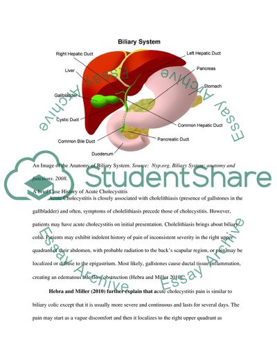
Anatomy of Biliary System Introduction Acute cholecystitis refers to an inflamed gallbladder, which results in pain, particularly in the right hypochondrium, usually with associated fever and leucocytosis (Keus, Broeders and Laarhoven 2006, 1040). It is a commonplace cause of pain in the right upper quadrant and normally affects younger women (less than 50 years of age) (Cheng, Ng and Shih 2004). Gallstones are more common in women compared to men and in both sexes, they become more common with age. Acute cholecystitis can arise in gallstone disease setting or in critically ill patients without gallstones (Health.allrefer.com 2010). The biliary system consists of the gallbladder, bile ducts, and associated structures, which are the organs and ducts concerned with bile production and transportation. Bile transportation follows a sequence: following its secretion by the liver cells, a system of ducts that ultimately drain into the common hepatic duct collect bile. These ducts flow from the liver all the way through the right and left hepatic ducts. The common hepatic duct connects with the cystic duct from the gallbladder forming the common bile duct, which runs to the duodenum from the liver. Not all bile, however, runs directly into the duodenum. The gallbladder first stores approximately 50% of the bile that the liver produces. This pear-shaped organ located directly below the liver. Subsequently, the gallbladder releases stored bile into the duodenum through contraction in order to help break down fats in foods (Nyp.org 2008). An Image of the Anatomy of Biliary System. Source: Nyp.org. Biliary System: anatomy and functions. 2008. A brief Case History of Acute Cholecystitis Acute Cholecystitis is closely associated with cholelithiasis (presence of gallstones in the gallbladder) and often, symptoms of cholelithiasis precede those of cholecystitis. However, patients may have acute cholecystitis on initial presentation. Cholelithiasis brings about biliary colic. Patients may exhibit indolent history of pain of inconsistent severity in the right upper quadrant of their abdomen, with probable radiation to the back’s scapular region, or pain may be localized or diffuse to the epigastrium. Most likely, gallstones cause ductal tissue inflammation, creating an edematous bile flow obstruction (Hebra and Miller 2010). Hebra and Miller (2010) further explain that acute cholecystitis pain is similar to biliary colic except that it is usually more severe and continuous and lasts for several days. The pain may start as a vague discomfort and then it localizes to the right upper quadrant as inflammation spreads, affecting the surrounding peritoneum. Often, a recent history of vomiting, nausea, a low-grade fever and anorexia is common in acute cholecystitis patients. Usually symptoms start approximately a week before presentation, although the patient may report years of the less aggravated chronic cholecystitis and biliary colic symptoms. On physical examination, the right upper quadrant of an acute cholecystitis patient is usually tender. Pain in the right upper quadrant, fever, and leukocytosis is the classic triad and it is indicative of the common bile duct obstruction. The patient may have a positive Murphy sign and abdominal guarding. Cholangitis may result from the inflammation of the ductal system and in half of these cases; the physician may find a Charcot triad, which is considered a medical emergency (Hebra and Miller 2010). Etiology The presence of gallstones in the gallbladder is the major cause of acute cholecystitis – evident in ninety percent of cases. Other causes include ischemia, severe illness, parasites, and protozoa, motility disorders associated with drugs; chemicals that enter biliary secretions; infections with microorganisms, allergic reactions and collagen disease. Factors associated with the onset of cholelithiasis include drugs such as oral contraceptives, age, obesity as well as, infrequently, gallbladder tumors may also cause cholecystitis (Longstreth 2010). Epidemiology Acute cholecystitis’ incidence is more or less alike in Western Europe as in the United States. However, the exact incidence globally is unknown. Owing to the close relationship between gallstones and acute cholecystitis, the distribution and incidence of acute cholecystitis follow that of cholelithiasis. Between 2007 and 2008, 15,916 cholecystitis cases were reported in the United Kingdom and approximately two-thirds of the cases were in females. Over twenty million Americans are projected to have gallstones, with half a million cholecystectomies being carried out per annum. A great percentage of patients with gallstones do not develop symptoms. Every year, roughly one to two percent of asymptomatic gallstones become symptomatic. Acute cholecystitis is the most common gallstones complication and occurs in ten percent of symptomatic patients (Bmj.com 2010). Clinical diagnosis Physicians usually use radiological tests to make the diagnosis of acute cholecystitis. Imaging tests that can reveal inflammation or gallstones include Abdominal CT scan, abdominal ultrasound, gallbladder radionuclide scan, cholecystoscintigraphy (HIDA), oral cholecystogram and abdominal x-ray. The most commonly used radiologic modalities in the diagnosis of acute cholecystitis are cholecystoscintigraphy and Ultrasonography (US) (Longstreth 2010). Ultrasonography and cholecystoscintigraphy, and the combination of the two can enhance acute cholecystitis’ clinical diagnostic accuracy. While cholecystoscintigraphy provides information on gallbladder nonvisualization, which signifies cystic duct obstruction, which is then used to diagnose acute cholecystitis, ultrasonography provides information regarding pericholecystic fluid, the gallbladder wall and presence of gallstones (Kalimi, et al. 2001). Role of Imaging Modalities in the Diagnosis of Acute Cholecystitis Sonographic examination of the gallbladder is the first imaging modality carried out (Cheng, Ng and Shih 2004, 131). However, due to these imaging modalities’ varying specificity (40%–95%) and sensitivity (40%–90%), these tests’ results can be inconclusive in some patients. Computed tomography (CT) has proved to have high specificity and sensitivity (ninety-five percent for both) for the diagnosis of acute cholecystitis although it has key limitations such as potential nephrotoxic effects from iodinated contrast media, limited soft-tissue contrast resolution and radiation exposure (Tsai et al. 2009, 575). Consequently, given its characteristic superior soft-tissue contrast resolution devoid of nephrotoxicity magnetic resonance or radiation exposure risks, magnetic resonance imaging may have a role to play in cholecystitis’ diagnosis. However, in the diagnosis of acute cholecystitis, few reports describe the use of magnetic resonance imaging (Altun et al. 2007).  Sonographic Picture of Acute Cholecystitis. Source: Ko, C. W. 2003. Biliary Sludge and Cholecystitis, 390. Cheng, Ng and Shih (2004) point out that in some cases with complicated presentation that may bring about a diagnostic dilemma or those with equivocal findings, CT may be necessary. Unal (2009) assert that in those areas where sonogram is unavailable, CT may act as the first or primary imaging tool for ruling out other causes of epigastralgia such as hepatitis, peptic ulcer disease, pancreatitis, among others.  CT scan of the abdomen showing the thickened gallbladder wall (arrow). Source: Unal, H. et al. 2009. Acute acalculous cholecystitis associated with acute hepatitis B virus infection. According to Keus, Broeders and Laarhoven (2006), a percutaneous suction of bile from the gallbladder might be diagnostic as well as therapeutic. In due course, the diagnosis is derived from a blend of clinical signs, laboratory findings in addition to imaging techniques. The diagnosis may not be easy, particularly in patients who are critically ill. To some extent, the role of the different imaging modalities in acute cholecystitis remains controversial. Riaz and Emran (2010) assert that generally, there is no single ideal imaging study. Cholescintigraphy, ultrasound and CT scan (the three primary imaging modalities) are often complementary. According to Mirvis and his co-authors (1986), computed tomography and sonography are over ninety-percent specific and sensitive for acute cholecystitis diagnosis, while cholescintigraphy is only thirty-eight percent specific despite being highly sensitive. The most important factors to take into account are early diagnosis and treatment with the intention of reducing morbidity as well as the high death toll of acute cholecystitis, which has a tendency of appearing during the first week and month following surgery (Lombao et al. 2007, 162). Pathology The underlying pathology of acute cholecystitis is cystic duct obstruction, giving rise to raised hydrostatic pressure in the gallbladder, necrosis and inflammation (Jonnalagadda 2009, 1). Acute cholecystitis leads to the trapping of bile in the gallbladder and the build-up of bile causes pressure and irritation within the gallbladder (Health.allrefer.com 2010). This can lead to perforation of the gallbladder and bacterial infection. Perforation of the gallbladder is one of the most severe acute cholecystitis’ complications and high mortality and morbidity associated with it have been reported (Tsai et al. 2009, 575). Acute cholecystitis brings about pain, particularly in the right hypochondrium, usually accompanied by leucocytosis and fever. The pain may be continuous and enduring, not colicky. Bilirubin and liver enzymes may be slightly higher. At times (in some patients), on the performance of an ultrasound, there may be a hypoechogenic rim (halo) and thickened gallbladder wall. Complications of acute cholecystitis include abscess formation/pericholecystic abscess or empyema, perforation of the gallbladder, progression to gangrene and fistula brought about by gallbladder wall ischaemia and infection (Spira et al. 2002, 64), (Fialkowski, Halpin and Whinney 2008). Some of the factors that contribute to acute cholecystitis development include biliary crystals, biliary sludge, supersaturated bile, bile-duct obstruction, vascular compromise and bacteria. Cystic duct obstruction, which leads to an increase in intra-luminal pressure and swelling, is deemed the most important factor. There ensues a disruption to normal defence mechanisms in the epithelium resulting in gallbladder wall’s exposure to bacteria. Another contributing factor may be vascular compromise, particularly in seriously ill patients exposed to hypotension episodes. Reperfusion injury, ischaemia and reduced blood flow lead to gallbladder damage, reducing its natural anti-inflammatory resistance (Keus, Broeders and Laarhoven 2006, 1040). Treatment Acute cholecystitis is a high burden in surgical departments and a frequent cause for emergency admission. Generally, patients with acute cholecystitis are admitted to the hospital where they receive supportive care, including pain control and hydration. Physicians administer antibiotics to most of them during hospitalization (Kanafan et al., 2005). Traditionally treatment of Acute cholecystitis patients has been either by instantaneous cholecystectomy, or conservatively with antibiotics until the infection subsides. In up to eighty-six percent of the cases, this has been successful. Cholecystectomy was applied to patients who failed to respond to the conservative medical treatment. For patients who were not fit to go through resection, physicians deemed operative cholecystostomy an extreme measure. While cholecystostomy was the definitive procedure in some patients, others underwent cholecystectomy later (Paran et al. 2006, 102). In the commencement of the era of laparoscopic cholecystectomy, gall bladder acute inflammation was considered laparoscopic resection’s contra-indication. Here, delayed elective surgery and conservative treatment of acute cholecystitis became an acceptable choice. Later with continued advancement in laparoscopic surgical skills, many authors reported excellent results with emergency laparoscopic inflamed gall bladder resection with a recent meta-analysis suggesting that in the treatment of acute cholecystitis, early operation is the best alternative. However, routine laparoscopic cholecystectomy in the emergency setting has proved to be a challenge. This is especially so in public hospitals, where theatre space is scarce and the services of a specialist are not always on hand. Additionally, the reported rate of conversion to open surgery is comparatively high even when a specialist-led service is readily accessible (Paran et al. 2006, 102). Generally, physicians use ultrasound guided percutaneous cholecystostomy (PC) as the standard treatment of acute cholecystitis. They often perform a percutaneous cholecystostomy on critically ill patients as a temporizing measure, in addition to administering antibiotics for management. This is before eventually performing definitive surgery. The management of critically ill patients with acute cholecystitis has been difficult as owing to their co-morbid conditions, many patients are poor operative candidates (Ko 2003, 389). In patients such as those with a terminal illness in whom a percutaneous tube is not acceptable or in cirrhotics awaiting liver transplant, there is a mounting experience with internal gallbladder drainage at the radiology suite or even bedside. This is because the aforementioned options are not desirable. Gallbladder drainage results in less pain, a shorter stay in hospital, in addition to increased chances for a successful conservative inflammation cooling down (Keus, Broeders and Laarhoven 2006, 1039). The inflamed gallbladder can be drained into the gut either transluminally during endoscopic ultrasonography (EUS) or at endoscopic cholangiography (ERCP) through the major papilla (transpapillary drainage). When successful, the latter has proved to be a safe and effective option. The experience with endoscopic ultrasonography-guided gallbladder drainage is limited and is linked with a higher risk of complications. Physicians should only perform it if necessary (Jonnalagadda 2009, 1). Concisely, in the treatment of acute cholecystitis, conservative treatment as well as delayed operation is still an acceptable option. Percutaneous cholecystostomy is a good, simple and effective procedure, especially when applied early by an expert laparoscopist. It has low morbidity and a high rate of success, and permits safe interval laparoscopic surgery. Elective delayed laparoscopic surgery following percutaneous gall bladder drainage is safe and has a low rate of conversion (Strasberg, 1997, 656). For patients whose symptoms fail to progress or respond following percutaneous cholecystostomy therapy, physicians are advised to consider emergent cholecystectomy. Physicians should also consider non-operative treatments for instance oral bile acid dissolution while treating patients who pull through the acute episode, but who are poor operative candidates (Ko 2003, 389). Prognosis In most of cases, acute cholecystitis’ prognosis is favourable. Even from gallstones with symptoms, death is very rare. Serious complications are also uncommon and if they occur, they usually develop after surgery or from stones in the bile duct (UMMC 2010). In patients below fifty years, cholecystectomy, whether open surgery or laparoscopic, is linked with a 0.1 percent mortality rate while in patients above 50 years, it is linked with a 0.8 percent mortality rate. a shorter hospital stay and recovery period, as well as less pain are characteristic of the less invasive laparoscopic procedure compared to the open procedure. In 75% - 90% of cases, cholecystectomy provides a full resolution of symptoms. Gallstones may occur in the bile ducts following cholecystectomy, a condition called choledocholithiasis (MDguidelines.com 2010). In cases in which cholecystitis is treated with medication only i.e. conservatively/nonsurgically, acute cholecystitis will recur in twenty-five percent of patients within a year, while it will recur in sixty percent of the patients within 6 years. Reports also indicate that depending on the specifics of the complication, complicated cases of cholecystitis such as acute cholecystitis, acalculous cholecystitis, gangrenous cholecystitis, sepsis or secondary pancreatitis, critically ill patients and those with perforated gallbladder have a less favourable prognosis, with death rate as high as 50% - 60% (MDguidelines.com 2010). Summary and Conclusion It is important to note that acute cholecystitis’ prognosis is favourable especially when experienced surgeons and appropriate support are available. As earlier discussed, it is apparent that high-density gallbladder wall sign is extremely sensitive for acute cholecystitis – it is a specific sign for acute cholecystitis. Recognition of this sign therefore assists in earlier detection of this condition. Early diagnosis and management of acute cholecystitis is of vital significance since delayed treatment makes it to progress rapidly to perforation or gangrenous cholecystitis, which have very high mortality. A timely diagnosis depends on properly interpreted imaging as well as a high index of suspicion in the appropriate patient, as well as the combined clinical findings results. Therefore, as Tsai and his co-authors suggest, in the management of acute cholecystitis, especially with gallbladder perforation (the most severe acute cholecystitis’ complications), early diagnoses as well as early surgical intervention are the key to decrease high mortality and morbidity associated with it. Bibliography Altun., et al. 2007. Acute cholecystitis: MR findings and differentiation from chronic cholecystitis. Journal of Radiology 244: 174-183. Bmj.com. Cholecystitis. 2010. < http://bestpractice.bmj.com/best-practice/monograph/78/basics/epidemiology.html>. Cheng, S. N., Suk-Ping, and S. Shin-Lin. 2004. Hyperdense gallbladder wall sign: An overlooked sign of acute cholecystitis on unenhanced CT examination.” Journal of Clinical Imaging 28: 128–131. Fialkowski, E., V. Halpin, and R.Whinney. 2008. Acute cholecystitis. http://clinicalevidence.bmj.com/ceweb/conditions/dsd/0411/0411_background.jsp (accessed October 8, 2010) Gakwaya, A.M., and J. Jombwe. 2006. Acute cholecystitis – current views. East and Central African Journal of Surgery 11(1): 99-101. Health.allrefer.com. 2010. Acute cholecystitis. http://health.allrefer.com/health/acute-cholecystitis-gallstones-info.html (accessed October 8, 2010) Hebra, A., and M. Miller. 2010. Cholecystitis. http://emedicine.medscape.com/article/927340-overview (accessed October 8, 2010) Jonnalagadda, S. 2009. Endoscopic therapy of acute cholecystitis. Techniques in Gastrointestinal Endoscopy 11: 13-18. Kalimi, R., et al. 2001. Diagnosis of acute cholecystitis: Sensitivity of sonography, cholescintigraphy, and combined sonography-cholescintigraphy. Journal of Am Coll Surg 193(6): 609-612. Kanafani, Z. A. 2005. Antibiotic use in acute cholecystitis: Practice patterns in the absence of evidence-based guidelines. Journal of Infection 51: 128–134. Keus, F., I. A. M. J. Broeders and C. J. H. M. Laarhoven. 2006. Surgical aspects of symptomatic cholecystolithiasis andacute cholecystitis. Best Practice & Research Clinical Gastroenterology 20(6): 1031-1051. Ko, C. W., 2003. Biliary sludge and cholecystitis. Best Practice & Research Clinical Gastroenterology 17(3): 383–396. Lombao, D., et al. 2007. Acute Cholecystitis as cause of death after surgery for lumbar canal stenosis. Rev Ortop Traumatol (Madr.) 51: 158-63. Longstreth, G. F. 2010. Acute cholecystitis. http://www.nlm.nih.gov/medlineplus/ency/article/000264.htm (accessed October 8, 2010) MDguidelines.com. 2010. Cholecystitis. http://www.mdguidelines.com/cholecystitis/prognosis (accessed October 8, 2010) Mirvis, S. E., et al., 1986. The Diagnosis of Acute acalculous cholecystitis: a comparison of sonography, scintigraphy, and CT. American Journal of Roentgenology 147(6): 1171-1175. Nyp.org. Biliary System: anatomy and functions. 2008. . Paran, H., et al. 2006. Prospective evaluation of patients with acute cholecystitis treated with percutaneous cholecystostomy and interval laparoscopic cholecystectomy. International Journal of Surgery 4: 101-105. Riaz, M. M., and F. Emran. 2010. Pitfalls of diagnosis of acalculous cholecystitis. case presentation and literature review. The Internet Journal of Surgery 23(2). Spira, R. M., et al. 2002. Percutaneous transhepatic cholecystostomyand delayed laparoscopic cholecystectomy in critically ill patients with acute calculus cholecystitis. The American Journal of Surgery 183: 62–66. Strasberg, S. M. 1997. Cholelithiasis and acute cholecystitis.” Bailliere’s Clinical Gastroenterology 11(4): 643-660. Tsai, M., et al. 2009. Can acute cholecystitis with gallblader perforation be detected preoperatively bycomputed tomography in ED? American Journal of Emergency Medicine 27: 574–581. UMMC. 2010. Gallstones and gallbladder disease - prognosis and complications. http://www.umm.edu/patiented/articles/how_serious_gallstones_gallbladder_disease_000010_3.htm (accessed October 8, 2010) Unal, Hakan, et al., 2009. Acute acalculous cholecystitis associated with acute hepatitis B virus infection. International Journal of Infectious Diseases 13: 310-312. Read More
Cite this document
  • APA
  • MLA
  • CHICAGO
(“Anatomy of Biliary System Essay Example | Topics and Well Written Essays - 2750 words”, n.d.)
Anatomy of Biliary System Essay Example | Topics and Well Written Essays - 2750 words. Retrieved from https://studentshare.org/health-sciences-medicine/1570674-acute-cholecystitis-case-study
(Anatomy of Biliary System Essay Example | Topics and Well Written Essays - 2750 Words)
Anatomy of Biliary System Essay Example | Topics and Well Written Essays - 2750 Words. https://studentshare.org/health-sciences-medicine/1570674-acute-cholecystitis-case-study.
“Anatomy of Biliary System Essay Example | Topics and Well Written Essays - 2750 Words”, n.d. https://studentshare.org/health-sciences-medicine/1570674-acute-cholecystitis-case-study.
  • Cited: 0 times

CHECK THESE SAMPLES OF Anatomy of Biliary System

Anatomy of Human Body, Liver

In human anatomy Liver, although an accessory organ, is the largest organ in our body.... It constitutes about 1/50 of the entire weight of the body.... The weight of liver in an adult is approximately 1500 gms.... Thousands of functional units called 'liver lobules' are there in liver....
8 Pages (2000 words) Essay

Holistic Physiotherapy Management

Nevertheless significant differences are also present. … One of the pathologies occurring in respiratory system is CF syndrome.... If CF disorder progresses it may cause also pancreatic insufficiency and focal biliary cirrhosis.... It is apparent that the case of a teenage cystic fibrosis (CF) patient admitted to hospital with a worsening productive cough has several similar traits with the case of elderly patient admitted with pneumonia....
9 Pages (2250 words) Essay

Treaty of Versaille and World War II

The culprit, Germany, was to make reparations and must pay for all the trouble it got the world into.... Germany was, of course, not happy about the… Many critics find the Treaty of Versailles flawed and weak. Until now, the issue: Was the Treaty of Versailles Responsible for World War II?...
8 Pages (2000 words) Essay

Leonardo Da Vinci's Influence on Western Civilization

“His four main areas of study resulted in what is known as his Treatises, on painting, architecture, mechanics, and human anatomy” (Mason, 2004: 21).... The essay "Leonardo Da Vinci's Influence on Western Civilization" evaluates the impact of the paintings by Leonardo Da Vinci on succeeding Western cultures....
7 Pages (1750 words) Essay

Cases of Bile Duct Blockage and Subsequent Acute Cholecystitis

This obstruction can lead to issues with the bile system, which involve a thickening of the bile and associated stasis.... The paper 'Cases of Bile Duct Blockage and Subsequent Acute Cholecystitis' presents acute cholecystitis which is an inflammation of the gallbladder that is characterized by a sharp pain in the upper right quadrant....
8 Pages (2000 words) Case Study

Global Change Threatens the Liberal Order

The system is now facing some sort of a crisis where there is confusion regarding power roles in maintenance of the international liberal order.... The optimist view for international liberal order goes forth to discuss that all states have the ability to overcome constraints with a view to cooperate, thereby resolving security issues, working towards collective action and consequently, developing a stable and open system (Steins, 2004).... The political system of the western world, including currency, technology, alliances and markets, has been fused with the liberal order for providing a base for liberal internationalisation....
5 Pages (1250 words) Assignment

Leonardo da Vinci: A Man of His Times or a Visionary

“His four main areas of study resulted in what is known as his Treatises, on painting, architecture, mechanics and human anatomy” (Mason, 2004: 21).... In the paper “Leonardo da Vinci: A Man of His Times or a Visionary?... rdquo; the author looks at a great painter who lived during the time of the Italian Renaissance, making significant contributions to the world of art....
7 Pages (1750 words) Assignment

The Peculiarities of Human Body

Ciliary muscles provide attachment to the irisIridocorneal angle contains meshwork of cells, it contains Schlemm's canal which carries out AH from the anterior chamber into the venous system, the process is called aqueous drainage.... This work "The Peculiarities of Human Body" describes the structure and function of the eye and ear, the changes that occur within a nerve cell, and across its membrane during the process of nerve impulse initiation....
6 Pages (1500 words) Assignment
sponsored ads
We use cookies to create the best experience for you. Keep on browsing if you are OK with that, or find out how to manage cookies.
Contact Us