StudentShare
Contact Us
Sign In / Sign Up for FREE
Search
Go to advanced search...
Free

Cases of Bile Duct Blockage and Subsequent Acute Cholecystitis - Case Study Example

Cite this document
Summary
The paper 'Cases of Bile Duct Blockage and Subsequent Acute Cholecystitis' presents acute cholecystitis which is an inflammation of the gallbladder that is characterized by a sharp pain in the upper right quadrant. It is usually caused by the presence of gallstones…
Download full paper File format: .doc, available for editing
GRAB THE BEST PAPER96.8% of users find it useful
Cases of Bile Duct Blockage and Subsequent Acute Cholecystitis
Read Text Preview

Extract of sample "Cases of Bile Duct Blockage and Subsequent Acute Cholecystitis"

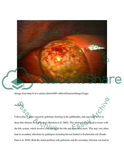
Acute cholecystitis is an inflammation of the gallbladder that is characterized by a sharp pain in the upper right quadrant. It is usually caused by the presence of gallstones which block the bile duct, leading to a build up of bile and subsequent inflammation (Sherlock et al, 2002). There may also be bacterial species present, including Escherichia coli or Salmonella spp (Eldar et al, 1997). The syndrome is usually mild and has a good prognosis, although complications can lead to gangrene and tissue necrosis, which are more dangerous and lethal (South-Paul et al, 2010). The disease is fairly common, with over 500,000 operations being performed each year in the United States on individuals presenting with more severe cases of bile duct blockage and subsequent acute cholecystitis. Acute cholecystitis is the case of this inflammation that occurs sporadically, with the similar chronic cholecystitis being characterised by a more constant inflammation (South-Paul et al, 2010). Acute cholecystitis is far more common in women than in men, and occurs in the population more frequently with age (Sherlock et al, 2002). Pregnancy can also increase the liklihood of getting acute cholecystitis due to the presence of hormones that increase the liklihood of developing gallstones and the subsequent blocking and inflammation (Lai et al, 1998). There are many associated symptoms which are usually vague, and include nausea and fever (Eldar et al, 1997). Acute cholecystitis can be diagnosed in a number of ways that will be discussed within the course of this essay, but imaging techniques are the most conclusive, with metabolic panels being used as the primary detection method in suspected cases (Sherlock et al, 2002). Imaging techniques are the only way to get a conclusive diagnosis of the affliction. Treatment is usually antibiotics, although more severe and aggressive treatments are necessary in cases where gangrene and tissue necrosis are present. Operations may be needed to remove the causative gallstones (Kiviluoto et al, 1998). This essay will discuss all of these features of acute cholecystitis in more detail. (Image from http://www.uninet.edu/cin2001-old/conf/moreno/Image18.jpg) Aetiology Colecystitis is often caused by gallstones forming in the gallbladder, and especially refers to those that obstruct the custic duct (Sherlock et al, 2002). This obstruction can lead to issues with the bile system, which involve a thickening of the bile and associated stasis. This may very often lead to secondary infections by pathogens including but not limited to Escherichia coli (South-Paul et al, 2010). Both the initial problem with gallstones and the secondary infection can lead to to the inflammation that is characteristic of cholecystitis, and especially cases of acute cholecystitis. It is interesting to note that the incidence is higher in females, and this has been linked to higher levels of progesterone during the pregnancy period (Eldar et al, 1997). Epidemiology Around 10-20% of Americans have gallstones (Sherlock et al, 2002) and this is mirrored in countries with a similar breakdown of age, ethnicity and wealth (Eldar et al, 1997). From these individuals, around 30% will develop acute cholecystitis (South-Paul et al, 2010) and it is more likely to affect female than males. In the United States, around 500,000 operations are carried out each year to remove the cholecysts (Ralls et al, 2005). The chances of getting acute cholecystitis increases with age, although the reason for this is not clear. This increasing of incidence with age is more pronounced in elderly men (Lo et al, 1998). Cholelithiasis (gallstones) is more pronounced in white people of Hispanic or Scandinavian descent, and less common in those from Asia or sub-Saharan Africa (Sherlock et al, 2002). A brief case history A 40-year old woman with a weight in the clinically obese category is complaining of pain in the right upper quadrant. She has a history of gallstones, and is running a mild fever of 38C. She is also complaining of nausea. On further investigation, it is evident that there is a mild case of associated jaundice (serum bilirubin levels of 62micromol/L). Due to the sex, age and history of this patient, a metabolic panel with bicarbonate is carried out, as recommended by Sherlock et al, (2002). All counts of aspirate aminotransferase, alanine aminotransferase, bilirubin and alkaline phosphate are normal, although this cannot be used to rule out acute cholecystitis. White blood cell count is 11,000 cells/ul, a common feature of a milder case of inflammation of the gallbladder. Due to these symptoms, the examining physician had a reason to suspect acute colecystitis. An ultrasound was carried out and there appeared to be gallstones and a mild inflammation of the gallbladder, although this was not conclusive from this test. Due to this, an abdominal CT scan was done on the patient, which confirmed the gallstones and the inflammation, leading to a diagnosis of acute colecystitis (Singer et al, 2008) A blood test was also carried out to check for symptoms of gangrene and sceptacemia, both of which were not present in the individual, although there was evidence of E. coli in the patient (Singer et al, 2008) The patient was presumed to have a mild case of acute cholecystitis from the diagnostic measures above. She was treated with a broad-spectrum antibiotic intravenously, as well as being administered intravenous hydration. The patient made a full recovery, and was sent home with instructions for bowel rest and a diet plan to help prevent a recurring case cholecystitis (Singer et al, 2008). Clinical diagnosis There is no conclusive diagnostic tests for acute cholecystitis. However, testing levels of white blood cells, aspirate aminotransferase, alanine aminotransferase, bilirubin and alkaline phosphate may show to be elevated in some cases (South-Paul et al, 2010). However, it must be noted that this does not give a conclusive diagnosis of acute cholecystitis. Singer et al (2008) have shown that there may also be elevated levels of hepatic 2,6-dimethyliminodiacetic acid in some cases, although again this cannot be used as a conclusive diagnostic test. Levels of amylase during testing may be up to three times higher in patients presenting with acute cholecystitis (Sherlock et al, 2002). All these levels can be examined using a metabolic panel with bicarbinate. From all these tests, it appears that the white blood cell count elevation is the most conclusive, with 61% of patients with acute cholecystitis having a count of 11,000 cells/ul. A count of 15,000 cells/ul could indicate gangrene or extreme inflammation (Eldar et al, 1997). The main symptom of acute cholecystitis is an intense pain in the abdomen that can radiate towards the shoulder blade. Other symptoms include fever, nausea, vomiting, loss of appetite and jaundice (South-Paul et al, 2010). The pain in the abdomen is usually enough for a physician to suspect acute cholecystitis, especially if it has lasted for more than 5 hours (which rules out biliary colic) (Yamashita et al, 2007). In patients presenting with this pain, several conditions should be ruled out to ensure that the diagnosis is correct. These include, but are not limited to, abdominal aneurism, pregnancy, eclampsia, hepatitis, gastroenteritis, myocardial infarction, appendicitis, cholangitis, an obstruction of the small bowel, urinary tract infections and pancreatitis (Sherlock et al, 2002). A conclusive diagnosis can also be helped by imaging, with ultrasonography and nuclear medicine being the two best imaging styles (Yamashita et al, 2007). These will usually show both the gallstones and the related inflammation of acute cholecystitis (Sherlock et al, 2002). CT scanning and abdominal radiography can also be used in patients that are suspected of the condition but do not show any signs using the aforementioned imaging techniques (Ralls et al, 2005). Anatomy of billiary system The biliary system includes the bile duct, the pancreas and the liver (Yamashita et al, 2007) and forms part of the digestive system. It is responsible for the absorption of nutrients and the disposal of waste (Eldar et al, 1997). There are also a number of ducts involved in the biliary system. Together, these work to produce and store bile for release into the duodenum (Ralls et al, 2005). The pathway of the system is as follows. The hepatocytes of the liver produce the bile from waste products. This then flows through the hepatic duct and through the cystic duct which leads from the gallbladder (Eldar et al, 1997). These join to form the common bile duct, which in turn joins the pancreatic duct to empty the bile into the duodenum, where it is used for the emulsification of fats in the small intestine (Sherlock et al, 2002). (Image from http://www.diabetesmonitor.com/images/biliary_system.gif) Pathology In acute cholecystitis the gallbladder wall is edematous and is infiltrated by neutrophils (Lo et al, 1998). This can be caused by a number of things, such as a blocking of the bile duct leading to build up of bile causing infection and inflammation (Eldar et al, 1997). The serosa is usually beige in colour and granulated due to the presence of transmural inflammation (Lo et al, 1998). Under the microscope, the mucosa can appear to be partially eroded (Borzellino & Cordiano, 2008). The gallbladder itself may be filled with blood, bile and gallstones, which lead to the inflammation and pain commonly associated with acute cholecystitis (Lo et al, 1998). Role of imaging modalities in the diagnosis of Acute cholecystitis As previously mentioned, imaging modalities are very useful in the diagnosis of acute cholecystitis. The primary method of detection is ultrasound (Bortoff et al, 2000), although numerous other technologies can be used. Ultrasound usually finds a highly reflective echo from the anterior surface of the gallstone, mobility of the gallstone on moving the patient, and marked posterior acoustic shadowing (Bortoff et al, 2000). Additionally, the older but still effective technique of oral cholecystography can be used in many cases (Sherlock et al, 2002). Imaging modalities are extremely useful in many cases because symptoms can be non-existant or vague, as mentioned above, and the only conclusive proof of acute cholecystitis is an image showing the inflammation (Yamashita et al, 2007). In detection of choledocholithiasis, endoscopic retrograde cholangiopancreatography and magnetic resonance cholangiopancreatography are superior to ultrasound (Bortoff et al, 2000) which leads to detection of the inflammation rather than just the gallstones which alone may not cause any symptoms. The image below shows an ultrasound image of acute cholecystitis. (Image from http://www.med-ed.virginia.edu/courses/rad/edus/text%20jpegs1/5b-AcuteChole-.jpg) Treatment and prognosis The treatment for mild cases of acute cholecystitis are generally a broad-spectrum antibiotic, usually administered intravenously. Bowel rest may also be recommended (Yamashita et al, 2007). Additionally, intravenous hydration and electrolyte treatment can be recommended. Operations to remove the offending gallstones is sometimes necessary in more severe cases of the illness, although this is rare (Sherlock et al, 2002). Analgesia is used to reduce the pain in the upper right quadrant that is caused by the affliction (South-Paul et al, 2010). Prognosis is generally very good for patients with acute cholecystitis. Full recovery is expected in the majority of patients with uncomplicated cases (Borzellino & Cordiano, 2008). It has a very low mortality rate (Yamashita et al, 2007). Most cases involve a complete remission within a few days (Sherlock et al, 2002). However, when there are complications, the prognosis is worse and mortality rates sharply rise, especially in cases involving gangrene or tissue necrosis (Borzellino & Cordiano, 2008). Complications may occur in up to 30% of cases, and of these the majority will require hospitalization and possible surgery (Eldar et al, 1997). Summary and Conclusion To summarize, acute cholecystitis is a fairly simple disease with a good prognosis. It is generally caused by a build up of bile due to a blocked bile duct from the presence of gallstones (Sherlock et al, 2002). It occurs more frequently in females than males, and becomes more common with increasing age (Eldar et al, 1997). It can usually be treated simply with antibiotics, although complications may require surgery and hospitalization (Eldar et al, 1997). Imaging techniques remain the only conclusive diagnosis method, although there are other ways to help rule out other diseases presenting with the same main symptoms. These symptoms are primarily pain in the upper right quadrant, although nausea, jaundice, vomiting and fever may also be present (Sherlock et al, 2002). References Bortoff, Gregory A., Michael Y. M. Chen, David J. Ott, Neil T. Wolfman, and William D. Routh. 2000. “Gallbladder Stones: Imaging and Intervention1.” Radiographics 20 (3) (May 1): 751 -766. Borzellino, Giuseppe, and Claudio Cordiano. 2008. Biliary Lithiasis: Basic Science, Current Diagnosis and Management. Springer. Eldar, S., E. Sabo, E. Nash, J. Abrahamson, and I. Matter. 1997. “Laparoscopic cholecystectomy for acute cholecystitis: prospective trial.” World journal of surgery 21 (5): 540–545. Gurusamy, K., K. Samraj, C. Gluud, E. Wilson, and BR Davidson. 2010. “Meta-analysis of randomized controlled trials on the safety and effectiveness of early versus delayed laparoscopic cholecystectomy for acute cholecystitis.” British journal of surgery 97 (2): 141–150. Kiviluoto, T., J. Sirén, P. Luukkonen, and E. Kivilaakso. 1998. “Randomised trial of laparoscopic versus open cholecystectomy for acute and gangrenous cholecystitis.” The Lancet 351 (9099): 321–325. Lai, PBS, KH Kwong, KL Leung, SPY Kwok, ACW Chan, SCS Chung, and WY Lau. 1998. “Randomized trial of early versus delayed laparoscopic cholecystectomy for acute cholecystitis.” British journal of surgery 85 (6): 764–767. Lo, C.M., C.L. Liu, S.T. Fan, EC Lai, and J. Wong. 1998. “Prospective randomized study of early versus delayed laparoscopic cholecystectomy for acute cholecystitis.” Annals of surgery 227 (4): 461. Lujan, JA, P. Parrilla, R. Robles, P. Marin, JA Torralba, and J. Garcia-Ayllon. 1998. “Laparoscopic cholecystectomy vs open cholecystectomy in the treatment of acute cholecystitis: a prospective study.” Archives of Surgery 133 (2): 173. Neugebauer, Edmund A., and Abe Fingerhut. 2006. EAES guidelines for endoscopic surgery: twelve years evidence-based surgery in Europe. Springer. Norrby, S., P. Herlin, T. Holmin, R. Sjödahl, and C. Tagesson. 1983. “Early or delayed cholecystectomy in acute cholecystitis? A clinical trial.” British Journal of Surgery 70 (3): 163–165. Ralls, PW, PM Colletti, SA Lapin, P. Chandrasoma, WD Boswell, C. Ngo, DR Radin, and JM Halls. 2005. “Real-time sonography in suspected acute cholecystitis. Prospective evaluation of primary and secondary signs.” Radiology 155 (3): 767. Schein, Clarence J. 1972. Acute cholecystitis. Medical Dept., Harper & Row. Sherlock, Sheila, Sheila Sherlock (Dame.), and James S. Dooley. 2002. Diseases of the liver and biliary system. Wiley-Blackwell. South-Paul, Jeannette, Samuel C. Matheny, Evelyn L. Lewis, and Evelyn Lewis. 2010. Current diagnosis & treatment in family medicine. McGraw-Hill Prof Med/Tech, November 23. Strasberg, S.M. 2008. “Acute calculous cholecystitis.” New England Journal of Medicine 358 (26): 2804–2811. Yamashita, Y., T. Takada, Y. Kawarada, Y. Nimura, M. Hirota, F. Miura, T. Mayumi, et al. 2007. “Surgical treatment of patients with acute cholecystitis: Tokyo Guidelines.” Journal of hepato-biliary-pancreatic surgery 14 (1): 91–97. Read More
Cite this document
  • APA
  • MLA
  • CHICAGO
(Cases of Bile Duct Blockage and Subsequent Acute Cholecystitis Case Study, n.d.)
Cases of Bile Duct Blockage and Subsequent Acute Cholecystitis Case Study. Retrieved from https://studentshare.org/health-sciences-medicine/1580633-acute-cholecystitis-case-study
(Cases of Bile Duct Blockage and Subsequent Acute Cholecystitis Case Study)
Cases of Bile Duct Blockage and Subsequent Acute Cholecystitis Case Study. https://studentshare.org/health-sciences-medicine/1580633-acute-cholecystitis-case-study.
“Cases of Bile Duct Blockage and Subsequent Acute Cholecystitis Case Study”. https://studentshare.org/health-sciences-medicine/1580633-acute-cholecystitis-case-study.
  • Cited: 1 times

CHECK THESE SAMPLES OF Cases of Bile Duct Blockage and Subsequent Acute Cholecystitis

Analysis of Brians and Alberts Tort Case and Jamess Dismissal and Subsequent Recourse

While some lawyers of other disciplines occasionally take on personal injury cases that appear promising, many focus on one or more specialty within the field”.... In advising Cretins Ltd.... this paper is an analysis of what happened and: i) what the outcome should be for the injuries sustained by Brian and Albert at the skating rink on 29th October; and ii) what the implications are for the dismissal of James....
8 Pages (2000 words) Case Study

Horaces Blockades -- To Rent or Buy

The dilemma is faced after the Horace Company made a bid for the provision of barricades for the new stretch of Interstate 752 that is planned to replace the FL highway 103 out… Barricades are required to provide blockage for a 10 mile length road that is to be built along one side of the highway and shall require shutting down one lane of the road so as to enable its construction per lane.... Barricades are required to provide blockage for a 10 mile length road that is to be built along one side of the highway and shall require shutting down one lane of the road so as to enable its construction per lane....
2 Pages (500 words) Case Study

The Reason One Experiences Pain in the Right Upper Quadrant

The gall bladder has the cystic duct through which bile flows into the duodenum.... A blockade of this duct means that bile cannot pass through.... Because of the existence of the gallstone on the cystic duct, the contraction causes pain (Crutchlow, Dudac, MacAvoy and Madara 213).... This is a storage pouch that keeps bile from the liver.... bile is a liquid used in food digestion.... The sack, which store bile, was trying to push the bile… The duodenum is the first part of the gut after the stomach....
1 Pages (250 words) Case Study

Myocardial Infarction - Mr Canton

Percutaneous Transluminal Coronary Angioplasty Improves Survival in acute Myocardial Infarction Complicated by Cardiogenic Shock.... These result from exhaustion of the heart, heart attack, artery blockage in the lungs, internal bleeding, pneumonia, dehydration, drug overdose causing heart function to reduce, and reduced blood pressure (Lee Linda, Bates, Pitt, & Walton, 2010).... uestion 2: In many cases, when oxygen lacks in a person's heart, particularly because of heart attack, the major pumping area, the left ventricle becomes damaged....
3 Pages (750 words) Case Study

Images of the Interior of the Body

The paper 'Images of the Interior of the Body' presents ultrasound imaging, also known as ultrasound scanning or sonography which is the viewing of images of the interior of the body by the use of high-frequency sound waves.... Ultrasound scanning is useful to visualize various organs of the body....
7 Pages (1750 words) Case Study

Specalist Radiographic Imaging

The subsequent liver tests undertaken on the patient are deranged, thus unable to give a clear indication of the decease the patient is suffering from.... The focus of this essay "Specialist Radiographic Imaging" is to determine the most suitable imaging strategy applicable for diagnosing this patient, which will also entail the evaluation of the appropriate diagnosis procedure and the applicable disease management measures....
6 Pages (1500 words) Case Study

Digestion System and Subsequent Diseases and Problems

hellip; Digestion is the process of refining large particles of food by physical and chemical refinement and subsequent absorption of resultant nutrients from the process.... The fluids secreted by the body includes the saliva, mucus, hydrochloric acid, enzymes, and bile.... The liver then produces bile which further helps in the breakdown of fats into molecules....
8 Pages (2000 words) Case Study

Conflict Resolution Approaches and the Subsequent Process

The conflict taking place in this paper "Conflict Resolution Approaches and the subsequent Process" came about due to the construction of a mosque near a residential area.... The parties involved are the residents, the Imam, and the architect building the mosque.... hellip; The approach used to resolve the arising conflict was mediation....
9 Pages (2250 words) Case Study
sponsored ads
We use cookies to create the best experience for you. Keep on browsing if you are OK with that, or find out how to manage cookies.
Contact Us