StudentShare
Contact Us
Sign In / Sign Up for FREE
Search
Go to advanced search...
Free

Microbiological infections - Essay Example

Cite this document
Summary
This paper discusses other microbiological infections that may have been considered before the microbiology laboratory results were available. It also, gives reasons for their consideration and reasons for their elimination. Based on the history and physical examination of the patient, my primary working impression for this particular case is Herpes zoster. …
Download full paper File format: .doc, available for editing
GRAB THE BEST PAPER98.4% of users find it useful
Microbiological infections
Read Text Preview

Extract of sample "Microbiological infections"

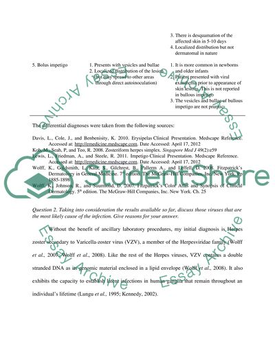
?Question Discuss other microbiological infections that may have been considered before the microbiology laboratory results were available. Give reasons for their consideration and reasons for their elimination. Based on the history and physical examination of the patient, my primary working impression for this particular case is Herpes zoster. However, other microbiological infections that can be considered as differentials are the following: Disease Entity Reasons for ruling in Reasons for ruling out 1. Herpes zoster 1. Unilateral grouped vesicles 2. Dermatomal presentation 3. Neuropathic pain localized on the affected dermatome prior to the development of vesicles 4. Pruritic lesions 5. With prodromal stage prior to appearance of vesicle (i.e. tired and run down) 2. Zosteriform Herpes Simplex Virus eruption 1.Grouped vesicles on an erythematous base 2. Dermatomal distribution 3. Pruritic vesicles 4. Neuropathic pain localized on the affected dermatome prior to the development of vesicles 5. Presence of constitutional signs and symptoms (i.e.tired and run down) 3. Contact Dermatitis 1. May present as acute vesicular allergy 2. Area affected may be painful or pruritic 1. No history of contact to irritants was elicited 2. Contact dermatitis does not present with neuropathic pain prior to the contact 3. Contact dermatitis does not present with viral prodrome 4. Erysipelas 1. Presents with vesicles and bullae 2. With prodromal symptoms prior to onset of lesions 3. Lesions may be itchy 1. Heralded by painful erythematous, well- demarcatedad plaques with advancing borders.(the vesicles develop on top of the erythematous plaques). 2. Presents with lymphadenopathy 3. There is desquamation of the affected skin in 5-10 days 4. Localized distribution but not dermatomal in nature 5. Bolus impetigo 1. Presents with vesicles and bullae 2. Localized distribution of the lesion (but may spread to other areas through direct autoinoculation) 1. It is more common in newborns and older infants 2. Patient presented with viral exanthema prior to appearance of skin lesions. This is not reported in bullous impetigo 3. The vesicles and bullae of bullous impetigo are not pruritic The differential diagnoses were taken from the following sources: Davis, L., Cole, J., and Benbenisity, K. 2010. Erysipelas Clinical Presentation. Medscape Reference. Accessed at: http://emedicine.medscape.com. Date Accessed: April 17, 2012 Koh, M., Seah, P, and Teo, R. 2008. Zosteriform herpes simplex. Singapore Med J 49(2):e59 Lewis, L., Friedman, A., and Steele, R. 2011. Impetigo Clinical Presentation. Medscape Reference. Accessed at: http://emedicine.medscape.com. Date Accessed: April 17, 2012 Wolff, K., Goldsmith, L, Katz, S., Gilchrest, B., Pallerm A., and Leffell, D. 2008. Fitzpatrick’s Dermatology in General Medicine. 7th edition. The McGraw-Hill Companies, Inc. New York. Pp 1885-1898 Wolff, K., Johnson, R., and Suurmond, D. 2007. Fitzpatrick’s Color Atlas and Synopsis of Clinical Dermatology. 5th edition. The McGraw-Hill Companies, Inc. New York. Ch. 25 Question 2. Taking into consideration the results available so far, discuss those viruses that are the most likely cause of the infection. Give reasons for your answer. Without the benefit of ancillary laboratory procedures, my initial diagnosis is Herpes zoster secondary to Varicella-zoster virus (VZV), a member of the Herpesviridae family (Wolff et al., 2007; Wolff et al., 2008). Like the rest of the Herpes viruses, VZV contains a double stranded DNA as its genomic material enclosed in a lipid envelope (Wolff et al., 2008). It also exhibits the capacity to establish latent infections in human ganglia that remain throughout an individual’s lifetime (Lungu et al., 1995; Kennedy, 2002). The clinical signs and symptoms of Herpes zoster include an exanthema of general weakness, headache, dysethesia and pain along the affected dermatome (Benbernou et al., 2011). According to Wolff and colleagues (2008), this pre-eruptive pain usually precedes the onset of vesicular lesions by several days. The vesicles are arranged in groups and distributed unilaterally along a dermatome. Since these signs and symptoms are consistent with what Mrs. M has presented in the case, it is possible that VZV is the causative agent. Another virus that can be considered as the etiologic agent of the patient’s disease is Herpes simplex virus (HSV), a double-stranded, enveloped DNA virus from the Herpesviridae family and is closely related to Varicella zoster virus (Fatahzadeh and Schwartz, 2007). Two serotypes, HSV-1 and HSV-2 are implicated in Herpes infection which clinically presents with mucocutaneous lesions (Wolff et al., 2008). Specifically, HSV-1 is more commonly associated with orofacial affectations while HSV-2 often manifests as genital herpes (Wolff et al., 2008). However, the two serotypes can infect both genital and orofacial areas. During the period of acute infection, the replication of the virus at the site of entry results in primary cutaneous lesions, which eventually spread to include the sensory ganglia where latency is established (Fatahzadeh and Schwartz, 2007). Similar to VZV, HSV carries the capability to reactivate when the host’s immune system is down (Fatahzadeh and Schwartz, 2007). More commonly, HSV-2 reactivates preferentially from the sacral ganglia where the rate of reactivation is directly correlated with the amount of viral load that specific ganglion harbors (Wolff et al., 2008). In females, genital herpes can occur in areas other than the genitals, such as the buttocks. The lesions presents as grouped vesicles with erythematous base with dermatomal distribution (Koh et al., 2008). Recurrent herpes infection is preceded by an exanthema of pain and tenderness at the site of affectation (Koh et al., 2008). Sometimes, the patient experience severe ipsilateral sacral neuralgia. Again, if we look at the case of our patient and base the diagnosis on history and physical exam alone, we cannot rule out HSV as a possible etiologic agent. Question 3. Following this result – what is the most likely viral cause of the rash? Describe the classification, morphology and genome of this virus Since the patient was negative for HSV detection test, Herpes simplex virus can now be ruled out as the cause of the rashes. Hence, the final etiologic agent is Varicella zoster virus (VZV). Varicella zoster virus is classified as a member of the Alphaherpesvirus group, under the genus Varicellovirus from the Herpesviridae family (Wauters et al., 2010). Similar to the other Alphaherpesviruses, VZV is fast-growing and cytolytic in nature. It is a large-encapsulated virus measuring 150-200 nm in diameter, with a linear double-stranded DNA genome that expresses approximately 70 proteins (Liesegang, 1999; Harvey et al., 2007; Kumar et al., 2010). In addition, its genome is only 125 kbp in length, making it the smallest among the Herpesviruses (Rahaus et al., 2006). An interesting feature of VZV DNA which is shared by the rest of the Herpesvirus is the presence of terminal and internal repeats of nucleotide sequences (Rahaus et al., 2006). Specifically, the virions consist of an icosahedral capsid encased in a lipoprotein envelope derived from the nuclear membrane of the infected host (Harvey et al., 2007; Jawetz et al., 2007). In addition, the envelope is studded with glycoprotein spikes approximately 8 nm long (Jawetz et al., 2007). Enzymes and transcription factors necessary for infecting the hosts are found in between the capsid and the envelope within a structure called the tegument (Harvey et al., 2007). VZV does not have any animal reservoir. Question 4. In regard to Mrs M; how long is the rash likely to last and what precautions should she take until the rash disappears and why? The natural course of the disease begins with a viral prodrome that lasts for about 1-4 days (Leisegang, 1999). Then, the vesicles begin to appear and peak after 3-5 days from its onset. The vesicles eventually become pustular and even hemorrhagic in some instances, which exhibit crusting in 7-10 days (Leisegang, 1999). Finally, the scabs gradually disappear in two to three weeks. It must be noted however that Mrs. M is contagious until the last group of vesicles has crusted (Wolff et al., 2008). Patients with Herpes zoster are less contagious compared to those suffering from primary Varicella infection (chickenpox) (Wolff et al., 2008). However, susceptible subjects such as infants and immunocompromised individuals without previous history of chickenpox can get infected from contact or exposure to Herpes zoster. In addition, the infection can spread through aerosols (Wolff et al., 2008). Hence, contact and airborne precautions must be instituted in patients with Herpes zoster. For the case of Mrs. M, she should avoid contact with individuals with no previous history of chicken pox infection until the last crop of vesicles has crusted. It is also better if she wears mask to minimize airborne transmission of the virus and to protect herself from acquiring secondary bacterial pneumonia (Wolff et al., 2008). Mrs. M should keep the infected area uncovered, not to scratch or touch the skin affected because this may lead to superimposed bacterial infections. Question 5. Discuss possible outcomes regarding this virus in relation to infection during the different stages of pregnancy and postnatally. Fetal involvement due to Varicella zoster virus (VZV) depends much on the time at which infection occurred during pregnancy. Specifically, the clinical manifestations and severity of neonatal involvement due to VZV presents differently depending on the age of gestation (AOG) at which maternal infection was established (Qureshi et al., 1996). The table below summarizes fetal outcome of VZV with respect to age of gestation. Age of Gestation (AOG) Fetal Outcome First trimester 1. Specifically, if infection occurs during the first 20 weeks AOG, varicella embryopathy can be acquired by the fetus. This syndrome presents with limb hypoplasia, limb aplasia, cutaneous scars, neurologic abnornalities, and skeletal malformations 2. Congenital Varicella Syndrome can also occur which manifests with cataract, chorioretinitis, deafness, psychomotor damage, and micropthalmia 3. Fetal growth retardation 4. Fetal loss Second trimerster 1. Congenital Varicella Syndrome. Two to three weeks before delivery 1. Neonatal Varicella Infection- the newborn will develop the lesions 1-4 days after birth Five days before or 2 days after delivery 1. The fetus will develop the Neonatal Varicella Infection presenting with lesions 5-10 days postpartum. If the infection occurs during this time, fetal mortality rate increases by as much as 20% because maternal antibody transfer against the virus is insufficient (5 days before) and the neonate’s immune system is not yet fully developed 2 days after delivery. 2. Sepsis 3. Encephalitis and meningitis Traditionally, fetal complications of VZV can be divided into three forms: Varicella embryopathy, which develops within the first 20 weeks of pregnancy; Congenital Varicella Syndrome, from 20 weeks AOG until term (but can also occur from the 13th to 20th week); and Neonatal Varicella Infection, which occurs when the mother becomes infected around the time of her delivery (Katz et al., 1995). Varicella embryopathy is characterized by hypolasia of the limbs, central nervous system involvement, skin scarring, and skeletal malformations resulting in multiple organ anomalies. (Katz et al., 1995) In general, fetal insults during the first trimester of pregnancy presents with multiple organ malformations because organogenesis (organ formation) is still taking place. The incidence of congenital anatomical anomalies secondary to VZV was reported to be 1.2%- 2% (Kandinov, 2005; James et al., 2006; Fauci et al., 2008). On the other hand, Congenital Varicella Syndrome presents with deafness, cataracts, chorioretnitis and microcephaly (Katz et al., 1995; Sauerbrei and Wutzler, 2007). These manifestations are due to central nervous system involvement. In addition, CVS can also cause scars, and liver and spleen problems (Katz et al., 1995) Neonatal Varicella usually results from an infection five days before delivery or two days after labor. The neonates develop rashes typical of Varicella infection 5-10 days postpartum (Stover et al., 1998). The infection is severe, with mortality reaching as high as 30% (Sauerbrei and Wutzler, 2001; Fauci et al., 2008). This increased mortality is accounted for by the fact that transplacental transfer of maternal antibodies to the fetus is not sufficient if the infection occurs five days prior to delivery (Stover et al., 1998). In addition, if the infection occurs two days after delivery, the fetus is not yet capable of mounting an effective immune response against the virus because of an immature immune system (Baba et al., 1982). In addition, VZV can also cause maternal complications with significant effect on the progression of pregnancy independent of the time of infection. The most feared maternal complication of VZV during pregnancy with implications on it’s the outcome is pnuemonitis (Qureshi et al., 1996; Fauci et al., 2008). Pneumonitis, which presents with fever, cough, tachypnea, pleuritic chest pains, cyanosis, and hemoptysis may result in termination of pregnancy or preterm labor (Stover et al., 1998; Fauci et al., 2008). Occasionally, maternal death occurs (Stover et al., 1998). Question 6. Discuss precautions/prophylaxis and any anti-viral treatments (with mechanism of action) in regard to this virus. Prophylaxis. Prophylaxis for post-VZV exposure includes passive immunization with Varicella zoster immunoglobulin (VZIG) (Stover et al., 1998; Wolff et al., 2008). The efficacy of this strategy in preventing or lessening the severity of VZV infections has been demonstrated way back in the mid-1960s in susceptible individuals administered with VZIG within 48 hours of contact. However, administration of VZIG to susceptible VZV-exposed individuals should not be delayed beyond 96 hours (Stover et al., 1998). A 125 units/1.25 mL preparation of VZIG contains 10-18% of immunoglobulin G, which must be injected intramuscularly (Stover et al., 1998). This will confer protection against the virus for 3 weeks (Stover et al., 1998). Candidates for VZIG immunization include immunocompromised children without prior chickenpox infection, infants born to mothers who had VZV infection within 5 days prior or 48 hours after delivery, hospitalized premature infant (< 28 weeks AOG) born to mothers without a history of chickenpox, among others (Wolff et al, 2008). Aside from VZIG administration, active immunization with live attenuated Varicella vaccine has also been reported to be an effective prophylaxis if given within 3 days of exposure (Wolff et al., 2008). The advantage of active immunization over giving VZIG is that it confers a long-lasting protection against VZV infection. Contact and air-borne infection prevention precautions should be instituted in patients without prior history of VZV infection, especially in immunocompromised individuals. Treatment. Anti-viral treatment for VZV includes acyclovir, famciclovir, valacyclovir, docosanol, and trifluridine, with acyclovir being the treatment of choice (Stover et al., 1998; Sampathkumar et al., 2009). Acyclovir is an acyclic guanosine derivative, the mechanism of which is to inhibit the replication of VZV (Katzung, 2006; Brunton et al., 2007; Sampathkumar et al., 2009). It requires three phosphorylation steps by viral thymidine kinase (TK) and host enzymes for its activation. Because the initial activation of acyclovir occurs only in the presence of viral TK, the bioactive metabolite selectively affects the infected cells (Brunton et al., 2007). Specifically, acyclovir inhibits viral DNA synthesis by virtue of: competing with deoxyGTP for the viral DNA polymerase and termination of chain synthesis after its incorporation into the VZV DNA (Katzung, 2006). Treatment of Herpes zoster with acyclovir has been found to prevent postherpetic neuralgia (PHN), avoid the spread of the virus to other dermatomes, and limit the extent, severity, and duration of neuropathic pain (Wolff et al., 2008). On the other hand, valacyclovir is an L-valyl ester derivative of acyclovir with 3-5 times greater bioavailability compared to the parent acyclovir (Katzung, 2006). Meanwhile, famciclovir is a diacetyl ester prodrug of 6-deoxypenciclovir, an acyclic guanosine analog capable of competitive inhibition of VZV DNA polymerase (Katzung, 2006). Docosanol is a long-chain saturated aliphatic alcohol that prevents the entry of VZV into the cells by preventing the fusion of the viral envelope and the host cell’s plasma membrane (Brunton et al., 2007). Trifluridine is a fluorinated pyrimidine nucleoside that competes with thymidine triphosphate for incorporation by the VZV DNA polymerase (Katzung, 2006). Hence, VZV DNA synthesis is terminated. Question 7. Would treatment be given to Mrs M? (Give reasons for you answer) For non-immunocompromised patients less than 50 years old like Mrs. M, the current FDA recommendations for the treatment of Herpes zoster is symptomatic management or administration of acyclovir. For supportive treatment, Mrs. M may be given antihistamine like diphenhydramine for pruritus. Anti-inflammatory drugs like prednisone (60 mg PO for 7 days, then lower the dose for the next two weeks), in combination with antivirals can be instituted to reduce the severity and duration of the acute symptoms (Sampathkumar et al., 2009). For pain medications, Tramadol or Oxycodone can be given (Sampathkumar et al., 2009). In addition to the symptomatic management, Mrs. M can be treated with acyclovir, 800 mg/PO 5x/day for 7 days (Wolff et al., 2008). Administration with acyclovir should be instituted within 72 hours of appearance of cutaneous lesions to decrease severity and duration of the rashes (Wolff et al., 2008). Question 8. Discuss persistent infections, latency and reactivation in regard to this virus. Herpes zoster results from reactivation of VZV from a dormant phase and the spread of the virus from a sensory ganglion to a particular dermatome or dermatomes (Dworkin and Portenoy, 1996). Herpes zoster represents the localized counter part of the systemic Varicella infection or chicken pox (Dworkin and Portenoy, 1996). According to James et al., 2006, during the course of the primary Varicella infection, the virus penetrates the sensory nerve endings from the cutaneous lesions and is eventually transported to the sensory ganglia. Here, the VZV establishes latency and remains dormant until its recrudescence (Lungo et al., 1995; Wolff et al., 2008; Sampathkuma et al., 2009; Kennedy and Cohrs, 2010). Although the mechanisms by which latent VZV reactivation is still unclear, its recrudescence has been associated with local trauma, immunosupppression such as hematologic malignancy and HIV infection, emotional stress, irradiation of the spinal column, and surgical manipulation of the spine (Wolff et al., 2008, James et al., 2006). However, the most important factor for reactivation of the virus is the decrease in VZV-specific cellular immunity. In addition, the cutaneous lesions of Herpes zoster appear in the dermatome which had the highest rash density during the chicken pox infection. The most common dermatomes affected are those innervated by the spinal sensory ganglia from T1-L2 causing thoracic lesions, and those innervated by the ophthalmic division of the fifth cranial nerve, causing Herpes zoster opthalmicus (James et al., 2006). Question 9. Discuss further tests that could be carried out to confirm the diagnosis? A rapid laboratory test for the presumptive diagnosis of VZV can be done by preparing a Tzanck smear, followed by microscopic examination of the specimens. A Tzanck smear is done by scraping the base of the vesicles and spreading the material unto a glass slide, which is then stained by hematoxylin-eosin, Giemsa, or Papanicolaou (Leisegang, 1999; Wolff et al., 2008). The presence of multinucleated giant cells and epithelial cells with intracellular inclusions upon microscopic examination is indicative of a positive Tzanck smear. However, this method has a low sensitivity (approximately 60%) and will not be able to differential VZV from HSV (Fauci et al., 2008). A more reliable laboratory test than Tzanck smear is punch biopsy. This procedure includes taking a sample of the infected skin in an attempt to demonstrate VZV DNA (Arvin and Gershon, 2000). Immunohistochemical staining and in-situ DNA hybridization are the most common methods by which VZV DNA is visualized under the microscope (Arvin and Gershon, 2000). Another method to confirm the diagnosis of VZV is by viral culture. Fluid in the vesicles can be aspirated and seeded in cultures of primary human cell lines for propagation. After 3-5 days of incubation, the infected host cells are harvested and examined under the microscope for the presence of cytopathic manifestations like multinucleated giant cells (Leisegang, 1999). A more specific approach is to detect VZV antigens on the cultures using monoclonal antibodies visualized through immunofluorescence techniques (Leisegang, 1999). Aside from confirming previous VZV infection, serologic testing also allows measurement of VZV-specific antibodies, making it useful in evaluating the patient’s susceptibility to Herpes zoster (Leisegang, 1999; Wolff et al., 2008). The most common serologic techniques used are complement fixation, immune adherence hemagglutination, fluorescent antibody to membrane antigen (FAMA) method, neutralization, and ELISA, among others (Krah, 1996). However, the most sensitive and specific diagnostic test for VZV is detection of viral DNA following its amplification using polymerase chain reaction (PCR) (Jacobs et al, 1999; Medelson et al., 2006). Once PCR is done, the amplicons are run in gel electrophoresis along with standards for VZV DNA. This way, viral DNA can be detected. Question 10. How would the vesicle fluid drop be processed in order to be examined by electron microscopy? The following procedures for EM specimen preparation was adapted from Vale et al., 2010. First, vesicle fluid is collected from the cutaneous lesions using a syringe. Then, the fluid is centrifuged at 1000-2000 rpm for 5 minutes to concentrate the virus particle. The resulting pellet is resuspended in 10-15 uL of distilled water. After this, the viral suspension is transferred unto a parafilm square in a Petri dish. Then, a formvar-coated grid is placed on top of the suspension, with the formar-coated side coming in contact with the specimen. Thereafter, the grid is immersed in 2% ammonium molybdate or 1% aqueous uranyl acetate for 1 minute. Following this staining step, the grid is allowed to dry for a few minutes before mounting unto the microscope. References: Arvin, A., and Gershon, A. 2000. Varicella-Zoster Virus: Virology and Clinical Management. 1st edition. Cambridge University Press. The Pitt Bldg. Trumpington St. Cambrige, UK. pp 125-126 Baba, K., Yabuuchi,H.,Takahashi, M., and Ogra, P.1982. Immunologic and epidemiologic aspects of varicella infection acquired during infancy and early childhood. J Pediatr 100:881– 885 Benbernou, A., Drolet, M., Levin, M., Schmader, K., Oxman, M., Johnson, R., Patrick, D., Camden, S., and Mansi, J. 2011. Association between prodromal pain and the severity of acute herpes zoster and utilization of health care resources. European Journal of Pain. 15: 1100–1106 Brunton, L., Lazo, J., and Parker, K. 2007. Goodman and Gilman’s The Pharmacologic Basis of Therapeutics. 11th edition. The McGraw-Hill Companies, Inc., California. Ch49 Davis, L., Cole, J., and Benbenisity, K. 2010. Erysipelas Clinical Presentation. Medscape Reference. Accessed at: http://emedicine.medscape.com. Date Accessed: April 17, 2012 Dworkin, R., and Portenoy R. 1996. Pain and its persistence in herpes zoster. Pain, 67 (1996) 241-251 Fatahzadeh, M., and Schwartz, R. 2007. Human herpes simplex virus infections: Epidemiology, pathogenesis, symptomatology, diagnosis, and management. J Am Acad Dermatol. 57:737-63. Fauci, A., Kasper, D., Longo, D., Braunwald, E., Hauser, S., Jameson, J., and Loscalzo, J. 2008. Harrison’s Principles of Internal Medicine. 17th edition. The McGraw-Hill Companies. Ch 173 Jacobs, J., Folkers, E., and Vreeswijk. 1999. Detection of varicella-zoster virus and herpes simplex virus by the polymerase chain reaction with degenerate primers. Journal of Virological Methods. 83:155–167 James, W., Berger, T., Elston, D. 2006. Andrew’s Diseases of the Skin: Clinical Dermatology. 10th edition. Elsevier, Inc., Canada. pp 367-420 Kandinov, L. 2005. Varicella Zoster Virus Infection During Pregnancy. Accessed at http://www.medscape.com. Date accessed: April 20, 2012 Katz, V., Kuller, J., McMahon, M., Warren, M., and Wells, S. 1995. Varicella during pregnancy. Maternal and fetal effects. West J Med. 163(5): 446–450 Katzung, B. 2006. Basic and Clincal Pharmacology. 10th edition. McGraw Hill Companies, Inc.. San Francisco. Ch49 Kennedy, P. 2002. Varicella-zoster virus latency in human ganglia. Rev Med Virol. 12(5):327-34. Kennedy, P., and Cohrs, R. 2010. Varicella-zoster virus human ganglionic latency: a current summary. Journal of NeuroVirology 16(6): 411-418 Koh, M., Seah, A., and Teo, R. 2008. Zosteriform herpes simplex. Singapore Med J. 49(2):e59 Kumar, R., Abbas, A., Delancey, A., and Malone, E. 2010. Robbins and Cotran Pathologic Basis of Disease. 8th edition. Saunders Elsevier. 1600 John F. Kennedy Blvd. Philadelphia. Ch8 Krah DL. Assays for antibodies to varicella-zoster virus. Infect Dis Clin North Am. 10:507- 527. Liesegang, T. 1999. Varicella zoster viral disease. Mayo Clin Proc. 74:983-998 Lewis, L., Friedman, A., and Steele, R. 2011. Impetigo Clinical Presentation. Medscape Reference. Accessed at: http://emedicine.medscape.com. Date Accessed: April 17, 2012 Lungo, O., Annuziato, P., Gershon A., Staugaitis, S., Josefson, D., LaRussa, P., and Silverstein, S. 1995. Reactivated and latent varicella-zoster virus in human dorsal root ganglia. Proc. Natl. Acad. Sci. USA. 92: 10980-10984 Mendelson, E., Aboudy, Y., Smetana, Z., Tepperberg, M., and Grossman, Z. 2006. Laboratory assessment and diagnosis of congenital viral infections: Rubella, cytomegalovirus (CMV), varicella-zoster virus (VZV), herpes simplex virus (HSV), parvovirus B19 and human immunodeficiency virus (HIV). Reproductive Toxicology. 21:350–382 Rahaus, M., Desloges, N., and Wolffm M. 2006. Molecular Biology of Varicella–Zoster Virus. Monogr Virol. 26:1–8 Sauerbrei, A., and Wutzler, P. 2001. Neonatal varicella. J Perinatol. 21:545–549 Sauerbrei, A., and Wutzler, P. 2007. Herpes simplex and varicella-zoster virus infections during pregnancy: current concepts of prevention, diagnosis and therapy. Part 2: Varicella- zoster virus infections. Med Microbiol Immunol (2007) 196:95–102 Wauters, O., Lebas, E., and Nikkels, A. 2010. Chronic mucocutaneous herpes simplex virus and varicella zoster virus infections. J Am Acad Dermatol. e1-e11 Wolff, K., Goldsmith, L, Katz, S., Gilchrest, B., Pallerm A., and Leffell, D. 2008. Fitzpatrick’s Dermatology in General Medicine. 7th edition. The McGraw-Hill Companies, Inc. New York. pp 1873-1874, 1885-1898 Wolff, K., Johnson, R., and Suurmond, D. 2007. Fitzpatrick’s Color Atlas and Synopsis of Clinical Dermatology. 5th edition. The McGraw-Hill Companies, Inc. New York. Ch. 25 Vale, F., Correia, A., Matos, B., Moura-Nunes, J., and de Matos, A. 2010. Applications of transmission electron microscopy to virus detection and identification.Microscopy: Science, Technology, Applications and Education. 128-136 Read More
Cite this document
  • APA
  • MLA
  • CHICAGO
(“Microbiological infections Essay Example | Topics and Well Written Essays - 3250 words”, n.d.)
Retrieved from https://studentshare.org/health-sciences-medicine/1397681-microbiological-infections
(Microbiological Infections Essay Example | Topics and Well Written Essays - 3250 Words)
https://studentshare.org/health-sciences-medicine/1397681-microbiological-infections.
“Microbiological Infections Essay Example | Topics and Well Written Essays - 3250 Words”, n.d. https://studentshare.org/health-sciences-medicine/1397681-microbiological-infections.
  • Cited: 0 times

CHECK THESE SAMPLES OF Microbiological infections

Sources of Meningococcal Infection

This research paper “Sources of meningococcal infection” is based broadly on the case study of a 19-year old student who was well, until the evening before admission to the A&E department, when he went to bed with a terrible headache.... hellip; The author explains that the student was taken to hospital where he appeared drowsy but orientated,with a temperature of 40°C and a heart rate of 126/min....
14 Pages (3500 words) Essay

MRSA-Caused Mortality Levels

Introduction Staphylococcus aureus and Methicillin-resistant Staphylococcus aureus (MRSA) colonization, results in severe infections in humans.... Discussion According to Klein et al, (2007), antimicrobial drug-resistant illnesses inflict larger costs as compared to the susceptible infections caused for instance infection caused by Staphylococcus... On the other hand the study carried out by Hardy et al, (2007) specify the microbiological identification of patients followed by the isolation of such cases to prevent cross-infection together with application of mupirocin and skin disinfectant in order to remove the chances of any carriage....
3 Pages (750 words) Research Paper

Mathicillin Resistant Staphylococcus Aureus

Name: Name of Instruction: MRSA (Methicillin resistant Staphylococcus aureus) Staphylococcus aureus is one of the most dominant and significant members of the bacteria family that are the source of local and invasive infections.... aureus has a very vast and major disease spectrum which includes skin and soft tissue infections, osteomyelitis, sepsis, muscle and visceral abscesses, pleural empyema, bloodstream infections, endocarditis and toxin-mediated syndromes such as scalded-skin syndrome and toxic shock syndrome and food poisoning (Crossley et al 272)....
4 Pages (1000 words) Research Paper

Community-Acquired Pneumonia Treatment

tularensis and Pseudomonas pseudomallei Fungi Coccidioides immitis, blasmomyces dermatitidis and Histiplasma Capsulatam A significant proportion of the CAP occurs as a result of mixed infections where a number of pathogen is involved.... Name: Tutor: Course: Date: University: Community Acquired Pneumonia (CAP) Introduction Community Acquired Pneumonia refers to a type of pneumonia acquired infectiously through normal social contacts as opposed to acquisition through hospitalization....
11 Pages (2750 words) Essay

Antibiotic Management of Common Respiratory Infections

Antibiotics are the drugs gifted to mankind as a "miracle drug" that can efficiently treat a variety of bacterial infections, which otherwise could be devastating.... It is evident that major respiratory infections viz.... lower respiratory tract infections (includes, pneumonia, acute bronchitis, lung abscess, emphysema) and upper respiratory tract infections (encompassing tonsillitis, laryngitis and sometimes bronchitis) are bacterial diseases entailing treatment with antibiotics (Hedrick, E)....
3 Pages (750 words) Essay

MRSA Infection

Additionally, MRSA is responsible for causing severe problems such as pneumonia, bloodstream infections, and surgical site infections.... aureus infections are rendered useless in case of MRSA.... HA-MRSA infections are generally acquired through invasive procedures or clinical devices such as artificial joints, surgeries, intravenous tubings, and catheters while CA-MRSA generally spreads through skin contact among child care workers, high school wrestlers and people living in crowded areas (mayoclinic....
8 Pages (2000 words) Research Paper

Molecular Biology & Mechanism of Disease

Considering the impending role of these micro-organisms in the disease-causing abilities and virulence they possess it is essential to consider the microbiological examination of consumables as a necessity.... This report "Molecular Biology & Mechanism of Disease" discusses the commodities which are used in day-to-day life indicated that there is a soaring requirement to microbiologically examine the consumables especially the food and water which are potent carriers of infectious micro-organisms....
6 Pages (1500 words) Report

The Infection Risks That Are Associated with the Second-Hand Clothing and the Household Linens

… The paper "The Infection Risks that are associated with the Second-Hand Clothing and the Household Linens' is a wonderful example of a case study on social science.... nbsp;The purpose of the dissertation is to examine the microbial noxious waste of the already used clothes and linen.... nbsp;60 samples of various used clothes such as pants, children wear and the women bra were sampled in different markets....
10 Pages (2500 words) Case Study
sponsored ads
We use cookies to create the best experience for you. Keep on browsing if you are OK with that, or find out how to manage cookies.
Contact Us