StudentShare
Contact Us
Sign In / Sign Up for FREE
Search
Go to advanced search...
Free

Surface Analysis and Materials Science - Essay Example

Cite this document
Summary
This essay "Surface Analysis and Materials Science" is about the primary objective of materials science is to develop materials that possess specific mechanical, electrical, magnetic, optical, thermal, and chemical properties. Such materials are required to be exhibit stability under normal environmental conditions. …
Download full paper File format: .doc, available for editing
GRAB THE BEST PAPER96.9% of users find it useful
Surface Analysis and Materials Science
Read Text Preview

Extract of sample "Surface Analysis and Materials Science"

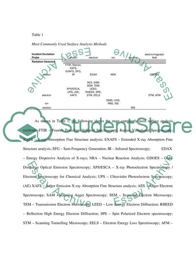
?Surface Analysis and Materials Science Introduction Klauber and Smart (2003) declared that the primary objective of materials science is to develop materials which possess specific mechanical, electrical, magnetic, optical, thermal, and chemical properties. Such materials are required to be exhibit stability under normal environmental conditions. The behaviour of a material’s surface as it reacts to its environment is important because it reveals how well-suited a particular materials performs its intended function. For example, corrosion in metal is prevented through the use of specific chemicals; various optical effects on lenses may be done through special coatings; and automobile emissions are significantly reduced through the unique chemical composition on the surface of an auto-exhaust catalyst. To achieve the desired function, the surface a material should be analyzed to determine its physical characteristics, chemical composition, chemical and atomic structure, electronic state, and molecular bonding (Vickerman, 2009). Methods Several probes may be applied on a solid surface to measure its response, namely: electrons, ions, neutrons, photons, and heat or field. Each probe has a specific response. The combination of probes and corresponding responses provides 36 basic classes of experimental techniques which may be utilized for surface analysis. Table 1 Most Commonly Used Surface Analysis Methods Incident Excitation Probe photon electron ion neutron electric/magnetic field Radiation Detected photon FTIR, Raman, XAFS, EXAFS, SFG, IR EDAX NRA GDOES electron XPS/ESCA, UPS, (AE) XAFS AES, SAM, SEM, TEM, LEED, RHEED, SPE, STM, EELS STM, AFM ion SIMS, LEIS, RBS, ISS neutron INS As shown in Table 1, the following shows the most commonly used surface analysis methods: FTIR – Fourier Transform Infrared Spectroscopy; Raman Vibrational Spectroscopy; XAFS – X-ray Absorption Fine Structure analysis; EXAFS – Extended X-ray Absorption Fine Structure analysis; SFG – Sum Frequency Generation; IR – Infrared Spectroscopy; EDAX – Energy Dispersive Analysis of X-rays; NRA – Nuclear Reaction Analysis; GDOES – Glow Discharge Optical Emission Spectroscopy; XPS/ESCA – X-ray Photoelectron Spectroscopy / Electron Spectroscopy for Chemical Analysis; UPS – Ultraviolet Photoelectron Spectroscopy; (AE) XAFS – Auger Emission X-ray Absorption Fine Structure analysis; AES – Auger Electron Spectroscopy; SAM –Scanning Auger Spectroscopy; SEM – Scanning Electron Microscopy; TEM – Transmission Electron Microscopy; LEED – Low Energy Electron Diffraction; RHEED – Reflection High Energy Electron Diffraction; SPE – Spin Polarized Electron spectroscopy; STM – Scanning Tunnelling Microscopy; EELS – Electron Energy Loss Spectroscopy; AFM – Atomic Force Microscopy; SIMS – Secondary Ion Mass Spectrometry; LEIS – Low Energy Ion Scattering spectroscopy; RBS – Rutherford Backscattering Spectroscopy; ISS – Ion Scattering Spectroscopy; and INS – Inelastic Neutron Scattering; Analysis Auger electron spectroscopy or AES is considered as a key chemical surface analysis tool for conducting material samples. The AES technique is based on the excitation of auger electrons which allow not only the imaging of atoms but for chemical identification as well. Information available through AES ranges between the first 2 to 10 atomic layers of the sample surface (Matheiu, 2009). Meanwhile, low energy electron diffraction or LEED works by bombarding a surface with beam of low energy electrons which enable the identification of the surface structure by electron diffraction (Vickerman, 2009). A beam of low energy electrons between 10 to 200 eV is used to determine crystallographic structure. A device called a Retarding Field Analyzer is utilized to detect diffracted electrons. Diffracted electrons appear as spots on a phosphorescent screen which move according to energy variations of electrons. The intensity of the spots also provides information regarding surface reconstructions (Walker, 2011). An auger electron spectroscopy analysis conducted by Palmberg and Rhodin (2009) was used to determine the energy spectra of auger electrons from clean gold (Au), silver (Ag), copper (Cu), palladium (Pd), and nickel (Ni) surfaces. The process of creating a uniform disposition of a metal onto another clear metal surface has paved the way for the development of depth assessment techniques and the identification of contributors to auger peaks in the secondary electron energy distribution characteristic. Results Results of the study revealed that the mean escape depth of auger electrons in gold ranges from 4 to 8 angstroms for energies of 72 and 362 eV. By the utilization of auger electron spectroscopy (AES) and low energy electron diffraction (LEED), it was also shown that a clean silver (Au) surface is reconstructed into a 1x5 structure. On the other hand, clean gold (Ag), copper (Cu), and palladium (Pd) surfaces are reconstructed by bulk atomic arrangement (Palberg & Rhodin, 2009). Impact on Materials Science The combinatory utilization of AES and LEED techniques has also been proven in the interpretation of structure and combination of copper-aluminium surface alloys (Palberg & Rhodin, 2009). Surface Analysis and Forensic Science Introduction The use of surface analysis in forensic science has aided law enforcement in solving numerous crimes. One surface analysis tool commonly used nowadays is infrared microspectroscopy, specifically Fourier Transform Infrared Spectroscopy or FITR, which is used to analyze hair samples. Another surface analysis method is X-ray Photoelectron Spectroscopy (XPS) which can help determine the elements present, concentration, and chemical state in a material surface. Methods Infrared spectroscopy uses infrared light to induce vibrations in molecules which allows the forensic scientist to determine structural information of the sample being analyzed. This technique is applicable to solid and liquid samples (Tilstone, Savage & Clark, 2006). IR spectroscopy is the tool of choice for the qualitative identification of paint, fibres, explosives, and drugs (Lee and Harris, 2006). Meanwhile, Fourier Transform Infrared Spectroscopy or FITR is almost similar to the aforementioned technique. The main difference is that FITR uses an interferometer instead of a monochromator. This increases sensitivity and reduces time spent on analysis, making the processing of evidence faster (Tilstone, Savage & Clark, 2006). A microscope sampling device in combination with a Fourier transform infrared spectrometer is able to perform an analysis of samples as small as 10 x 10 microns (Lee and Harris, 2006). Analysis Infrared microspectometry is a non-destructive surface analysis technique which only requires a minimal amount of sample material. These qualities make it suitable for use in forensic investigations since samples come in very small quantities most of the time. In the case of hair analysis, evidence samples may be have already undergone degradation thru burial or exposure to the elements, making it hard to identify surface features. Infrared microspectrometry however, allows investigators to determine the nature of hair fibres, chemical treatments done to the hair, frequency of treatments, extent of chemical damage, natural weathering, and residues left by hair sprays and conditioners. A typical hair analysis via IR microspectrometry starts by cutting a single hair fibre to a length of 100 microns and flattening it out on a clear glass slide with a roller knife. The sample is then transferred to the bottom of a potassium bromide (KBr) salt plate in a micro-compression cell. A small crystal of KBr is also added into the salt plate. Another KBr salt plate is positioned on top of the bottom plate and the micro-compression cell is tightened until optical contact between the fibre and salt plate is established. A reference or background spectrum is acquired from the KBr crystal on the plate while the sample spectrum is secured via the hair fibre sample. Using a microscope and a FITR spectrometer, both spectra were collected with a 4cm-1 resolution and 64 scans were added for each spectrum. The resulting sample size was 50 x 100 microns (Thermo Fisher Scientific, 2007). The spectrum is a graph showing the wave numbers on the x-axis and the percentage of light transmitted on the y-axis. By examining the bands formed when the amount of transmitted light drops, a forensic analyst can ascertain the structural characteristics of a specific sample and determine its identity (Tilstone, Savage & Clark, 2006). Aside from hair samples, infrared spectroscopy is also used in the analysis of bone or skeletal remains. The work of Nagy et al. (2008) on the chemical analyses of bone samples from various burial environments via Fourier transform infrared spectroscopy revealed that using the crystallinity index and carbonate-phosphate index may be used as a new method of distinguishing recent and anthropological, archaeological bone samples. Another application of infrared spectroscopy in forensic analysis is through the identification of multiple fingerprints on a specific surface. IR spectroscopy is a non-invasive alternative to earlier methods which has the tendency to destroy the original condition of the surface in question (Bhargava, Perlman, Fernandez, Levin, and Bartick, 2002). X-ray Photoelectron Spectroscopy (XPS) is a non-destructive qualitative and quantitative analysis tool which can detect composition, concentration and state of a material via soft x-rays (CERAM Surface and Materials Analysis, 2011). Results Bertino and Bertino (2009) enumerated several case studies wherein infrared spectroscopy was utilized to analyze hair fibres found on crime scenes and evidence. The murder of Eva Shoen provided proof that hair fibre analysis is essential to forensic work. The victim was shot in the head and the police were able to recover the bullet. However, a ballistic match was impossible because the gun used in the murder was not recovered. After three years, police were able to arrest a suspect, Frank Marquis, based on a tip. A gun was recovered but was already tampered with, eliminating the possibility of a successful ballistic match. Further investigation revealed that the suspect was in the area the day Shoen was murdered and that the suspect dumped two bundles on the side of the road while he was driving to Arizona. The police were able to recover the bundles which contained clothes. A hair strand was found and sent for analysis. The hair sample matched the victim’s hair based on its colour and structure. The police were able to get a confession from the suspect afterwards. A case study conducted by CERAM Surface and Materials Analysis (2011) demonstrated the ability of XPS in qualitative and quantitative surface analysis. Two powder samples were used in the study. Figure 1 below shows the XPS survey spectrum for the first powder sample. Figure 1. XPS survey spectrum for talcum powder. As shown in the figure above, the elements magnesium, silicon, oxygen, and carbon were detected. The concentration of each element is also indicated on the spectrum survey result. Based on the chemical composition and concentration it was therefore identified as magnesium silicate or talcum powder. Further quantitative analysis makes it possible for XPS to determine specific types of talcum powder based on the elemental ratios of its components. Figure 2 shows the survey spectrum for the second powder sample. The spectrum survey detected the following elements: magnesium, silicon, oxygen, carbon, fluorine, titanium, potassium, and aluminium. Based on the composition and concentration, the second powder sample was identified as face powder. Comparative testing between the two samples would show the difference between the two samples in terms of composition and concentration. This proves useful in forensic investigations since some samples occur only in trace amounts or thin layers on surfaces. Figure 2. XPS survey spectrum for face powder. Impact on Forensic Science Forensic science has benefited greatly from the aforementioned techniques since typical evidence material is available only in the form of thin films, smudges, or residues on surfaces which are easily contaminated. Earlier methods result in the destruction or degradation of samples, which creates more problems whenever evidence material is in short supply. Non-destructive surface analysis techniques are a welcome addition to a forensic investigator’s arsenal. References Bertino, A. J. & Bertino, P. N. (2009). Forensic science: Fundamentals and investigations. Mason, OH: Cengage Learning. Bhargava, R., Perlman, R. S., Fernandez, D. C., Levin, I. W. & Bartick, E. G. (2002). Non-invasive detection of superimposed latent fingerprints and inter-ridge trace evidence by infrared spectroscopic imaging. Analytical and Bioanalytical Chemistry, 394(8), 2069-2075. CERAM Surface and Materials Analysis (2011). The Role of surface analysis in forensic science. Retrieved from http://www.csma.ltd.uk/legal/forensic.htm. Klauber, C. & Smart, R. St. C. (2003). Solid surfaces, their structure, and composition. In O’Connor, Sexton, B. A. & Smart, R. St. C. (Eds.), Surface analysis methods in materials science (3-65). Heidelberg, Germany: Springer. Lee, H. C. & Harris, H. A. (2006). Physical evidence in forensic science. Tuczon, AZ: Lawyers and Judges Publishing. Matheiu, H.J. (2009). Auger electron spectroscopy. In Vickerman, J. C. & Gilmore, I. (Eds.), Surface analysis: The principal techniques (9-46). West Sussex: John Wiley and Sons. Nagy, G., Lorand, T., Patonia, Z., Montsko, G., Bajnoczky, I., Marcsik, A. & Mark, L. (2008). Analysis of pathological and non-pathological human skeletal remains by FT-IR spectroscopy. Forensic Science International, 175(1), 55-60. Palmberg, P. W. & Rhodin, T. N. (2009). Auger electron spectroscopy of fcc metal surfaces. Journal of Applied Physics, 39(5), 2425-2432. Thermo Fisher Scientific (2007). Infrared microspectrometry in forensic science, hair fibre analysis. Retrieved from http://www.thermo.com/eThermo/CMA/PDFs/ Articles/articlesFile_2278.pdf. Tilstone, W. J., Savage, K. A. & Clark, L. A. (2006). Forensic science: An encyclopaedia of history, methods, and techniques. Santa Barbara, CA: ABC-CLIO. Vickerman, J. C. (2009). Introduction. In Vickerman, J. C. & Gilmore, I. (Eds.), Surface analysis: The principal techniques (1-8). West Sussex: John Wiley and Sons. Walker, C. (2011). An Introduction to LEED – Low Energy Electron Diffraction. Retrieved from http://www.uksaf.org/tech/leed.html. Read More
Cite this document
  • APA
  • MLA
  • CHICAGO
(“Surface analysis Essay Example | Topics and Well Written Essays - 2000 words”, n.d.)
Retrieved from https://studentshare.org/environmental-studies/1409373-surface-analysis
(Surface Analysis Essay Example | Topics and Well Written Essays - 2000 Words)
https://studentshare.org/environmental-studies/1409373-surface-analysis.
“Surface Analysis Essay Example | Topics and Well Written Essays - 2000 Words”, n.d. https://studentshare.org/environmental-studies/1409373-surface-analysis.
  • Cited: 0 times

CHECK THESE SAMPLES OF Surface Analysis and Materials Science

Spectrophotometry

hellip; Spectrophotometry is used in different fields and sciences like chemistry, biochemistry, physics, and materials, etc.... During the analysis of every glass, the file is clicked with the scanning option from 400 nm wavelength to 900 nm wavelength.... Then after analysis is switched on which performance summary information is selected.... All three analyses are made on P polarization When our analysis for P polarization is completed then any sample is selected for S polarization so a conclusion can be drawn by varying polarization....
8 Pages (2000 words) Essay

Earth and Space Science

Instruments connected to aircrafts and space based platforms of satellite devises perform the function of remote sensing with the analysis of the electronic data recorded during the emission of such radiation for the establishment of forecast ideas.... MMC or migrated means clustering classifier is the tools used in this model for labeling each pixel to unknown cluster centers with intend to move the pixel form one cluster center to another for accurate analysis of the image (ibid)....
8 Pages (2000 words) Essay

Forensic Material Identification Techniques, Spectroscopy

The functions of this department of forensic science are to visit crime scenes, search for, enhance, collect and examine finger, palm and footprint for evidence and to make identifications via the comparison and recovering of latent prints and recognized offender prints (Skoog, 2007).... Whether working under document Examination, armory, photography, scene of crime, electronic crime laboratory or under environmental science and research, forensic officers operate analytical laboratories where they analyse samples related to biology (DNA), physical evidence such as glass, fibres and paint, illicit drugs, toxicology, breath/ blood alcohol and workplace drug testing among others (Skoog, 2007)....
8 Pages (2000 words) Coursework

Atomic force microscopy

ifferent imaging techniques are used to picture the surface or for different surface analysis, they include the contact mode, non-contact mode and tapping mode.... This tip is attached to the free end of a The material to be scanned is placed below the tip, as the tip moves across the surface it is attracted or repelled by different forces, the atomic deflection from rest is recorded and an imaging software is used to process the results.... The results normally appears as a topographical image of the surface imaged2....
5 Pages (1250 words) Lab Report

Earth and Space Science

This paper 'Earth and Space science'' tells that Remote sensing of the atmosphere is essential for the risk-free navigation of airways and weather forecasting of a region.... hellip; IInstruments connected to aircraft and space-based platforms of satellite devices perform remote sensing with the analysis of the electronic data recorded during the emission of such radiation for the establishment of forecast ideas.... Thus, the photo data vary in their pixel depth and is separately considered for different spectral classes for quantitative analysis....
9 Pages (2250 words) Report

Failure Criteria of Aluminium

nbsp;In failure assessment, fracture surface analysis is hardly used in seclusion.... In this paper, an analysis of the failure criteria for aluminum is given.... In other instances, a wide array of methods and in-depth analysis is needed.... It is pertinent to have a deep understanding of the elements that impact the topography and analysis of pictures of fracture surfaces (Hull, 1999).... It also involves the review of the magnitude of the application of the component in conformity to terms, stress assessment, non-destructive assessment of the botched parts, chemical review, and fractographic analysis....
8 Pages (2000 words) Essay

Analysis of Surfaces Using an Atomic Force Microscope

AFMs have a wide range of applications including; biology, material science, data storage devices, semiconductors, polymers, and optics for measurement of nano-mechanical, nanoscale topography, nano-scale chemical mapping, and nano-electrical.... This lab report "analysis of Surfaces Using an Atomic Force Microscope" presents the AFM instrument that produces a specimen's image with a very high resolution by use of a mechanical probe.... Three carbon nanotubes surface AFM images were selected and analyzed to determine the average width and height using the Nanoscope analysis software....
8 Pages (2000 words) Lab Report

In Vitro Synthetic Biology: Surface Modified, 3D Printed Molecular Machines

This paper “In Vitro Synthetic Biology: surface Modified, 3D Printed Molecular Machines” discusses 3D printing mainly on the filament fabrication print technology based on Marker Replicator 2X experimental 3D printer.... The printer heats the filament while squeezing it out via the nozzle onto a heated surface and creates a solid object by applying layers upon layers through the process called Fused Filament Fabrication (FFF) (Mertz 2013)....
15 Pages (3750 words) Coursework
sponsored ads
We use cookies to create the best experience for you. Keep on browsing if you are OK with that, or find out how to manage cookies.
Contact Us