StudentShare
Contact Us
Sign In / Sign Up for FREE
Search
Go to advanced search...
Free

Gas Exchange and Transport - Coursework Example

Cite this document
Summary
This work called "Gas Exchange and Transport" describes the structure of the respiratory system. The author takes into account the structure and functions. From this work, it is clear about respiratory and circulatory systems disorders, composition and functions of blood components…
Download full paper File format: .doc, available for editing
GRAB THE BEST PAPER93.8% of users find it useful
Gas Exchange and Transport
Read Text Preview

Extract of sample "Gas Exchange and Transport"

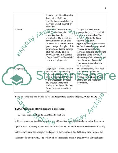
GAS EXCHANGE AND TRANSPORT Department TAQ Structure of the Respiratory System Respiratory System Component Structure Function(s) Nose The nasal cavity lining consist of lining of hair, cilia, blood capillaries and mucus The function of the nose is to filter all debris as well as warm and moisten the passing air Pharynx Consist of nasopharynx, oropharynx and laryngopharynx This is where the throat separates with the trachea and separates air from food Larynx It consists of cartilages: epiglottis at the upper, thyroid at the front, paired arytenoids horizontally and cricoids cartilage T he larynx function is to protect entry of particles into the trachea through the production of a strong cough reflex in case any solid particle pass through the epiglottis Trachea/ Windpipe It is a tube made up of ringed cartilages known as hyaline at sides and front to keep it open always. The ringed cartilages are not complete at the back because of the position of the esophagus to allow expansion of esophagus during swallowing food. The inner membrane known as mucosa is covered in tinny hairs called cilia and consists of mucus. The trachea is 10-16 cm long and 20-25mm wide The function of trachea is to carry air from the throat into the lungs. The other function of trachea is to sweep foreign particles from the lungs to the pharynx through the movement of cilia and trapping by mucus within the inner membrane Bronchi Bronchi are mainly two sets of tubes branching from the trachea. Left bronchi that is narrower, horizontal and longer enters the left lung while the other enters the right lung. The walls of bronchi are smooth and surrounded by irregular rings of cartilage. The bronchi divides further into tertiary bronchi in the lungs Carry air from the trachea into bronchioles and distribute it. It also removes particles from the bronchioles to the trachea Bronchioles Bronchioles are formed from the branching of tertiary bronchi. They are narrower than the bronchi and less than 1 mm wide. Unlike the bronchi, trachea and pharynx the walls are not covered by cartilages Distribute air into several alveoli for exchange Alveoli Alveoli are very narrow but numerous hollow tubes branching from the bronchioles. The alveoli are also surrounded by several capillary networks into which gas exchange takes place. It is approximated that an average adult has about 3 million alveoli. Alveoli also consists of type I and Type II epithelial cells, macrophage cells Oxygen diffusion occurs through the type I cells which are the primary cells of the alveoli wall into the dense network capillaries. Type II epithelial cells reduces surface tension by secretion of primary surfactant hence increases diffusion and prevent collapsing of the airway. Microphage Cells also referred to as the dust cells remove microorganisms and debris from the lungs Diaphragm Diaphragm is a dome-shaped sheet of muscle separating chest and abdomen. It is made up of a broadband of muscles found underneath the lungs and attached to sternum, lumbar spine, lower ribs thus forms the thoracic cavity’s base The diaphragm together with other muscles within the thoracic cavity enables breathing in and out through contraction and relaxation respectively. Table 1: Structure and Functions of the Respiratory System (Rogers, 2011 p. 19-28) TAQ 2: Mechanism of breathing and Gas exchange a. Processes Involved In Breathing In And Out Different organs are involved in the process of breathing in and out. As shown in the diagram in figure 1, when breathing in, the Intercostals muscles and pectoralis minor muscle contract leading to the expansion of the ribcage. The diaphragm then contracts thus flattens so as to increase the volume of the chest cavity. The activity of the intercostals muscles together with the diaphragm leads to reduction in pressure within the chest cavity thus air is sucked into the lungs. When breathing out, the opposite activities occur; the intercostals muscles and pectoralis minor muscle relax hence the ribcage contracts. The diaphragm then relaxes leading to the reduction in the volume of the lungs. This increases pressure in the lungs hence air is pushed out of the lungs. During breathing, oxygen and other gases are taken in when inhaling while carbon dioxide and other gases are exhaled from the lungs. Figure 1 shows how intercostal muscles and diaphragm aid the process of breathing in and out (Rogers, 2011 p. 41-50). Breathing in Breathing Out Figure 1: Process involved in breathing in and out (Rogers, 2011 p.41-50) b. How Gaseous Exchange Takes Place in the Lungs In the lungs, we have the tiny alveoli air sacs just at the end of bronchial tubes. The alveoli are mainly connected to a dense surrounding of capillaries which are connected to the blood stream. The alveoli are made up of specialized thin surfaces that allow gas exchange. Gas exchange takes place at the alveoli cells of the lungs through diffusion where molecules move from a region of high concentration to the region of low concentration. The blood capillaries surrounding alveoli have lower concentration of oxygen compared to the inhaled air within the alveoli cells. Oxygen will therefore diffuse across the tiny walls of alveoli into the blood capillaries. Consequently, the blood capillaries contain blood with higher concentration of carbon dioxide than the inhaled air. Carbon dioxide with therefore diffuse from the blood capillaries across the walls of alveoli and is exhaled out through breathing mechanism (Dendo et al, 1998 p.382). TAQ 3: Regulation of Breathing Figure 2: Regulation of the breathing System (Rogers, 2011 p. 45) Breathing is regulated by the levels of H+ ions and CO2 in the blood. There are two types of chemoreceptors that strongly detect different levels of concentration of H+ ions and CO2 in the blood; peripheral and central chemo-receptors. For peripheral receptors, the amounts of H+ and CO2 in the blood are measured in two points in the body; carotid arteries and at the exit of aorta which have swollen areas called sinuses. Sinuses do not only measure the concentration of Carbon dioxide and H+ in the body but also measures blood pressure There are nerves leading from these parts to the hind-brain (hypothalamus) which controls many aspects of the body homeostasis. Central chemo receptors are found on the surface of medulla oblongata and respond indirectly to blood concentration. CO2 diffuses across to the brain and specifically to cerebral spinal fluid but HCO3- and H+ cannot but are formed from Co2 reaction with H2O. Increase in the concentration of H+ cause stimulation of the chemoreceptors and results into relay of information and causes contraction of muscles and increase in ventilation (Whittemore, 2009 p.31) & Dendo et al 1998 p.385). Figure 2 shows the regulation of the breathing and how changes in CO2 concentration triggers relay of information to medulla oblongata which controls muscle contractions (Rogers, 2011 p.45). TAQ 4: Disorders of the Respiratory and circulatory Systems Disease Cause Symptoms Treatments Prognosis for future of patient Bronchitis Bronchitis is an inflammation of the bronchi caused by irritants like air pollution, smoke, or infections Bronchitis is manifested by the inflammation of the lining of the mucus membrane, high mucus production, reduced movement of cilia, reduced diameter. There is also reduction in the diameter of bronchi thus impairs ventilation Chronic bronchitis can be treated by administering mucolytics medication in order to make mucus easier to cough. There is also an exercise program known as pulmonary rehabilitation that is necessary for individuals to cope up with their symptoms. Stopping smoke is very important as well Even though this disease is progressive and chronic, Individuals who stop smoking and avoid chemicals, airborne dusts and other irritating situation normally have good prognosis for many years Asthma Asthma is a disorder of the respiratory caused by allergic responses to animal dander, dust, pollen and other substances within the inhaled air Asthma symptoms include contractions of bronchial smooth muscle periodically resulting into restriction in movement of air into and out of the lungs. Treatments of asthma include administering a drug that reduce inflammation and relax the bronchiole smooth muscles. Another treatment that may be administered is a drug to reduce the sensitivity of the immune system to those things that stimulate asthma attack (Acosta, 2003 p. 531) The prognosis of asthma is quiet good. The treatments administered often work so well to bring asthma into a control. While viral infections may result into severe episode, management is easily achieved. However, it may take more time for severe cases though not associated with long term consequences Atherosclerosis Atherosclerosis when the inner layers of arteries are damaged by accumulation of plagues leading to hardening of the arteries. The hardening of the arteries may be caused by smoking, high blood presser, high blood sugar, high amounts of cholesterol and more importantly low density lipoproteins (Sauer et al, 2007: 446) Angina which is exhibited by pressure or squeezing in the heart and may also be felt in around the arms, shoulders, jaw, neck or back. Rhythm of the heart beat is also expected to increase. When Carotid arteries are affected, individuals experience sudden weakness, breathing problems, confusion and loss of consciousness The first medication for atherosclerosis is the mandatory change in lifestyle. Other treatments available are surgery and medicines to relieve symptoms (Sauer et al 2010: 446). There are also dugs that are capable of reversing or slowing or just slow the rte of atherosclerosis. E.g. cholesterol medication Prognosis for atherosclerosis is very poor. While there are no specific drugs for atherosclerosis, management and reliving impact of this disease can be achieved. Moreover avoiding smoking, and consumption of alcohol can improve prognosis Myocardial infarction (MI) The primary cause of MI is the rapture of the high-risk plague within coronary arteries thus blood clot obstructs coronary artery completely resulting to death of heart muscles due to lack of blood supply. The risk factors include tobacco, increased blood pressure, diabetes, high level cholesterols The most common symptom of MI is chest pain, shortness of the breath, diaphoresis, weakness and ventricular failure and The treatments for MI emphasizes on stent placement. The treatments are mainly aimed at reopening the blockage of the coronary arteries as soon as possible to prevent further damage to the hearts and improve prognosis. Administering antiplatelet, clot dissolving drugs, anticoagulants, beta blockers, angiotensin converting enzyme inhibitors, aspirin and effective dieting (Whittemore, 2009 p. 41). Prognosis after Myocardial infarction (MI) is very poor and survivors are very susceptible to further cardiovascular events such as mortality though prognosis varies depending on whether there are risk factors or not. Table 2: Respiratory and Circulatory Systems disorders TAQ 5: Composition and functions of blood components The blood forms about 7-8% of the body weight and plays an important function of carrying and transporting nutrients and oxygen to the body tissues while at the same time getting rid of carbon dioxide and other wastes. It also plays a key role in maintaining the immune system and maintains body temperature as well. It consists of over four thousand components but the most important ones are; white cells, plasma, red blood cells and platelets (Houghton, 2007 p.19). Red Blood cells Red blood cells also referred to as erythrocytes make up 40% of the blood and are relatively microscopic cells that have no nuclei. The red blood cells are made of hemoglobin 95% and each cell contains over 270 million iron-rich hemoglobin molecules. The function of the red blood cells is to transport oxygen from the lungs to the body tissues and take carbon dioxide from the body tissues back to the lungs to be exhaled (Houghton, 2007 p.19-21). White blood cells White blood cells also referred to as leukocytes form only 1% of the blood volume and are not limited to blood alone. It is also found in other parts including the spleen, the liver and lymph glands. It consists of different types namely lymphocytes, granulocytes and macrophages Lymphocytes respond to the immune system by seeking, identifying and binding to the alien protein on viruses, bacteria or fungi so that they can be removed. The granulocytes as well as macrophages destroy the alien cells. These cells also remove from the body asbestos, dusts and dead blood cells. It is therefore important to note that the major function of white blood cells is to protect the body (Houghton, 2007 p.20). Platelets Platelets also referred to as thrombocytes are made up of fragments without nuclei, 1/3 the size of red blood cells with a lifespan of 9-10 days. Their main function is to clot blood and prevent loss of blood during bleeding. Platelets are capable of releasing coagulating chemicals that are used to cause formation of clots. Clots are formed when platelets cells adhere unto the walls of blood vessels thereby plugging the rapture in the vascular wall and also function in stimulating the immune system (Ward et al 201 p.1: 9 & Houghton, 2007 p.21).. Plasma Plasma is the yellow and relatively clear water and constitutes over 92% of the blood. It consists of fat, sugar, salt and protein and carries the red cells, platelets and white cells. The function of plasma is to bring nourishment unto organs such as the heart while it’s pumping and remove metabolic wastes from that organ as well. Plasma also contains lipids, minerals, enzymes, vitamins, antibodies, hormones and other proteins which are vital for metabolism and maintenance of body ion potential (Houghton, 2007 p.20). TAQ 6 a. The diagram of the heart Figure 3; Structure of the human heart (Phibbs, 2007 p. 1-6) b. Functions of the heart including the cardiac cycle The function of the heart is to supply blood to all parts of the body. Blood is needed by the body tissues since it is the carrier for nutrients and oxygen that is needed by the body tissues. The cardiac cycle is a complete heart beat and describes all the activities of the heart that allow a circular and continuous flow of blood in and out of the heart. The major activities of the heart involve contraction and relaxation of the heart atria and ventricles referred to as systole and diastole respectively (Acosta, 2003 p.518). The systolic and the diastolic activities results into changes in blood pressure and volumes within the heart. Blood enters the heart when there is relaxation of the volumetric venricular muscles. This is the time the ventricles are relaxed but the Atrioventricular and semilunar valves remain closed and the volume of the ventricles remains unchanged (Macía et al 2012 p.459). When the AV valves open, the blood fills the ventricles thus the ventricles remain in a diastole mode for the whole period of filling. As the blood fills the empty ventricles rapidly, the volumes of the ventricles increase rapidly as well. However, the rapid filling reduces slowly with the increase in the volume of the ventricles a process known as diastasis. Atrial systole as well as the P-wave of the electrocardiogram occurs which results into the emptying of all remaining blood from the atria into the ventricles resulting into the end-diastolic volume which is the blood at the end of this interval (DeVault et al 2008 p.889). The process of emptying the blood from the heart begins by the contraction of the ventricles a process referred to as ventricular systole. This starts as action potential of the Atrioventricular node enters the ventricles, the QRS complex is observed on electrocardiogram and the ventricles depolarize. QRS complex refers to a series of deflections within the electrocardiogram resulting from electrical activity due to depolarization of ventricular before the contraction of the muscles. This process occurs in the following phases. Isovolumetric contraction occurs as a result of closure of the atrioventricular valves but the semilunar valves remains closed thus the volume of the ventricles remains unchanged. This process takes a very short time. The contraction of the ventricles continues thereby forcing the semilunar valves to open and the blood is ejected out of the ventricles (Kokkinos, 2010 p.144). This process continues and ends when the ventricles start relaxation as well as the closure of the semilunar valves. The closure of the semilunar valves normally results into an increase in the blood pressure. There is small amount of blood remaining within the ventricles and this is called end-systolic volume. This cycle continues and there is inflow and outflow of blood from the heart. Figure 3 shows the various parts of the heart and how blood flows in and out of the heart (Acosta, 2003 p.518). c. Blood Vessels, Structures And Functions Blood vessel Structure of the blood vessel Functions Arteries The walls of arteries are made up of three major layers namely the outer layer and are made of fibrous tissues. These fibrous tissues normally merge and connect to the outer tissues to which arteries are connected. The middle layer is the second layer and is made up of thick and elastic tissues as well as involuntary muscle tissues. The middle layer is also supplied with nerves that controls relaxation and contractions of the arteries thus narrow or expand. The innermost layer consist of flat continuous epithelial cells packed together with the endorcadium of the heart and make the inner lining of arteries smooth to avoid friction between the inner lining and the blood (Kokkinos, 2010 p.144) The function of arteries is to carry blood from the heart to other body organs. All arteries carry oxygenated blood from the heart to the rest of the body except the pulmonary arteries which carry deoxygenated blood to the lungs Veins The veins consist of the three layers like the arteries. However, the middle layer of veins is much thinner than that of arteries. The veins also consist of semi-lunar valves that help in preventing the backflow of blood. This is because the pressures of blood in the veins are much reduced. The functions of veins is to carry blood from the body organs, tissues and cells back to the heart, carry waste matter especially carbon dioxide and by products of metabolism from the organs and tissues. The pulmonary vein function is to carry oxygenated blood from the lungs to the left atrium. Capillaries Blood capillaries are much smaller in size and form a network within the body tissues. They connect both veins and arteries through the arterioles. The walls of capillaries are much thinner and consist of only one layer of endothelial cells. The function of capillaries is to provide a structure where gaseous exchange between the tissue fluids and the blood vessels. It also provide a medium for exchange of nutrients and waste products like carbon dioxide between the tissues and the capillaries. Aorta Is the largest artery as well as the principal artery within the body. It is from the aorta where other arteries branch leading to other parts of the body. The structure is similar to that of arteries discussed above Aorta is the main blood vessel where the blood leaves the heart to other parts of the body Coronary artery Coronary artery is the first branch of aorta. Its structure is similar to the structure of all arteries The function of the coronary artery is to supply the heart tissues with oxygen and nutrients Pulmonary Artery It connects from the upper right hand corner of the right ventricle. It brunches into the left and right pulmonary arteries leading to left and right lungs respectively. The wall structure is similar to that of arteries discussed above. Pulmonary artery carry deoxygenated blood from the heart to lungs for oxygenation and release of carbon dioxide. Superior vena cava The structure is similar to that of veins but superior vena cava is much bigger in size. It carries deoxygenated blood from the head, thorax and arms to the right atrium Inferior Vena Cava Connects with the other veins originating from the lower part of the body and is much bigger in lumen due to the quantity of deoxygenated blood it carries The functions of the inferior vena cava are to carry deoxygenated blood from the legs, abdomen and the lower part of the body to the right atrium. Coronary Vein The structure is similar to that of other veins discussed above. The semi-lunar valves prevent backflow of deoxygenated blood to the heart muscles This blood vessels transports deoxygenated blood from containing mainly waste materials and carbon dioxide from the heart muscles back to the heart through the right atrium Pulmonary Veins They are two one from each lung. They are the only vein that carry oxygenated blood from the lungs to the right atrium Hepatic Portal Vein It leads from small intestine and other internal organs to the liver. The main function of the hepatic portal vein is to carry absorbed food from the small intestine to the liver where some food is deposited and stored and clarification is done on nutrients; glucose, amino acids, fatty acids before they are taken to other body tissues. Table 3: Blood Vessels’ structure and functions (Kokkinos, 2010 p.144-145) References Acosta, D, 2003, Cardiovascular toxicology: Third Edition, London: Taylor and Francis Publishers. Dendo, R.I, Phalen, R.F, Mannix, R.C & Oldham, M.J, 1998, "Effects of breathing parameters on sidestream cigarette smoke deposition in a hollow tracheobronchial model", American Industrial Hygiene Association Journal, vol. 59, no. 6, pp. 381-387. DeVault, K., Gremaud, P.A., Novak, V., Olufsen, M.S., Vernières, G. & Zhao, P. 2008, "Blood Flow in the Circle of Willis: Modeling and Calibration", Multiscale Modeling & Simulation, vol. 7, no. 2, pp. 888-22. Houghton, G, 2007, Blood: the Circulatory System. New York: Rosen Publishing Group Kokkinos, P, 2010, Physical activity and cardiovascular disease prevention, Sudbury, MA: Jones and Bartlett Publishers. Macía, I., Graña, M. & Paloc, C, 2012, "Knowledge management in image-based analysis of blood vessel structures", Knowledge and Information Systems, Vol. 30, no. 2, pp. 457-491. Phibbs, B, 2007, The Human heart: A basic guide to heart disease (Second Edition), Philadelphia, PA: Lippincott & Wilkins Rogers, K, 2011, The Human Body: The Respiratory System, New York: Britannica Educational Publishers, Inc Sauer, H, Shah, A. J., Laurindo, F.R.M, 2010, Studies in Cardiovascular Diseases, New York: Springer. Ward, J.P.T, Ward, J, Leach, R. M, 2011, The Respiratory System at a Glance: Third Edition, New York: Wiley-Blackwell. Whittemore, S, 2009, The Human Body How it Works: Respiratory System, New York: The Info base Publishers. Read More
Cite this document
  • APA
  • MLA
  • CHICAGO
(Gas Exchange and Transport Coursework Example | Topics and Well Written Essays - 3500 words, n.d.)
Gas Exchange and Transport Coursework Example | Topics and Well Written Essays - 3500 words. https://studentshare.org/biology/1812945-human-biological-science-gas-exchange-and-transport
(Gas Exchange and Transport Coursework Example | Topics and Well Written Essays - 3500 Words)
Gas Exchange and Transport Coursework Example | Topics and Well Written Essays - 3500 Words. https://studentshare.org/biology/1812945-human-biological-science-gas-exchange-and-transport.
“Gas Exchange and Transport Coursework Example | Topics and Well Written Essays - 3500 Words”. https://studentshare.org/biology/1812945-human-biological-science-gas-exchange-and-transport.
  • Cited: 0 times

CHECK THESE SAMPLES OF Gas Exchange and Transport

The Mechanism of Infection of the Hantavirus

The endothelial cells are responsible for various physiologic functions of the body such as the regulation of edema, vascular repair and gas exchange (Mackow & Gavrilovskaya, 2009).... The young man in the case study exhibited shortness of breath and labored and rapid breathing because the hantavirus has already invaded the endothelial cells of his lungs, which are responsible for gas exchange.... Thus, the physiological ability of his lungs to handle gas exchange is impaired....
2 Pages (500 words) Essay

Biology Unit 4 IP

Due to smoking, the alveoli in his lungs had been destroyed due to the loss of the surface area of gas exchange and airway flexibility.... Moreover, alveoli are destroyed due to the destruction of gas exchange surface area and airway flexibility.... The latter is a nucleotide that can transport energy throughout cells.... Subsequently, adenosine triphosphate gets transformed into energy by means of electron transport phosphorylation....
2 Pages (500 words) Research Paper

Explain how oxygen and carbon dioxide are transported by the blood

Physical transfer in which oxygen dissolves in the blood's plasma only transport a limited amount of oxygen in the body, while the major transportation is aided by haemoglobin.... The two processes occur in the gaseous exchange process that I explain in this paper, the process by which Transportation of oxygen and carbon dioxide in the blood The circulatory system and the respiratory system are some of the major characteristics of larger sized animals, whose mass to surface area ratios are small and therefore require specialized organs and systems to meet cellular needs....
2 Pages (500 words) Assignment

Natural Gas - Good Source of Energy

There are a variety of opinions surrounding natural gas.... Natural gas is a fossil fuel just like coal and oil.... Natural gas primarily constitutes methane which is the lightest… Natural gas neither has odor nor color and yields a lot of energy when burned, which makes it a good source of energy for all sorts of domestic and industrial purposes.... However, natural gas is not renewable since it is a fossil fuel....
4 Pages (1000 words) Essay

Parts and Functions of the Conducting Zones and Respiratory Zones

These parts are involved in gas exchange.... Their main role is to ensure there is gas exchange.... The transport of CO2 takes place in three ways.... About 7-10 percent of the gas is dissolved through this process.... The second way in which the gas is transported is getting attached to the globin of hemoglobin.... It is estimated that about 20 percent of the gas is transported by this route....
2 Pages (500 words) Essay

Liquefied Natural Gas

This essay analyzes that the capital costs are divided into the various machines that are required.... The first division is on the pumps and compressors.... The pump purchased was needed to increase the pressure of butane to the specifications of the pipes.... hellip; This essay discusses that for purposes of the capital cost, it refers to the one-time costs that are incurred to start the production....
7 Pages (1750 words) Essay

Transportation Industry in the United States of America

The transportation industry is one of the most important industries that aids in the economic growth of any industry and any economy.... In the USA for example the movement of goods has played… The movement of the goods to the prospect of the overall US economy has increased from 12% in the year 1990 to 23% in the year 2008....
5 Pages (1250 words) Essay

Human Activity and Climate Change

It is also used as energy to transport goods, to generate electricity and for many other purposes.... hellip; As the paper outlines, carbon dioxide has been described as one of the most anthropogenic greenhouse gas.... Carbon dioxide has been described as one of the most anthropogenic greenhouse gas.... Greenhouse gas emissions that has been attributed to the change in the way land is used has been found to be significantly less when compared to greenhouse gas emissions from carbon dioxide at a rates of 1....
2 Pages (500 words) Assignment
sponsored ads
We use cookies to create the best experience for you. Keep on browsing if you are OK with that, or find out how to manage cookies.
Contact Us