StudentShare
Contact Us
Sign In / Sign Up for FREE
Search
Go to advanced search...
Free

Parasites Prevalence in Malham Field - Lab Report Example

Cite this document
Summary
The author of this following paper "Parasite's Prevalence in Malham Field" sought to determine the parasite fauna inhabiting two of the common rodents in the Malham Tarn area – Wood mouse (Apodemus sylvaticus) and rabbit (Oryctolagus cuniculus)…
Download full paper File format: .doc, available for editing
GRAB THE BEST PAPER95.2% of users find it useful
Parasites Prevalence in Malham Field
Read Text Preview

Extract of sample "Parasites Prevalence in Malham Field"

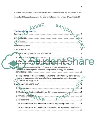
? Malham Field report 9th April Summary This report sought to determine the parasite fauna inhabiting two of the common rodents in Malham Tarn area – Wood mouse (Apodemus sylvaticus)) and rabbit (Oryctolagus cuniculus). We assessed the population of endoparasites present in the two rodents by dissecting them and observing the parasites. Common helminth parasites present included cestodes and trematodes. The rodents served as either intermediate or definitive host of the parasites. Examination of human faecal for the parasites yielded related parasites in terms of their biology and pathogenesis. Parasites prevalence correlated with rodent sex in addition to the rodent weight or length. Females harboured more parasites than males and a single host could have multiple infections of different endoparasites. In our results common parasites population we found include; Graphidium strigosum, Cittotaenia lobata, Passalurus spp. and Taenia pisiformis in rabbit (O. cuniculis and in wood mouse, Heligsomoides spp., Plagiorchis spp, Syphacea and Capillaria spp. Rainfall data for the period coinciding with the collection of the parasite data was also gathered to assess the influence of seasonal changes in weather against the parasites population. A comparison of the biology of endoparasites fauna present in the rodents in Malham Tarn and gastrointestinal parasites of humans was also undertaken. DNA extraction from Cittotaenia lobata (tapeworm) using affinity methods where DNA was allowed to bind to a membrane and then eluted using appropriate buffer was done. The purity of the recovered DNA was determined by taking absorbance at 260 nm and at 280 nm and comparing the ratio to the known ratio of pure DNA which is 1.8 Table of Contents Summary 2 List of figures 4 List of tables 7 Acknowledgement 8 1.INTRODUCTION: 9 1.1.Brief background to site: Malham Tarn 9 1.2.Aims/objectives of field course 9 1.3.Summary of host and parasite fauna from Malham via available literature/references; common parasites in woodmice, rabbits 9 1.4.Gastrointestinal parasites of humans; common parasites in underdeveloped regions; possible comparative biology for Malham parasites species 11 1.5.Importance of diagnostic tests in humans and veterinary parasitology, relative importance/usefulness of different approaches e.g. microscopy coprotests, serology, PCR 13 2.MATERIALS AND METHODS: 14 2.1.Microscopy 14 2.2.Preparation/staining blood films, thin smear faeces 14 2.3.Trapping rodents 14 2.4.Dissections; 14 2.4.1.Examination and dissection of rabbit (Oryctolagus cuniculus) 14 2.4.2.Examination and dissection of wood mouse (Apodemus sylvaticus) 16 2.5.Parasites Data Collection 16 2.6.Data Analysis 17 2.7.DNA extraction and concentration, protein evaluation 17 3.RESULTS: 18 3.1.Apodemus (wood mouse) parasites levels found in after dissection rabbit 23 3.2.Parasites levels found in wood mouse after dissection 30 3.3.DNA extraction and purity on OD280/260 convert to ug/ml 48 4.DISCUSSION: 49 REFERENCE 51 List of figures Figure 1: histogram showing the distribution of Graphidium parasites in rabbit host sampled between 1992 and 2011 in Malham Tarn area 24 Figure 2: Prevalence of Graphidium infections from 1992-2011 25 Figure 3: Histogram showing T. pisiformis parasites population from 1992-2011. Parasites populations was highest in 1999 as can been seen above. 26 Figure 4: Prevalence of Taenia pisiformis population from 1992-2011 at Malham Tarn 27 Figure 5: histogram showing mean Passalurus infections from 1992-2011 27 Figure 6: Line graph showing the prevalence level of Passalurus parasites among rabbit hosts from 1992 to 2011 28 Figure 7: Histogram of the mean Cittotaenia intensity from 1992 to 2011 28 Figure 8: Prevalence of Cittotaenia infections from 1992 to 2011 29 Figure 9: Histogram showing the changes in Trichostrongylus population from 1992 to 2011 29 Figure 10: Graph of the prevalence of Trichostrongylus parasites from 1992 to 2011 30 Figure 11: The graph shows the mean Heligmosomoides parasites found in wood mouse from 1993 to 2011 30 Figure 12: Line graph on the prevalence of Heligmosoides infections from 1993 to 2011 in wood mouse 31 Figure 13: intensity of Plagiorchis parasites in wood mouse 32 Figure 14: Prevalence of Plagiorchis infection in wood mouse from 1992-2011 32 Figure 15: Histogram of the Syphacea parasites population from 1993 to 2011 33 Figure 16: Line graph of the prevalence of Syphacea infection 33 Figure 17: Histogram of Capillaria intensity from 1992 to 2011 34 Figure 18: Capillaria parasites population distribution from 1993 to 2011 in wood mouse host in Malham Tarn 34 Figure 19: histogram of rabbit population sampled form 1992 to 2011 39 Figure 20: Wood mouse population sampled in Malham Tarn area from 1993 to 2011 40 Figure 21: Histogram of the comparison of the rabbit frequency vs. 40 Figure 22: Histogram of the rabbit population and intensity of T. pisiformis from 1992 to 2011 41 Figure 23: Histogram of the rabbit intensity and Passalurus ambigus population 41 Figure 24: Rabbit population and the mean Cittotaenia intensity per year from 1992 to 2011 42 Figure 25: Rabbit population and the mean Trichostrongylus intensity 42 Figure 26: Wood mouse population and the parasite intensity from 1993 to 2011 43 Figure 27: Histogram of Plagiorchis parasites population and the mean parasites population 43 Figure 28: Histogram of the wood mouse population and the Mean Syphacea parasites 44 Figure 29: Histogram showing a comparison of the host (wood mouse) and the intensity of Capillaria parasites 44 Figure 30: Graph showing the average seasonal rainfall (mm) received in Malham tarn for each month 46 Figure 31: Eggs of helminth parasites found in human faeces 47 Figure 32: intestinal amoebae - appearance of encysted forms 48 List of tables Table 1: Graphidium parasites population distribution in rabbits (1992 -2011) 23 Table 2: Host intensity and parasite population 25 Table 3: Prevalence vs. Mean weight 35 Table 4: Mean length Vs. Prevalence 36 Table 5: Differences in the prevalence of Graphidium infection in male and female rabbits 37 Table 6: Differences in the prevalence of Heligmosoides parasites in male and female mice 38 Table 7: wood mouse population: males vs. female 39 Table 8: Table showing the monthly rainfall received in Malham Tarn area from 1992 to 2011 44 Acknowledgement 1. INTRODUCTION: 1.1. Brief background to site: Malham Tarn The Malham Tarn Field centre falls under the field studies council and is located on the north shore of Malham Tarn. The Fields Studies Council is an independent educational charity which promotes environmental understanding for all across Britain. This site which is locate at an altitude of 381m stands at a magnificent limestone scenery at the Yorkshire Dales National Park and is owned by the national trust which has leased it to the field studies council. The tarn is unique as being one of the eight upland alkaline lakes in Europe spanning an area of 0.6 km2 with an average depth of 3 meters (Fryer, 1991). 1.2. Aims/objectives of field course Observation of parasites fauna in rodents - Rabbit (Oryctolagus cuniculus), wood mouse (Apodemus sylvaticus) – in Malham Tarn area 1.3. Summary of host and parasite fauna from Malham via available literature/references; common parasites in woodmice, rabbits Various studies have been mounted to ascertain the parasites infesting rabbits (Oryctolagus cuniculus) among which are helminth parasites such as anoplocephaline cestodes for instance Cittotaenia pectinata which infects 25% of rabbits in the British Isles (Allan et al., 1999; Mead-Briggs and Page, 1975) and common nematode parasites such as stomach worm (Graphidium strigosum). The anoplocephaline cestodes belong to the phylum Platyhelminthes (flatworms) and are common endoparasites of small vertebrates such as rodents. The larvae of the parasites occur in intermediate invertebrates’ hosts while the adult is widespread in small mammals (Georgiev et al., nd). Cestodes occurring in small mammals fall in the order Cyclophyllidea with the Catenotaeniidae family having host range limited to rodents (Quentin, 1994). Various literature sources have reported on the influence of weight, sex and age on the helminth fauna in infesting O. cuniculus (Dudzinski and Mykytowcz, 1963; Dunsmore, 1966a, b, c; Boag and Kolb, 1989). Another rodent which can be found in Malham region is wood mouse (Apodemus sylvaticus) which harbours a variety of nematodes, trematodes and cestodes. Corrigia vitta which is the most common trematode (20-100% prevalence) occurs in the pancreas while Brachylaemus recurvum is an intestinal trematode in wood mouse (Rogan et al., 2007). Helminths of A. sylvaticus exhibit seasonal variation in infection (Lewis and Twigg, 1972; Langley and Fairley, 1982; Montgomery and Montgomery, 1988, 1989; Abu-Madi et al., 2000). However a study undertaken by Rogan et al., (2007) has reported that climate may influence the prevalence of Plagiorchis muris, a trematode affecting A. sylvaticus which was associated with wetter spring/summer periods. P. muris is of interest in public health since it is also infective to humans and its transmission is unique because transmission to definitive host occurs via ingestion of insects or their aquatic larvae (Hong et al., 1996; Chai and Lee, 2002). Aquatic snails (fresh water snails) serve as the first intermediate host of Plagiorchis spp. with around 25 species of these species having being reported in Malham Tarn area alone (Norris, 2003). In the first intermediate host, fresh water snail, the trematode produces sporocysts and xiphidiocercariae which emerge from the snail and penetrate the second intermediate host and then proceed into the metacercariae stage. Several insects serve as second intermediate host and they include larvae of dragonflies, mosquitoes and chironomids (McMullen, 1973a; Jansen and Bock, 1990; Hong et al., 1996). In the virtue of the fish being mentioned as aquatic second intermediate host of Plagiorchis, metacercariae stage, they may be possible route of human infection (Hong et al., 1996). Rogan et al (2007) has reported an overdispersed pattern of P. muris in A. sylvaticus with very few rodents harbouring significant populations of this trematode to ascertain the worm burden of P. muris reported. This distribution in area adjacent to Malham Tarn, coincides with the typical distribution of parasites in the wild where extrinsic factors such as seasonal differences, density of intermediate host and intrinsic factors such as age, sex and variation in susceptibility come into play (Behnke et al., 1999) 1.4. Gastrointestinal parasites of humans; common parasites in underdeveloped regions; possible comparative biology for Malham parasites species Various parasites inhabit the gastrointestinal tract of humans and may also be present in rodents such as mice and rabbits. These include nematodes and trematodes. Among these parasites the soil-transmitted helminths such as hookworm, Trichuris trichura and Ascari lumbricoides are the most prevalent affecting almost a sixth of the world population (Harhay et al., 2010). Nematodes A small subset of nematode worms account for the human infections and among these Acylostoma doudenale, Necator americanus, Trichuris trichura and Ascaris lumbricoides which are all classified as soil-helminths are the most prevalent (Harhay et al., 2010). Cestodes Cestodes (tapeworm) are normally transmitted via infected meat products and therefore may not be common gastrointestinal parasites in poverty ridden communities such as immigrants and refugees as in the case of nematodes (Hall et al., 2008). Trematodes Intestinal flukes (trematodes) may be acquired by humans through consumption of food and water contaminated with the intermediate hosts. Common intermediate host of trematodes include invertebrates such as fresh water snails. Schistosoma mansoni, S. haematobium and S. japonicum are blood flukes which inhabit blood vessels with their eggs penetrating the intestines resulting in intestinal damage, spleen and liver enlargement and portal hypertension. The intensity of gastrointestinal (GI) parasites determines the general symptoms observe following infections in humans. These GI parasites cause harm to their human host by feeding on host tissue leading to a deficiency of iron and proteins for instance the hookworms. Others may cause maldigestion and/or malabsorption of nutrients and may also elicit inflammatory responses from the immune system which may affect the food intake and metabolism of key nutrients such as iron (Harhay et al., 2010). 1.5. Importance of diagnostic tests in humans and veterinary parasitology, relative importance/usefulness of different approaches e.g. microscopy coprotests, serology, PCR The techniques for detection of cestodes such as Taenia spp. in humans relies on the detection of parasites components such as proglottides and/or eggs in faeces. Albeit inexpensive, the procedures may be prone to low insensitivity and specificity (Allan et al., 2003). This method has also been adopted in the detection of these parasites in veterinary. This method is may also underestimate the prevalence of taeniasis since it relies on the intermittent phenomenon of egg excretion which may not actually be representative of the parasites population (Hall et al., 1981; Allan et al., 1996a). For instance destrobilisation occurs it may lead to a massive release of eggs and then stoppage of release of eggs in faeces. Moreover observation of eggs under a microscope may prove a daunting task as different species of some parasites such as Taenia may present eggs which appear identical. Molecular biology techniques involving DNA extraction and PCR have become vital tools in biomedical procedures especially in the helminthology laboratory where they assist in characterization and determination of species to subspecies and strain levels and even are detect genetic mutation contributing to antihelminthic drug resistant (Humbert et al., 2001). 2. MATERIALS AND METHODS: 2.1. Microscopy Parasites eggs were observed in saline under a microscope and their measurements taken in order to assist in the identifying the parasites. This was achieved by preparing thin smears from human faeces and blood smears from rabbits and wood mouse and observing carefully under a microscope as the measurement were being taken. 2.2. Preparation/staining blood films, thin smear faeces Human faeces collected were prepared for thin smears to observe the various gastrointestinal parasites present under a microscope. Thin smears were carefully prepared on a microscope slide. 2.3. Trapping rodents Rabbits and rabbits were trapped in Malham Tarn area site for observation of common ectoparasites and dissection to observe endoparasites. 2.4. Dissections; 2.4.1. Examination and dissection of rabbit (Oryctolagus cuniculus) We weighed the rabbit provided and checked the body surface for the presence of lumps (e.g. larval cysts of Taenia serialis and also checked the fur for the presence of ectoparasites. Any ectoparasites found such as Spilopsyllus cuniculi and Psoroptes communis was transferred into a capped tube with a cotton wool impregnated with chloroform. The arthropods were examined in a Petri dish using a dissecting microscope and then transferred to a microscope and observed in high magnification under a compound microscope. While the blood was fresh we examined blood for possible presence of moving flagellates such Trypanosoma spp. A dissection was made between the outer and inner body wall down the ventral mid-line as sex confirmation was made by observation of either testes or paired uterus. An observation of the body cavity was made for the presence of helminths e.g. Taenia pisiformis on pelvic space and stomach mesenteries. Liver examination The liver was prepared for the observation of cysts and lesions by cutting out small lesion and squashing between two microscope slides and checking for the presence of white spots of Eimeria or Taenia Spp. cysts. Larger cysts were observed under a Petri dish. Bile ducts were examined for flukes of Fasciola or Dicrocoelium. Gastrointestinal tract examination The stomach and intestines –small intestines (SL) and large intestines (LI) were removed separately and the location of parasites noted. The stomach was cut open over a dish and count of the worm population made. The sex of Graphidium strigosum (nematode) was made by identifying bursa and spicules in males. Measurement of the length a sample of either sex was also taken. 2.4.2. Examination and dissection of wood mouse (Apodemus sylvaticus) The sex of the dead mouse was noted and measurement from the anus to the nose taken. We examined the fur for common ectoparasites such as ticks (Ixodes), fleas, mites and lice and washed the eyes in saline so as to examine for the presence of nematodes such as Pelodera. To expose the live and intestines for the examination of endoparasites, we made a longitudinal slit in the skin and body wall. The liver was examined for the presence of cysts or any abnormal tissue with cysts being removed and placed in saline. A small incision was made on the cyst wall to reveal the cestodes larva for instance for Taenia taeniaeformis or T. mustela and a squash preparation was made for any observed abnormal liver tissue. The intestines were removed intact by cutting the oesophagus and rectum and constraining mesentery while ensuring no damage to the pancreas. The examination for the presence of the nematode, Corrigia was done in the pancreatic duct and we then placed the intestine in saline. We examined the pancreas for the presence of dark streaks which are indication of trematode-infected pancreas and freed any trematode present by making a small hole in the infected pancreatic duct carefully. We identified and made a tally of the parasites present and entered the data in a table for further analysis. The stomach was cut at the pyloric sphincter and we embarked to examine the parasites present in a separate dish. 2.5. Parasites Data Collection The data collected from parasites counted after dissection of wood mouse and rabbit was entered into a Microsoft© Excel sheet for further analysis. Microscopic observation of the parasites from human faeces was presented as diagrams as illustrated in the results chapter 2.6. Data Analysis The mean and prevalence levels of each parasite in the period spanning from 1992 to 2011 was calculated in Microsoft© Excel and charts (Histograms and line graphs) obtained to present this data. 2.7. DNA extraction and concentration, protein evaluation Cittotaenia was cut into small pieces and using a pestle and a mortar it was ground into pulp and 180µl buffer ATL added. The sample was lysed with 20µl proteinase K and then mixed thoroughly by vortexing and incubated in a water bath at 56 C until all the tissues were completely lysed. Vortexing was pulsed for and a further 200µl buffer AL added with thorough mixing. 200µl of absolute (100%) ethanol was added and the mixture thoroughly. The mixture was pipetted including the precipitate into a DNeasy Mini spin column and then placed into a collection tube. Centrifugation was done at more than 6000 x g for a minute and the flow-through discarded. The DNeasy Mini spin column was placed in a new 2 ml collection tube and 500µl buffer AW2 added with a further centrifugation for 3 minutes at more than 20,000 x g the flow-through and the collection tube discarded. DNA was eluted by pipetting 200µl of buffer AE directly onto the DNeasy membrane. It was incubated at room temperature for 1 min and then centrifuged for 1 min at 600 x g to elute. DNA recovery was maximized by repeating the addition of 200µl of buffer AE with centrifugation and incubation. Confirmation and determination of DNA purity UV spectroscopy was used to assess the purity of recovered DNA by taking the absorbance at 260 nm and at 280 nm. The absorbance should range between 0.1-1.0. 3. RESULTS: Parasites observed in the human faeces; Ascaris lumbricoides Taenia spp. Paragonimus eggs Trichuris eggs Diphyllobothrium eggs Fasciola spp. Hymenolepsis nana Hookworms Entamoeba histolytica Entamoeba coli Giarda spp. Schistosoma japonicum S. haematobium S. mansoni Iodamoebabutilli Enteroboius eggs Opisthorchid stronglyloides Lifecycles in brief of parasites observed in human faeces Dwarf tapeworm (Hymenolepsis nana) Humans serve as both the definitive and intermediate hosts where the adult tapeworm inhabits the gut lumen. It is transmitted via hand to mouth or by ingestion of insect intermediate host. The worm inhabits the upper ileum and ha the ability to autoinoculate itself a situation which may lead to huge worm burden especially in children. Hookworms (e.g. Ancylostoma duodenale) Eggs of hookworm s are passed in faeces and hatch in warm, moist soils realising a rhabditiform larvae which takes a few days to develop into a filariform larvae. The filariform larva invades the skin and migrates through the venous blood to the heart and then lungs. In the lungs, they penetrate into the alveoli and migrate via the trachea into the gastrointestinal system. In the intestines, the larvae mature into adults and attach themselves to the jejuna and duodenal mucosa where they suck blood. Entamoeba histolytica Pathogenic strains of this protozoon penetrate the epithelial tissue of colon resulting in ulcerations. The amoeba may spread to the liver via the portal blood stream and produce hepatic amoebiasis. Giardia lamblia This flagellate parasite exists in trophozoite and cysts forms with the cysts form being resistant to desiccation. Infection to this protozoon is limited to the biliary tract and intestine. It is transmitted through contaminated food/water and other faecal-oral routes. Schistosoma spp. Fresh water snails are the intermediate host of these trematodes. The cercariae stage of the parasite penetrates the skin and migrate through the blood stream via the lungs to the colon or venous plexus draining the bladder (haematobium). Stronglyloides Spp. (roundworm) Rhabditiform larva excreted in faeces moults after a couple of days into infective filariform larvae which penetrates the human skin and migrate via the blood streams as in hookworm and to the GI. Larvae develop into adults in the upper parts of the ileum. However in some environmental conditions the rhabditiform larvae may develop into free living sexually mature male and female adults in the soil. Whipworm (Trichuris trichiura) A warm, moist and shaded soil environment facilities the release of the unhatched of the infectious stage larvae which is produced after a period of 3 weeks. Upon ingestion of the egg by human, the activated larva escapes via the weakened egg shell in the upper small intestine and penetrates an intestinal villus. Albeit the Trichuris inhabits the cecum, it can also be found in appendix and lower ileum. Parasites observed after dissection of rabbit i. Trichostronglus retorformis ii. Passalurus ambigus iii. Cittotaenia Spp. iv. Taenia pisiformis Lifecycles in brief Trichostrongylus retorformis Trichostrongylus are commonly found in wild rabbits and inhabit the small intestines. Rabbits become infected from food such as hay and fresh vegetables contaminated with the parasite larvae. In the host eggs from female parasites are released in faeces and within a period of 10-11days they develop into an infective larva. These parasites have direct lifecycle with the absence of an intermediate host (Praag, 2003) Passalurus ambigus The parasite infects both domestic and wild rabbits. Adult worms are located in the anterior part of the caecum and in colon of rabbits with juvenile stages being found in the mucosa of small intestines and caecum. Like Trichostrongylus retorformis, this parasite has a direct lifecycle where a larva emerges from eggs and develops in the mucous layer of small intestine and caecum to mature adults. Cittotaenia Spp. Cittotaenia, a cestode, has a definitive host and an intermediate host which is a component of the definitive host food chain. Eggs of the parasites are released to the environment in faeces and contain an embryo (oncosphere). Eggs are ingested by an intermediate host where it hatches and penetrate the intermediate host gut. Taenia pisiformis Rabbits are definitive host of T. pisiformis with canines serving as the definitive host of this parasite. Adult tapeworm releases their eggs through the faeces of canines to the environment and through contaminated food the rabbit takes up the eggs. The eggs hatch in the rabbit gut into larvae which then migrates to the liver and within 15-30 days it develops into cysticercus which is a fluid-filled cyst. The immature tapeworm in the cysticercus only develops in the canid. Parasites observed after dissection of wood mouse i. Heligmosomoides spp. ii. Plagiorchis spp. iii. Syphacea spp. iv. Capillaria spp. Lifecycles Heligmosomoides spp. Rodents such as the wood mice are the definitive host of these parasites. Eggs released by females pass into faeces and hatch into L1 larvae which moult to L3 larvae. L3 larvae may penetrate the intestinal mucosa of a new host and moult into L4 larvae. The L4 larvae then moults into and adult which migrate to the small intestine at the pylorus and again begin to lay eggs to continue the cycle. 3.1. Apodemus (wood mouse) parasites levels found in after dissection rabbit Table 1: Graphidium parasites population distribution in rabbits (1992 -2011) year Host intensity Mean parasite intensity 1992 14 107.79 ± 165.75 1993 21 94.67 ± 279.90 1994 28 46.46 ± 316.33 1995 11 26.55 ± 292.01 1996 21 17.05 ± 293.40 1997 15 23.60 ± 218.81 1998 8 13.91 ± 282.86 1999 18 5.67 ± 233.40 2000 11 2.18 ± 294.27 2001 13 1.54 ± 294.30 2002 4 5.25 ± 163.30 2003 27 2.81 ± 176.24 2004 15 2.93 ± 223.15 2005 21 0.24 ± 195.39 2006 23 0.58 ± 222.87 2007 22 2.73±166.53 2008 27 6.89 ± 228.29 2009 20 1.35 ± 121.36 2010 14 6.07 ± 219.57 2011 35 0.59 ± 242.76 Figure 1: histogram showing the distribution of Graphidium parasites in rabbit host sampled between 1992 and 2011 in Malham Tarn area Figure 2: Prevalence of Graphidium infections from 1992-2011 Table 2: Host intensity and parasite population year Host intensity Mean parasite intensity 1992 14 107.79 ± 165.75 1993 21 94.67 ± 279.90 1994 28 46.46 ± 316.33 1995 11 26.55 ± 292.01 1996 21 17.05 ± 293.40 1997 15 23.60 ± 218.81 1998 8 13.91 ± 282.86 1999 18 5.67 ± 233.40 2000 11 2.18 ± 294.27 2001 13 1.54 ± 294.30 2002 4 5.25 ± 163.30 2003 27 2.81 ± 176.24 2004 15 2.93 ± 223.15 2005 21 0.24 ± 195.39 2006 23 0.58 ± 222.87 2007 22 2.73±166.53 2008 27 6.89 ± 228.29 2009 20 1.35 ± 121.36 2010 14 6.07 ± 219.57 2011 35 0.59 ± 242.76 Figure 3: Histogram showing T. pisiformis parasites population from 1992-2011. Parasites populations was highest in 1999 as can been seen above. Figure 4: Prevalence of Taenia pisiformis population from 1992-2011 at Malham Tarn Figure 5: histogram showing mean Passalurus infections from 1992-2011 Figure 6: Line graph showing the prevalence level of Passalurus parasites among rabbit hosts from 1992 to 2011 Figure 7: Histogram of the mean Cittotaenia intensity from 1992 to 2011 Figure 8: Prevalence of Cittotaenia infections from 1992 to 2011 Figure 9: Histogram showing the changes in Trichostrongylus population from 1992 to 2011 Figure 10: Graph of the prevalence of Trichostrongylus parasites from 1992 to 2011 3.2. Parasites levels found in wood mouse after dissection Figure 11: The graph shows the mean Heligmosomoides parasites found in wood mouse from 1993 to 2011 Figure 12: Line graph on the prevalence of Heligmosoides infections from 1993 to 2011 in wood mouse Figure 13: intensity of Plagiorchis parasites in wood mouse Figure 14: Prevalence of Plagiorchis infection in wood mouse from 1992-2011 Figure 15: Histogram of the Syphacea parasites population from 1993 to 2011 Figure 16: Line graph of the prevalence of Syphacea infection Figure 17: Histogram of Capillaria intensity from 1992 to 2011 Figure 18: Capillaria parasites population distribution from 1993 to 2011 in wood mouse host in Malham Tarn Rabbit data: Mean weight vs. Graphidium prevalence Correlation = 0.3365 Table 3: Prevalence vs. Mean weight Year Mean weight Prevalence 1992 1385.71 92.86 1993 1585.24 95.24 1994 1290.54 82.14 1995 1360.91 63.64 1996 1413.14 47.62 1997 1362.67 73.33 1998 1509.09 62.50 1999 1561.67 72.22 2000 1421.82 27.27 2001 1334.62 23.08 2002 1367.13 25.00 2003 1392.85 22.22 2004 1438.67 46.67 2005 1410.24 90.48 2006 1264.58 21.74 2007 1552.27 45.45 2008 1374.07 59.26 2009 1393.00 35.00 2010 1306.43 50.00 2011 1430.44 14.29 Mouse data: mean length vs. Heligmosoides prevalence Correlation = 0.6322 Table 4: Mean length Vs. Prevalence Year Mean length (cm) prevalence 1993 8.55 78.57 1994 8.56 69.23 1995 8.61 82.35 1996 8.08 70.83 1997 8.00 91.67 1998 9.03 86.67 1999 8.04 50 2000 8.59 88 2001 8.34 82.35 2002 8.91 81.25 2003 8.03 50 2004 8.28 62.5 2005 8.11 87.5 2006 8.49 66.67 2007 8.16 64.71 2008 8.01 60.87 2009 8.36 62.5 2010 7.33 37.5 2011 8.07 66.67 Tables line graphs and histograms Male vs. female prevalence; intensity (means) Differences in the prevalence of Graphidium infection in male and female rabbits Testing the hypothesis: i. Null hypothesis (H0) : there is no difference in the prevalence of Graphidium strigosum between males and females ii. Alternate hypothesis (H1) : there is a significant difference between Graphidium prevalence in between males and females Table 5: Differences in the prevalence of Graphidium infection in male and female rabbits Rabbits Total Observed IF Expected IF Males 154 60 77 Females 236 114 118 total 390 174 195 : Key IF: infected After testing the two hypothesis P value =0.05 we accepted the alternate (H1) hypothesis (p Read More
Cite this document
  • APA
  • MLA
  • CHICAGO
(“Malham Field Lab Report Example | Topics and Well Written Essays - 9500 words”, n.d.)
Retrieved from https://studentshare.org/biology/1396538-malham-field-course-report
(Malham Field Lab Report Example | Topics and Well Written Essays - 9500 Words)
https://studentshare.org/biology/1396538-malham-field-course-report.
“Malham Field Lab Report Example | Topics and Well Written Essays - 9500 Words”, n.d. https://studentshare.org/biology/1396538-malham-field-course-report.
  • Cited: 0 times

CHECK THESE SAMPLES OF Parasites Prevalence in Malham Field

The Specialization and Parasites

Factors such as 'jack of all, master of none' and freedom from natural enemies, have been used to justify the prevalence of specialists.... Factors such as “jack of all, master of none” and freedom from natural enemies, have been used to justify the prevalence of specialists, however; it has also been postulated that in spite of these advantages, specialization would gradually lead to loss of genetic variations, thus disabling capacity of adaptation to altered set of environmental conditions....
11 Pages (2750 words) Term Paper

Commensal and Parasitic Barnacles

Barnacles are both commensals and parasites.... tm Certain Lepadomorpha barnacles, which act as commensals, are usually found in the teeth of the pygmy killer whale or Feresa attenuata, the sperm whale or Physeter macrocephalus, the goosebeak whale or Ziphius cavirostris, and the Antillean beaked whale or Mesoplodon europaeus, as well as other cetaceans that thrive the deep marine waters (Mignucci-Giannoni, “Metazoan parasites”).... “Metazoan parasites”)....
6 Pages (1500 words) Term Paper

How Parasites Affect the Spread of Malaria

The present paper "How parasites Affect the Spread of Malaria" has identified that malaria is a disease caused by one of the four protozoan species of the genus Plasmodium.... This paper deals with the cause and spread of malaria, and its association with parasites and protozoa.... The mosquito then bites the human and transmits the parasites in the human blood which account for the onset of the symptoms (Cdc.... They are given the attenuated malarial parasites that effectively immunize them for the shorter duration....
5 Pages (1250 words) Research Paper

Policy paper-fixing health care (Zoonotic disease)

The overall prevalence of D.... Table -1 shows the prevalence of round worm infestation in the Caribbean and Table 2 shows the prevalence of whip worm with the sample sizes given in parenthesis.... Table – 1 prevalence of round worm Country 0-5 Years M & F6-14 Years M & F15+ Years M15-44 Years F45+ Years FJamaica6.... Table – 2 prevalence of HookwormCountry 0-5 Years M & F6-14 Years M & F15+ Years M15-44 Years F45+ Years FJamaica6....
2 Pages (500 words) Research Paper

Molecular Mechanisms Used by Adult Schistosoma Worms to Survive in the Bloodstream

Possible interventions are proposed to counter the effect of these adaptations so as to make the parasites vulnerable to the host's immune system and possible elimination.... They are parasites that live in the vascular system and mature as separate-sex entities in the veins of birds and mammals.... They are parasites that live in the vascular system, and mature as separate-sex entities in the veins of birds and mammals.... These parasites experience remarkable physiological and morphological changes throughout their life as a means of survival and adaptation to their varying living conditions in different hosts....
11 Pages (2750 words) Essay

Parasitic Parasites

The study would help in generating genetic markers which may be used for characterization and distribution of the sub-populations of the parasites.... Malaria maybe caused by one of the four species of Plamodium parasites-Plasmodium falciparum, P.... Parasitic parasites s ... Malaria maybe caused by one of the four species of Plamodium parasites-Plasmodium falciparum, P.... iotto et al conducted an experiment to understand the pattern of genome variation in the parasites....
3 Pages (750 words) Essay

Commensal and Parasitic Barnacles

Barnacles are both commensals and parasites.... Barnacles are both commensals and parasites.... Certain Lepadomorpha barnacles, which act as commensals, are usually found in the teeth of the pygmy killer whale or Feresa attenuata, the sperm whale or Physeter macrocephalus, the goosebeak whale or Ziphius cavirostris, and the Antillean beaked whale or Mesoplodon europaeus, as well as other cetaceans that thrive the deep marine waters (Mignucci-Giannoni, “Metazoan parasites”). ...
5 Pages (1250 words) Term Paper

Characterization of a Novel Protein of the Animal Parasite, Toxoplasma Gondii

he systematic procedure also entailed determination of the gene is conserved in various apicomplexan parasites alongside in a diversity of eukaryotes.... The paper "Characterization of a Novel Protein of the Animal Parasite, Toxoplasma Gondii" is focused on toxoplasma gondii refers to an obligate parasite (which is intracellular) and has the capability of posing a substantial threat to the health of humans, in the form of toxoplasmosis....
5 Pages (1250 words) Case Study
sponsored ads
We use cookies to create the best experience for you. Keep on browsing if you are OK with that, or find out how to manage cookies.
Contact Us