StudentShare
Contact Us
Sign In / Sign Up for FREE
Search
Go to advanced search...
Free

Role of GLUT4 Glucose Transporter - Essay Example

Cite this document
Summary
This paper 'Role of GLUT4 Glucose Transporter' tells us that glucose is the major energy and carbon source for organisms like human beings and yeast. These organisms can derive the other monosaccharides required for glycan (which refers to the assembly of sugars either in free form) biosynthesis from these sources…
Download full paper File format: .doc, available for editing
GRAB THE BEST PAPER94% of users find it useful
Role of GLUT4 Glucose Transporter
Read Text Preview

Extract of sample "Role of GLUT4 Glucose Transporter"

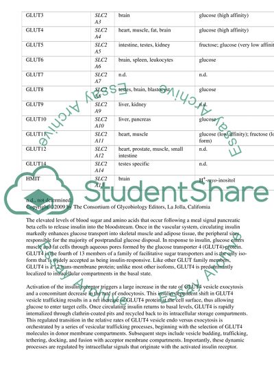
Role of GLUT4 glucose transporter in glucose homeostasis and cellular signaling pathways that regulate its endogenous expression and membrane recycling. Glucose and fructose are the major energy and carbon sources for organisms like human beings and yeast. These organisms can derive the other monosaccharides required for glycan(which refers to the assembly of sugars either in free or in attached form) bio-synthesis from these sources. Initially these monosaccharides have to be activated to high energy donor molecules to act as precursors for glycan synthesis. This activation process requires nucleoside tri phosphates(like UTP or GTP) and a glycosyl-1-phosphate and is catalysed by a kinase. The generalised form of this reaction is written as follows Sugar + NTP ---------> Sugar-P. Sugar-P + NTP---------> Sugar-NDP +PPi There are three types of sugar transporters that carry sugars across the plasma membrane in to the cells. First are the energy independent facilitated diffusion transporters such as the glucose transporters family(GLUT) of hexose transporters seen in yeast and in mammalian cells. These proteins are encoded by SLC2A genes(solute carriers 2A). Second are the energy dependent transporters for example sodium dependent glucose transporters(SGLT) encoded by SLC5A genes in the intestine and in kidney epithelial cells. The third type of transporters couple ATP dependent phosphorylation with sugar import and are seen in bacteria. Glucose Transporters family This family of glucose transporters were first discovered in yeast where 18 genes have been identified. Humans have 14 GLUT homologs. All of the yeast glucose transporters are of the same size(40-55 Kilodaltons) and have similar structures containing 12 membrane spanning domains. These domains form a barrel with a small pore for the sugar to pass through. The only "sugar transport signatures" are a few widely scattered glycine and tryptophan residues and one PET tri-peptide sequence Monosaccharide transporters Protein Gene Localization Primary sugar transported GLUT1 SLC2A1 erythrocytes, brain, ubiquitous glucose GLUT2 SLC2A2 liver, pancreas, intestine, kidney glucose (low affinity); fructose; glucosamine GLUT3 SLC2A3 brain glucose (high affinity) GLUT4 SLC2A4 heart, muscle, fat, brain glucose (high affinity) GLUT5 SLC2A5 intestine, testes, kidney fructose; glucose (very low affinity) GLUT6 SLC2A6 brain, spleen, leukocytes glucose GLUT7 SLC2A7 n.d. n.d. GLUT8 SLC2A8 testes, brain, blastocyst glucose GLUT9 SLC2A9 liver, kidney n.d. GLUT10 SLC2A10 liver, pancreas glucose GLUT11 SLC2A11 heart, muscle glucose (low affinity); fructose (long form) GLUT12 SLC2A12 heart, prostate, muscle, small intestine n.d. GLUT14 SLC2A14 testes specific n.d. HMIT SLC2A13 brain H+-myo-inositol n.d., not determined. Copyright '2009 by The Consortium of Glycobiology Editors, La Jolla, California The elevated levels of blood sugar and amino acids that occur following a meal signal pancreatic beta cells to release insulin into the bloodstream. Once in the vascular system, circulating insulin markedly enhances glucose transport into skeletal muscle and adipose tissue, the peripheral sites responsible for the majority of postprandial glucose disposal. In response to insulin, glucose enters muscle and fat cells through aqueous pores formed by the glucose transporter 4 (GLUT4) protein. GLUT4 is the fourth of 13 members of a family of facilitative sugar transporters and is the only iso-form that is widely accepted as being insulin-responsive. Like other GLUT family members, GLUT4 is a 12 trans-membrane protein; unlike most other isofoms, GLUT4 is predominantly localized to intracellular compartments in the basal state. Activation of the insulin receptor triggers a large increase in the rate of GLUT4 vesicle exocytosis and a concomitant decrease in the rate of endocytosis. This insulin-dependent shift in GLUT4 vesicle trafficking results in a net increase of GLUT4 protein at the cell surface, thus allowing glucose to enter target cells. Once circulating insulin returns to basal levels, GLUT4 is rapidly internalized through clathrin-coated pits and recycled back to its intracellular storage compartments. This regulated transition in the relative rates of GLUT4 vesicle endo versus exocytosis is orchestrated by a series of vesicular trafficking processes, beginning with the selection of GLUT4 molecules in donor membrane compartments. Subsequent steps include vesicle budding, trafficking, tethering, docking, and fusion with acceptor membrane compartments. Importantly, these dynamic processes are regulated by intracellular signals that originate with the activated insulin receptor. As a result of these processes, elevated glucose levels are rapidly returned to normal even after huge caloric ingestions, and they are maintained at only slightly lower levels during long-term starvation. Such control prevents severe dysfunctions such as loss of consciousness due to hypoglycemia and toxicity to peripheral tissues in response to the chronic hyperglycemia of diabetes. The GLUT4 glucose transporter is thus a major mediator of glucose removal from the circulation and a key regulator of whole-body glucose homeostasis. Structural Features of the Insulin-Regulated GLUT4 Glucose Transporter Protein The unique sensitivity of GLUT4 to insulin-mediated translocation appears to derive from sequences in the N-terminal and COOH-terminal regions. These sequences are involved in rapid internalization and sorting of GLUT4 in intracellular membranes termed GLUT4 storage vesicles (GSV). Such unique sequences in the N- terminal and COOH-terminal cytoplasmic domains that direct its characteristic membrane trafficking capability are shown in figure 2. Figure2. Structural Features of the Insulin-Regulated GLUT4 Glucose Transporter Protein (The GLUT4 Glucose Transporter, Shaohui Huang and Michael P. Czech) Cell Metabolism, Volume 5, Issue 4, 237-252, 4 April 2007. These include a distinctive N-terminal sequence with a potentially critical phenylalanine residue as well as dileucine and acidic motifs in the COOH terminus. These motifs govern kinetic aspects of both endocytosis and exocytosis in a continuously recycling trafficking system. The COOH terminus LL and acidic motifs are also present in an amino peptidase protein (IRAP) that in adipocytes is similarly sequestered within GLUT4-enriched intracellular membranes and highly responsive to insulin action. These considerations place GLUT4 at the interface of two exciting fields of biology-insulin signaling and membrane trafficking. GLUT4 Is a Key Determinant of Glucose Homeostasis A central role for GLUT4 in whole-body metabolism is strongly supported by a variety of genetically engineered mouse models where expression of the transporter is either enhanced or ablated in muscle or adipose tissue or both. It has been demonstrated that conditional depletion of GLUT4 in either adipose tissue or skeletal muscle causes insulin resistance and a roughly equivalent incidence of diabetic animals .These tissue-specific depletions of GLUT4 have profound metabolic effects on other tissues. For example, mice with muscle-specific GLUT4 deficiency display decreased insulin responsiveness in adipose tissue and liver, while those with adipose-specific GLUT4 depletion exhibit muscle and liver insulin resistance .In the muscle-specific GLUT4 knockout mice, this is at least partially mediated through elevated blood glucose levels that occur in the conditional knockout animals, which secondarily impairs insulin signaling. More recently, it has been reported that a retinol binding protein (RBP4) is released into the serum from GLUT4-deficient adipose tissue and that RBP4 may contribute to the insulin resistance of obese and diabetic individuals . Even though cell surface GLUT4 is highly dependent on insulin in vitro, muscle-specific (MIRKO) or adipose-specific (FIRKO) insulin receptor knockout mice produce surprisingly mild metabolic phenotypes compared to the conditional GLUT4 knockout mice. Analysis of the above mouse models also highlights the potential dissociation between glucose uptake versus triglyceride synthesis and storage in the adipocyte. The adipose-specific GLUT4 deficient mouse has normal fat mass and adipocyte size. In the absence of robust glucose import and glycolysis, triglyceride synthesis in adipocytes can proceed via enhanced glyceroneogenesis based on substrates derived from liver and muscle. On the other hand, lack of the lipogenic and antilipolytic actions of insulin in adipocytes of the FIRKO mouse leads to reduced fat mass. These observations are consistent with the concept that adipocytes are the major site for energy (triglyceride) storage but not glucose disposal, which mostly occurs in muscle. Control of Endogenous GLUT4 Expression The profound effects on whole-body glucose homeostasis observed in mouse models of GLUT4 deficiency or overexpression heighten the potential physiological importance of changes in endogenous GLUT4 expression in different states. Examples of particular interest are the down regulation of GLUT4 expression in adipose tissue in obesity and the up regulation of GLUT4 expression in skeletal muscle in response to exercise or thyroid hormone. Also, GLUT4 expression is greatly decreased in muscle atrophy and denervation consistent with decreased energy requirements in these conditions. Signals from neuronal cells in the form of neuregulins may mediate some of these effects .These changes in GLUT4 protein levels could translate into alterations in glucose tolerance if the effects observed in transgenic mice apply to normal physiology. Thus, therapeutic strategies based on enhancing GLUT4 expression may facilitate drug discovery. Tissue-specific expression of GLUT4 in adipose tissue, skeletal muscle, and cardiac muscle, as well as its regulation by fasting and re-feeding, is conferred within a 2.4 kb DNA segment at the 5' region of the GLUT4 gene . For skeletal muscle-specific expression, a region between -522 and 420 has been inferred to be important in transgenic mice. This region contains an apparent myocyte enhancer factor (MEF)2 binding domain at -466 to -457 that is critical for specifying tissue expression and increased GLUT4 expression during muscle regeneration. Near this same region it has been proposed that thyroid hormone receptor forms a complex with MEF2 to regulate GLUT4 expression. Another domain that has been implicated in tissue-specific expression is termed Domain I and includes the region -742 to -712 relative to the initiation site for transcription . Signaling Mechanisms that Acutely Regulate GLUT4 Both insulin and exercise acutely stimulate GLUT4 recruitment to the cell surfaces of muscle and adipose cells independent of transcription or translation. These two physiological stimuli initiate distinct signaling mechanisms that lead to enhanced GLUT4 translocation and glucose uptake as shown in figure 3. The insulin signaling pathway to GLUT4 is triggered by activation of the insulin receptor (IR) tyrosine kinase leading to tyrosine phoshphorylation of insulin receptor substrate proteins (IRS) and their recruitment of PI 3-kinase, which catalyzes conversion of phosphatidylinositol (4,5)P2 to phosphatidylinositol (3,4,5)P3 (denoted PIP3). PIP3 in turn triggers the activation of the protein kinase Akt through the actions of two intermediate protein kinases, PDK1 and Rictor/mTOR . The requirements of these overall reactions have largely been confirmed in vivo using genetically engineered mouse models and more recently by siRNA knockdown experiments in cultured cells. It has been established that Akt2 isoform appears to control GLUT4 trafficking in adipose and muscle cells as well as mediate insulin signaling to control glucose output in liver while the Akt1 isoform appears to control cell and body size and Akt3 controls brain size . Figure 3 Convergence of Signaling Pathways Initiated by Insulin and Exercise Leading to GLUT4 Translocation. (The GLUT4 Glucose Transporter, Shaohui Huang and Michael P. Czech). Cell Metabolism, Volume 5, Issue 4, 237-252, 4 April 2007. Insulin signaling through the PI 3-kinase pathway and muscle contraction through both elevated AMP/ATP ratios and intracellular [Ca2+] leads to activation of downstream protein kinases (Akt, aPKC, AMPK, CaMKII cPKC) that phosphorylate putative effectors that modulate steps in the GLUT4 trafficking pathways. AS160 is one such substrate that appears to negatively regulate an early step in GLUT4 exocytosis. Negative regulation of these pathways by fatty acids, cytokines, and endoplasmic reticulum stress responses are observed in obesity and diabetes, contributing to insulin resistance. Dashed lines imply hypothetical pathways not yet experimentally verified. Muscle contraction also increases AS160 phosphorylation, but it may do so by a PI 3-kinase independent mechanism. Contraction-induced AS160 phosphorylation is mediated through the AMPK pathway, providing a potential convergence of insulin and exercise-mediate signaling to GLUT4. It has been observed that calmodulin binds to AS160 in a Ca2+-dependent manner and treatment of T cells with calcium ionophore increases AS160 mRNA levels. However, AS160 is a target of metabolic disease related to insulin resistance. Insulin-stimulated AS160 phosphorylation is impaired in skeletal muscle of type II diabetic patients. A proposed PI 3-kinase-independent pathway including component proteins APS, c-Cbl, CAP, CrkII/C3G, and TC10 has also been proposed to operate in parallel to the PI 3-kinase pathway as a required mechanism that promotes GLUT4 translocation. Hence it is clear that APS and CAP proteins do associate with insulin receptor complexes .The PI 3-kinase pathway mediates insulin signaling to regulate GLUT4 in skeletal muscle as well. In addition, muscle contraction acutely increases GLUT4 transporter levels in sarcolemma and T-tubules, and is additive to the effect of insulin stimulation. Muscle contraction and insulin stimulation were suggested to target separate pools of intracellular GLUT4-containing membranes. Two cellular consequences of muscle contraction, a transient increase in intracellular calcium concentration ([Ca2+]i) and an increase in [AMP]/[ATP] ratio, are thought to contribute to enhanced GLUT4 translocation to the cell surface as shown in the figure3 through activations of the protein kinase CaMKII and protein kinase C. AMPK acts as a cellular energy gauge that is allosterically activated by increasing [AMP]/[ATP] ratios. This transforms AMPK into a better substrate for at least one of its upstream activator kinases, e.g., LKB1, or a potentially poorer substrate for one of its phosphatases. Muscle glycogen is a potent negative regulator of AMPK activity thus providing a negative feedback mechanism for AMPK-mediated glucose uptake. A variety of other cellular stress signals also enhance glucose uptake in skeletal muscle. Hypoxia and inhibitors of cellular metabolism (e.g., glycolysis inhibitor arsenate; electron transport inhibitor rotenone; oxidative phosphorylation uncoupler 2,4-dinitrophenol) signal at least partially through AMPK by decreasing the cellular energy supply and increasing AMP/ATP ratios. Hyperosmolality also promotes GLUT4 translocation by activating AMPK in muscle or by activating a Gab-1 dependent signaling pathway in adipocytes. Thus impaired responsiveness of GLUT4 to the insulin signaling pathway occurs in obesity and diabetes through the actions of fatty acids cytokines and the endoplasmic reticulum stress response. These negative regulators appear to act in part through the activation of stress protein kinases that phosphorylate IRS proteins on serine residues, thus attenuating the tyrosine phosphorylation of IRS by insulin. Regulated Steps in the Membrane Recycling of GLUT4 Newly synthesized and glycosylated GLUT4 enters a continuously recycling pathway that concentrates most of the GLUT4 in intracellular membrane systems in unstimulated adipocytes or muscle cells. Insulin markedly stimulates GLUT4 exocytic pathways while also significantly inhibiting its endocytosis from the PM which together cause the overall redistribution of GLUT4 to the cell surface. Recently, it has been reported that endocytosis of GLUT4 in unstimulated cells occurs mostly through a cholesterol-dependent, clathrin adaptor AP-2-independent pathway, while insulin selectively inhibits this. Thus,GLUT4 recycling in insulin-stimulated adipocytes and muscle may be a specialized case of the clathrin-dependent recycling pathway which occurs in all cells, best studied in reference to the transferrin receptor. Iron-bound transferrin (Tf) is taken up by TfR and endocytosed through a clathrin-mediated mechanism in the plasma membrane (PM) which requires the Rab5 GTPase. Newly endocytosed vesicles merge with each other to form sorting endosomes/early endosomes (SE/EE) or are delivered directly to existing SE/EE. Proteins or lipids can be rapidly recycled back to the cell surface through narrow tubular structures derived from SE/EE (thin blue dotted line in Figure 4). SE/EE are transient structures that appear to gradually mature into later endosomes (LE) to form lysosomes (LY). During this process, ferric ions dissociate from Tf-TfR in the increasingly acidic luminal environment, while the receptor is sorted to the endosomal recycling compartment (ERC) through tubular-vesicular structures. ERC is a stable cellular organelle, from which TfR escapes (thin blue solid line in figure4 ) with a slower rate than its influx rate. In the case of adipocytes, a highly insulin-responsive, specialized GLUT4 sequestration compartment (GSV) is thought to be downstream of the ERC (figure 4). Skeletal and heart muscle have uniquely structured membrane systems through which insulin-sensitive GLUT4 trafficking proceeds, but the mechanisms for stimulation of GLUT4 translocation to the cell surface may be very similar . Figure 4 Combinatorial Regulation by Insulin of Multiple Steps in the Membrane Trafficking Pathways of GLUT4. (The GLUT4 Glucose Transporter, Shaohui Huang andMichael P. Czech). Cell Metabolism, Volume 5, Issue 4, 237-252, 4 April 2007. As shown in figure 4, GLUT4 continuously recycles through several membrane systems of adipose and muscle cells and accumulates in sequestration vesicles (GSV) and perhaps other intracellular membranes (e.g., ERC, TGN) due to very slow exocytosis rates in the absence of stimulation. Insulin stimulates exocytosis of GSV (arrow 1) and ERC (arrow 2) and may also enhance a rapid short circuit pathway from the sorting endosome/early endosome (SE/EE) to the plasma membrane (PM) (dashed arrow 3). Recent data indicate that insulin signaling also directly enhances the fusion of docked GLUT4-containing vesicles by decreasing docking duration (arrow 5), and a preceding tethering step may also be insulin regulated (dashed arrow 6). In addition, insulin signaling attenuates GLUT4 endocytosis (arrow 4), which in combination with its stimulatory effects on exocytic steps promotes redistribution of GLUT4 to the cell surface. Insulin-responsive Akt (figure 3) may operate in the early steps of exocytosis (arrows 1 and 2, figure 4), while other PI 3-kinase-dependent mediators may operate at the fusion step (arrow 5 in figure 4). Dashed arrows emanating from insulin indicate hypotheses that await definitive experimental approaches. Insulin may also regulate the interchange (blue dashed bidirectional arrows) between membranes deriving from endosomal recycling compartment (ERC), the trans-Golgi (TGN), and GSV (circle 7). Hence figure 4 confirms that that insulin signaling directly modulates the GSV (arrow 1) and to a lesser extent the ERC (arrow 2) to promote translocation of GLUT4 and other recycling proteins to the PM. Multiple studies have established GSVs as major targets of insulin-mediated mobilization in insulin-sensitive tissues. Also GLUT4 continuously recycles through this compartment, albeit slowly in the basal state, as opposed to being stored in a static site that is not in equilibrium with the PM. Biochemically, GSVs are a subpopulation of GLUT4-containing vesicles that can be isolated based on lack of cellugyrin content and are mostly sensitive to insulin mobilization. Electron microscopy shows a sizable GLUT4 population is located in endosomes and TGN. It has been proposed that TGN interacts with recycling endosomes to generate GSVs. After passing through endosomal compartments in the 3T3-L1 adipocyte, GLUT4 apparently transits into perinuclear vesicular-tubular structures that are partially labeled by endosome/TGN t-SNAREs syntaxins 6 and 16 . These syntaxins appear to be important for transporting GLUT4 to GSVs, and an acidic targeting motif near the dileucine on the COOH terminus of GLUT4 is proposed to be the sorting signal involved in this process. On the other hand, endocytosed GLUT4 localizes poorly with TGN38 and furin, suggesting that TGN plays a minimal role in intracellular GLUT4 recycling. It is clear from figure 4 that the TGN has been involved in GLUT4 transit to the GSV compartment. Golgi-localized Arf binding proteins (GGAs) are a family of adaptors for clathrin coat assembly that define a sub-population of transport vesicles involved in TGN-endosome trafficking. These GGA proteins apparently function in effective sorting of newly synthesized GLUT4 and IRAP into GSVs in adipocytes, which occurs before these molecules enter the GLUT4 recycling pathway. Acquiring GSVs is an important component of adipocyte differentiation, and is assisted by sortilin, a resident membrane protein on GSVs. Furthermore, GGAs may also be involved in regenerating GSVs following insulin stimulation Cargo proteins recognized by GGAs have a dileucine sorting motif on their cytoplasmic domains consistent with the functional dileucine motif in IRAP and GLUT4. Also, p115, a member of the Golgin family of proteins associated with the Golgi, colocalizes with GLUT4 in peri-nuclear structures through interaction with the N terminus of IRAP. Over expression of the p115 N terminus disperses GLUT4 peri-nuclear localization and inhibits insulin-stimulated GLUT4 translocation. More recently, Golgin-160 has been shown to function at a step in the GLUT4 bio-synthetic pathway prior to GGA-dependent sorting into GSVs. The Eps15-homology (EH) domain containing proteins are important components of the molecular machinery regulating intracellular membrane sorting and trafficking. Mammalian cells contain a subset of these proteins with the EH-domain located on the COOH terminus (EHD1-EHD4). EHD2 resides on GLUT4-containing membranes in primary adipocytes and EHD1 partially colocalizes with GLUT4 in perinuclear structures of 3T3-L1 adipocytes. EHD2 functions in clathrin-mediated Tf-TfR and GLUT4 endocytosis while EHD1 is important for perinuclear sequestration of GLUT4 and may also play a role in generating GSVs. These effects are in part mediated by EHBP1 (EH-domain binding protein 1), which binds to actin bundles through its CH domain and to EHD1/EHD2 through NPF motifs. Importantly, RNAi knockdown of EHBP1 completely inhibits insulin-stimulated GLUT4 translocation in cultured adipocytes producing an inhibitory phenotype that is stronger than Akt knockdown in the same cells.EHD1 is also required for exocytosis of major histocompartibility complex I and cystic fibrosis trans-membrane conductance regulator. It has been established that EHD1 interacts with the Rab11 effector Rab11-FIP2 , and Rab11 is known to function in GLUT4 exocytosis . Thus, EHD1, Rab11 and Rab11-FIP2 may coordinate intracellular sorting and trafficking of endocytosed GLUT4 in adipocytes. Role of RAB 11 in GLUT4 exocytosis The Rab proteins are evolutionarily conserved GTPases which control the intracellular traffic events in yeast and mammalian cells. Recent studies have shown that a direct connection has been established between insulin signaling and GLUT4 trafficking through Rab activity control. There is a co-localization of Rab 11 and the glucose transporter GLUT4 in cardiac muscle and an insulin stimulated increase of Rab 11 in GLUT4 containing vesicles in this tissue. By using a compartment-specific fluorescence-quenching assay, that GLUT4 is equally distributed between two intracellular pools: the transferrin receptor-containing endosomes and a specialized compartment that excludes the transferrin receptor. These pools of GLUT4 are in dynamic communication with one another and with the cell surface. Insulin-induced redistribution of GLUT4 to the surface requires mobilization of both pools. These data establish a role for the general endosomal system in the specialized, insulin-regulated trafficking of GLUT4. Trafficking through the general endosomal system is regulated by rab11.Recent experiments support the fact that rab11 is required for the transport of GLUT4 from endosomes to the specialized compartment and for the insulin-induced translocation to the cell surface. TIRF Imaging Applied to GLUT4 Recycling The concept that insulin signaling causes recruitment of GLUT4 to regions near the PM from the intracellular GSV and ERC as shown in figure 4 has recently been supported by total internal reflection fluorescence (TIRF) microscopy. This technique images a region of about 100-250nm from the outside surface of the PM into the cytoplasm and reveals that insulin stimulates total GLUT4 levels in this region of the adipocyte by 2- to 3-fold .This increased GLUT4 in the TIRF zone includes GLUT4 fused into the PM as well asGLUT4 in membrane vesicles underneath or docked to the PM. High resolution TIRF microscopy has now clearly consolidated the concept that insulin signaling directly modulates the PM membrane fusion mechanism (arrow 5 in figure 4) that completes GLUT4 exocytosis in response to insulin . The insulin-mediated fusion step (arrow 5 in figure 4) and insulin-stimulated recruitment/docking steps (arrows 1 and 2) are both dependent on PI 3-kinase activity. Akt inhibition greatly decreases GLUT4 in the TIRF zone, reflecting its presumed role in mediating the recruitment/docking of GLUT4 containing vesicles in response to insulin. Also there is continuing debate about the requirement of Akt for the fusion step of GLUT4 trafficking to the PM. The rate of membrane fusion of GLUT4-containing vesicles relative to the amount of GLUT4 in the TIRF zone appeared to be greater when Akt was inhibited than when PI 3-kinase was inhibited in a recent study. However, these results are based on the use of small molecule inhibitors and the analysis must be further confirmed by additional experiments. Indeed, reconstitution of GLUT4-containing vesicle fusion to the PM in a cell free system does seem to require Akt . It should also be noted that the mechanisms that may contribute to the increased GLUT4 in the TIRF zone in response to insulin include intracellular mobilization of GSVs , movements on microtubule or actin , motor proteins or even the formation of new GLUT4-containing membranes . Another potential mechanism for intracellular retention of GLUT4 is insulin-sensitive tethering through TUG protein. These potential mechanisms all require further investigation to rigorously define their contributions to GLUT4 recruitment to the TIRF zone or docking prior to the fusion step. A particularly appealing approach to unraveling the mechanisms of GLUT4 recruitment into the TIRF zone (arrows 1 and 2 in figure 4) is to identify additional putative Akt substrates that may mediate these effects. The mechanisms that regulate membrane fusion of GLUT4 containing vesicles with the PM remain elusive. The PM contains both the machinery for initiation of insulin signaling and the sites for exocytic GLUT4 vesicle fusion and clathrin-dependent and cholesterol-dependent GLUT4 endocytosis as shown in figure 4. The SNARE core proteins (i.e., VAMP2, syntaxin 4 and SNAP23) mediating exocytic GLUT4 vesicle fusion with the PM have been well characterized, and a few SNARE-associated proteins potentially regulating SNARE core formation have been identified (i.e., Munc18c, synip, tomosyn). In addition, the exocyst complex has been proposed to function in exocytic GLUT4 vesicle tethering. Recently, a two-color TIRF microscope that is capable of imaging membrane trafficking events at 10 frames per second (fps) and at a spatial resolution approaching the optical limitation (i.e., 259 nm at 488 nm laser excitation) has been used to image GLUT4 docking and fusion events. Novel image-analysis tools have been developed to process thousands of TIRF images obtained from a single experiment. Analysis of hundreds of single-vesicle fusion events showed that insulin stimulates GLUT4 vesicle fusion frequency by 4 fold while significantly shortening vesicle tethering/docking durations prior to membrane fusion (arrow 5 in figure 4). These conclusions have also been recently reported by using a different type of TIRF-based analysis. Insulin action is also associated with immobilization of GLUT4-containing structures in the TIRF evanescence field which is largely due to endocytic GLUT4 accumulation in static clathrin coats on the PM. These evidences strongly support multiple direct effects of insulin on the movement of GLUT4 into proximity of the PM as well as direct effects of insulin on one or more steps of the docking and fusion process at the PM as seen in figure 4. Conclusion The ability to transport glucose across the plasma membrane is a feature common to nearly all cells, from simple bacteria through to highly specialised mammalian neurones. Facilitative sugar transport is mediated by members of the GLUT transporter family, which form an aqueous pore across the membrane through which sugars can move in a passive (i.e., energy-independent) manner. The major cellular mechanism that diminishes blood glucose when carbohydrates are ingested is insulin-stimulated glucose transport into skeletal muscle. Skeletal muscle both stores glucose as glycogen and oxidizes it to produce energy following the transport step. The principal glucose transporter protein that mediates this uptake is GLUT4, which plays a key role in regulating whole body glucose homeostasis. GLUT4 is the main glucose transporter activated by insulin in skeletal muscle cells and adipocytes. GLUT4 storage vesicles (GSVs) traffic in endocytic and exocytic compartments. In the basal state, GLUT4 compartments are preferentially sequestered in perinuclear deposits wherein stimuli including insulin and non-insulin factors can increase GLUT4 vesicle formation, its exocytosis, and fusion to plasma membrane. Recent studies have elucidated the insulin signal transduction pathways and GLUT4 vesicle trafficking components that are necessary for insulin-stimulated glucose uptake and GLUT4 translocation in adipoctyes . The role of GLUT4 as a dominant regulator of whole-body glucose homeostasis is now well established, based on many genetically engineered mouse models of GLUT4 over expression or deficiency. These data reinforce the concept that either acute or long-term changes in the abundance of GLUT4 on the cell surface of adipose or muscle cells could provoke systemic changes in glucose disposal in vivo. Such changes include decreased GLUT4 expression in adipocytes in obesity and increased GLUT4 expression in adipocytes and muscle cells in response to exercise. Some transcription factors involved in GLUT4 mRNA regulation are identified, but their exact roles within the various physiological states that alter GLUT4 expression need to be further clarified within transgenic mouse models. Other transcriptional regulators also likely play important roles and remain to be discovered. Significant progress in understanding the membrane recycling pathway of GLUT4 has been made, including the identification of a RabGAP substrate (AS160) of the insulin-sensitive protein kinase Akt that may help restrain GLUT4 exocytosis in the basal state. Application of new imaging techniques to GLUT4 recycling has also yielded important new insights, including the hypothesis that both Akt-dependent and Akt-independent steps downstream of PI 3-kinase may be involved. Importantly, it is now clear that multiple steps in the GLUT4 trafficking pathway are regulated by insulin, and that the overall response of GLUT4 to insulin is combinatorial. For example, TIRF microscopy has demonstrated direct effects of insulin signaling on both the movement of GLUT4 from intracellular sites to distances within about 200nm of the PM as well as on the kinetics of GLUT4-containing vesicle docking and fusion. The ability to visualize GLUT4 recycling and docking/fusion steps by such imaging technology reveals the exciting complexity we face as each of the many trafficking steps are dissected and analyzed in future studies. REFERENCES 1.Hudson H. Freeze and Alan D. Elbein. Glycosylation Precursors, Copyright '2009 by The Consortium of Glycobiology Editors, La Jolla, California. 2.Robert T. Watson , Alan R. Saltiel , Jeffrey E. Pessin , Makoto Kanzaki. Endocrinology,Madame Curie Bioscience Database.Subcellular Compartmentalization of Insulin Signaling Processes and GLUT4 Trafficking Events 3.Shaohui Huang and Michael P. Czech. The GLUT4 Glucose TransporterCell Metabolism, Volume 5, Issue 4, 237-252, 4 April 2007. doi:10.1016/j.cmet.2007.03.006 4.Blot,V., and McGraw,T.E. (2006). GLUT4 is internalized by a cholesterol-dependent nystatin-sensitive mechanism inhibited by insulin. EMBO J. 25, 5648-5658. 5.Martin etal, 1996 J., Livingstone,C., Slot,J.W., Gould,G.W., and James,D.E. (1996). The glucose transporter (GLUT-4) and vesicle-associated membrane protein-2 (VAMP-2) are segregated from recycling endosomes in insulin-sensitive cells. J. Cell Biol. 134, 625-635. CrossRef/PubMed 6.Karylowski etal., 2004 Karylowski,O., Zeigerer,A., Cohen,A., and McGraw,T.E. (2004). GLUT4 is retained by an intracellular cycle of vesicle formation and fusion with endosomes. Mol. Biol. Cell 15, 870-882.CrossRef | PubMed 7.Naslavsky etal., 2006 Naslavsky,N., Rahajeng,J., Sharma,M., Jovic,M., and Caplan,S. (2006). Interactions between EHD proteins and Rab11FIP2: a role for EHD3 in early endosomal transport. Mol. Biol. Cell 17, 163177. CrossRef | PubMed 8. Naslavsky,N., and Caplan,S. 2005. C-terminal EH-domain-containing proteins: consensus for a role in endocytic trafficking, EH'. J. Cell Sci. 118, 40934101. CrossRef | PubMed 9.Gonzalez,E., and McGraw,T.E.2006. Insulin signaling diverges into Akt-dependent and -independent signals to regulate the recruitment/docking and the fusion of GLUT4 vesicles to the plasma membrane. Mol. Biol. Cell 17, 44844493. CrossRef | PubMed 10.Bai etal., 2007 Bai,L., Wang,Y., Fan,J., Chen,Y., Ji,W., Qu,A., Xu,P., James,D.E., and Xu,T. (2007). Dissecting multiple steps of GLUT4 trafficking and identifying the sites of insulin action. Cell Metab. 5, 47-57 11.Huang etal., 2007 Huang,S., Lifshitz,L.M., Jones,C., Bellve,K.D., Standley,C., Fonseca,S., Corvera,S., Fogarty,K., and Czech,M.P. (2007). Insulin stimulates membrane fusion and GLUT4 accumulation in clathrin coats on adipocyte plasma membranes. Mol. Cell. Biol. 12. V. Kaddai, Y. Le Marchand-Brustel and M. Cormont. Institut National de la Sant' et de la Recherche M'dicale INSERM U568 Rab proteins in endocytosis and Glut4 trafficking.Facult' de M'decine, Universit' de Nice-Sophia Antipolis, Nice Cedex, France 13. Mathias Uhlig, Waltraud Passlack and J'rgen Eckel. Functional role of Rab11 in GLUT4 trafficking in cardiomyocytes .. Institute of Clinical Biochemistry and Pathobiochemistry, German Diabetes Center, Auf'm Hennekamp 65, D-40225 D'sseldorf, Germany 14.Anja Zeigerer,Michael A. Lampson,Ola Karylowski,David D. Sabatini, Milton Adesnik, Mindong Ren, and Timothy E. McGraw. GLUT4 Retention in Adipocytes Requires Two Intracellular Insulin-regulated Transport Steps .Department of Biochemistry and 'Program in Physiology, Biophysics and Molecular Medicine, Weill Medical College of Cornell University, New York 15.Research article summary (published 31 May 2006). How many signals impinge on GLUT4 activation by insulin' www.find-health-articles.com 16.Research article summary (published )30 March 2007.The GLUT4 glucose transporter. www.find-health-articles.com 17.Robert T. Watson and Jeffrey E. Pessin. Intracellular Organization of Insulin Signaling and GLUT4 Translocation . Department of Physiology & Biophysics, 51 Newton Road, The University of Iowa. 18.InterPro: IPR002441 Glucose transporter, type 4 (GLUT4). http://www.ebi.ac.uk/interpro/DisplayIproEntry'ac=IPR002441 19.Hurley etal., 2005 Hurley,R.L., Anderson,K.A., Franzone,J.M., Kemp,B.E., Means,A.R., and Witters,L.A. (2005). The Ca2+/calmodulin-dependent protein kinase kinases are AMP-activated protein kinase kinases. J. Biol. Chem. 280, 2906029066. Read More
Cite this document
  • APA
  • MLA
  • CHICAGO
(“GLUT4 glucose transporter Essay Example | Topics and Well Written Essays - 2500 words”, n.d.)
Retrieved from https://studentshare.org/science/1500709-glut4-glucose-transporter
(GLUT4 Glucose Transporter Essay Example | Topics and Well Written Essays - 2500 Words)
https://studentshare.org/science/1500709-glut4-glucose-transporter.
“GLUT4 Glucose Transporter Essay Example | Topics and Well Written Essays - 2500 Words”, n.d. https://studentshare.org/science/1500709-glut4-glucose-transporter.
  • Cited: 0 times

CHECK THESE SAMPLES OF Role of GLUT4 Glucose Transporter

Treatment Proposals on Type 2 Diabetes Mellitus

The reduction of insulin activity removes the glucose transporter in the urine samples out of the body.... The phosphorylation of glut4 is mediated by the cyclic cAMP- dependent protein kinase inhibiting the glucose transport.... The increase in the insulin receptor will increase the binding of the insulin and makes the GLUT4 transporter gene translocation from the intracellular level to the plasma membrane.... This concludes that glucose level monitoring and the Vitamin D level monitoring is very important for the type 2 Diabetes patients....
4 Pages (1000 words) Research Proposal

The Biochemical Cause for the Defective Muscle Glycogen Synthesis

The paper describes hypotonia, poor feeding.... In an effort to diagnose his condition, you examined mitochondria in muscle cells obtained from a biopsy and detect a deficiency in oxidative metabolism.... The patient's mitochondria consumed oxygen normally when incubated with pyruvate and malate....
5 Pages (1250 words) Essay

Diabetes and Its Affects to the Body

Physical therapists who wish to provide their patients with the best possible care should make it a practice to stay current in research studies that may have an affect on the way in which they work with those patients.... In working with diabetic patients, there are many advances… happening all the time as researchers learn more about how diabetes affects the normal functioning of the body and how exercise and physical therapy play a part....
6 Pages (1500 words) Essay

How Diabetes Works and Diabetes Statistics Alarming

Diabetes, a disease which alters the body's capability to utilize glucose effectively, plays a significant role in the deaths of millions of people worldwide and the number is growing exponentially.... Insulin is a chemical the body produces naturally to mange the induction of glucose into the system.... When the body produces too little amounts of insulin, greater amounts of glucose are allowed to enter the bloodstream thereby causing the symptoms of the disease called diabetes....
12 Pages (3000 words) Essay

Cells, Tissues, Movement of Molecules & Action Potential Physiology

The role of skeletal muscle in glucose transport, glucose homeostasis, and insulin resistance: implications for physical therapy.... This hormone acts as a transporter of glucose among cell membranes which then utilize them according to the cell's needs and function (Sinacore & Gulve, 1993)ReferencesSinacore, D.... Describe the effects this would have on your immediate blood glucose levels and the osmotic movement of water between water compartments within the body....
2 Pages (500 words) Essay

Control of Neuronal Environment by Astrocytes

Until 25 years ago, the role of neurones was not much known and scientists thought that the purpose of the presence of these cells in abundance in the brain tissue was to provide only support.... However, research on various mice models has led to the understanding of various… It has been reported that astrocytes play a major role in the synaptic transmission of messages, in the process of repair and regeneration, maintenance of extracellular ionic balance In this article, the role of astrocytes in maintaining the neuronal environment in the brain has been explored with reference to health and disease in human beings....
12 Pages (3000 words) Essay

Regulating Endocytic Trafficking from the Late Endosome

The glucose load in the type 2 diabetics can thus be handled in the manner indicated, and hence this could be a possible signal mediated trafficking mechanism that may be involved in the control of extracellular glucose levels in these individuals.... Most beta-granules reside in an internal storage pool, but in response to a physiological stimulus such as elevated blood glucose, a sub-population of these granules is transported to the plasma membrane and undergo exocytosis, resulting in the extracellular secretion of insulin (Kalina et al....
15 Pages (3750 words) Assignment

Clinical and Metabolic Consequences of Type 1 and 2 Diabetes

Type 2 diabetes manifests in adults as impaired fasting glucose on account of progressive beta-cell failure.... The Adult Treatment Panel III (ATP III, 2001) Report guidelines have suggested the working definition of the metabolic syndrome as the presence of at least 3 of the following features: abdominal obesity, atherogenic dyslipidemia or elevated triglycerides, reduced levels of HDL cholesterol, small low-density lipoprotein [LDL] particles, high blood pressure, insulin resistance and high fasting glucose, and prothrombotic and proinflammatory states (Reaven, 1988)....
14 Pages (3500 words) Dissertation
sponsored ads
We use cookies to create the best experience for you. Keep on browsing if you are OK with that, or find out how to manage cookies.
Contact Us