StudentShare
Contact Us
Sign In / Sign Up for FREE
Search
Go to advanced search...
Free

BLIP Echo Planar Imaging Method - Report Example

Cite this document
Summary
This paper 'BLIP Echo Planar Imaging Method' tells that  FSE (Fast Spin Echo), EPI is an imaging method that reduces the imaging time as it acquires multiple phase encoding steps in each repetition time (TR). It is a fast and flexible method of imaging that provides good contrast and resolution for the images…
Download full paper File format: .doc, available for editing
GRAB THE BEST PAPER94.7% of users find it useful
BLIP Echo Planar Imaging Method
Read Text Preview

Extract of sample "BLIP Echo Planar Imaging Method"

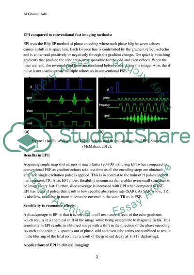
Q1. BLIP Echo Planar Imaging (EPI) method- Principles of EPI: EPI, like FSE (Fast Spin Echo), is an imaging method that reduces the imaging time asit acquires multiple phase encoding steps in each repetition time (TR). It is a fast and flexible method of imaging that provides with good contrast and resolution for the images. This method of imaging is used in functional MRI and rapid whole brain imaging. The time required to acquire an image is dependent on Tacq = Nacq x Ny x TR Where Nacq is the no. of acquisitions, Ny is the no. of phase encoding steps and TR is the repetition time. Sampling of k-space is different in EPI that enables high quality of images acquired. EPI uses quickly switching gradients to produce its echo train as against use of RF pulses in FSE. EPI frequency encoding gradient oscillates from positive to negative to create odd and even echoes. Each read gradient echo corresponds to one k-space with the positive and negative gradients changing the direction of the line that is read. The changing of direction of the line read results in a phase ‘blip’ causing a shift in Ky and this method of phase encoding is referred to as blip EPI. Characteristics of EPI: EPI has three distinct characteristics in terms of speed, the variety of contrast and the flexibility in terms of available resolution for imaging small structures like the pituitary gland. EPI offers higher speeds (6 to 10 times) of imaging of short temporal events under motion. It provides with high image quality by collecting more averages with increased slice frequency per TR. The signal to noise ratio of the images is high and the resolution is also high. Its usefulness depends on the MR system used and the efficiency is determined by the ability to perform 1282 or 2562 in a very short time to avoid large off resonance artifacts. It means that high quality images of moving patients can be obtained by using the speed of EPI. A variety of tissue contrasts (T1, T2 and T2*) are allowed by EPI for image acquisition and small structures are imaged using the flexibility in resolution where the field view can be reduced, the echo train length can be increased or geometric distortions can be reduced to increase the resolution. Also, just half of the data of the image can be gathered that can be further synthesized through conjugation to obtain the remaining data. Flexibility in contrast can be used to produce an image similar in standards to that of an SE image with the same TE and TR by using an excitation pulse in the front part followed by a 1800 pulse (T2* dephasing) to create the first echo. EPI compared to conventional fast imaging methods: EPI uses the Blip EP method of phase encoding where each phase blip between echoes causes a shift in k-space line. Each k-space line is contributed by the gradient refocused echo and is either read positively or negatively through the gradient change. The quickly switching gradients that produce the echo train are responsible for the odd and even echoes. When the lines are read, the reversed read lines are reordered before constructing the image. Also, the rf pulse is not used to create multiple echoes as in conventional FSE. Figure 1: (a) Pulse diagram of FSE, 4 echoes and (b) pulse diagram of EPI. Source: (McMahan, 2012). Benefits in EPI: Acquiring single snap shot images is much faster (20-100 ms) using EPI when compared to conventional FSE as gradient echoes take less time as all the encoding steps are obtained after one single excitation pulse is applied. This is in contrast to the train of rf pulses applied that increases TR. Also, EPI allows flexibility in contrast that enables even small structures to be imaged very fast. Further, slice coverage is increased with EPI when compared to FSE. EPI has fewer rf pulses that result in low specific absorption rate (SAR). As SAR is low, TR is also low, resulting in more slices to be covered in the same TR as in FSE. Sensitivity to resonance effects: A disadvantage in EPI is that it is sensitive to off resonance effects of the echo gradients which results in a chemical shift of the image while being susceptible to magnetic fields. This sensitivity in EPI results in a blurred image with a shift in the direction of the phase encoding. As each echo train in k-space is out of phase, odd and even echo trains are combined to result in the blurring of the final result as a result of the gradient decay at T2* (T2* dephasing). Applications of EPI in clinical imaging: EPI can be used in clinical imaging for faster scans where T2 weighted (Spin Echo or SE-EPI) is replaced by T2* (Gradient Echo or GE EPI) (see figure 2) and helps obtain functional brain images as the high speed enables imaging in motion and of small structures. Apart from this, EPI can also be used for perfusion imaging, diffusion imaging, abdominal imaging, cardiac imaging, imaging of Lung function and Musculoskeletal function. Figure 2: Pulse sequence diagrams for SE-EPI, GE-EPI and inversion recovery. Source: (McMahan, 2012). Q2. EPI and Nyquist ghosting. The off resonance effect induced signal decay is corrected using Fourier transform applied to the odd and even echo trains which are then reconstructed. The reconstructed image from the dataset of odd and even echoes commonly suffers from ghosting which is the result of aliasing of the images along the phase encode axis at half the field of view (FOV). In an ideal EPI, a ghost free image is obtained as the ghosts formed by the two datasets cancel out each other perfectly as these datasets are sampled at exactly half the Nyquist rate in the phase encode direction. However, the zero and first-order phase differences in the sampled odd and even images result in a ghost formed due to the imperfect balancing of the phases of the samples. This imbalance in imaging results in Nyquist ghosting in the image, whose intensity can be determined by point-by-point phase differences that exist between the odd and even sampled images. The Nyquist criterion for sampling states that the associated Nyquist frequency is given by  fc=1/(2dt)  where, fc is the critical Nyquist frequency and dt is the time interval between consecutive samples. The Nyquist criterion for sampling is related to the ability to take Fourier transforms of data and reconstruct that data accurately and while frequencies below fc can be captured in the reconstruction of the data, frequencies above fc are not necessarily captured in the reconstruction of the data, so this requires that sampling rate is adjusted to acquire correct images. By altering the time between the samples, the bandwidth represented in the reconstruction can also be decreased or increased. Further, the gradient coils of Fourier Transformed images produce small Eddy currents called cross-term Eddy currents that are in directions other than the principle gradient axis. These cross-terms when uncorrected alongside the small encoding blips in EPI have serious affects on the image quality as there is a high potential for image ghosting. Since cross-term Eddy currents last just about a millisecond (1 ms) it becomes difficult to eliminate the ghosting using Eddy current compensation techniques but can be sufficiently removed by adjusting the gradient propagation and ADC associated delays to result in minimal Eddy current in the signal phase accumulation. Two approaches, one of introducing compensation blips to reverse the accumulated cross-terms and the other of post processing the dataset, are used to reduce Nyquist ghosting in EPI (Grieve, Blamire and Styles, 2002). Figure 3 shows the N/2 (Nyquist) ghosting in an EPI image: Figure 3: Nyquist ghosting in an EPI image. Source: (McMahan, 2012). The above image is the result of two shifted Fourier Transform signals in k-space that are superimposed in such a way that the N/2 ghosting is seen in the direction of phase encoding which are 1800 out of phase with the original image. This ghost image is formed in half the FOV of the actual image. Different extents of Nyquist ghosting are shown in Figure 4: Figure 4: Different extents of N/2 ghosting in EPI. Source: (McMahan, 2012). Q3. In EPI, the oscillating frequency gradient results in every second k-space line being reversed. These gradient echoes are refocused every TR either by producing the initial echo with a spin or gradient or by subjecting the gradient to an inverse recovery method. However, the echo train is susceptible to off resonance effects like chemical shift and magnetic susceptibility and this affects the contrast in EPI. Also, the amplitude of the echo train decreases at the rate of T2* and the EPI image is blurred with the decay spreading out in the direction of phase encoding. There is consistent effective bandwidth in both directions as the data along ky is also uniformly sampled along with the line in the direction of phase encoding. This result in uniform positional shifts due to the chemical shift and magnetic susceptibility produced due to irregular sampling in time along both ky and kx directions, causing blurring of the image. To correct such errors in imaging, repeated reconstruction of raw data acquired at various frequencies is used to focus the image data at multiple reconstructions. Figure 5: Signal decay in EPI due to off resonance effects. Source: (McMahan, 2012). The extent to which the image spreads is characterized by the point spread function (PSF). When PSF is less than one pixel width, the blurredness will not be obvious in the image. The signal decay rate, T2* is a result of the echo train’s chemical shifting and magnetic susceptibility which can be corrected by refocusing of the image data from repeated reconstructions (using Fourier Transform) of the raw data at various frequency offsets while starting the data acquisition at the origin of k-space which helps in avoiding reconstruction artefacts. Figure 6: Reconstruction of image data. Source: (McMahan, 2012). Q4. EPI is commonly used in functional MRI (fMRI) as it is a rapid imaging technique that is capable of imaging moving organs like the heart and brain activation. Neural activity within the brain can be captured by imaging the changes in deoxyhemoglobin levels in the capillary veins based on blood oxygen-level dependent (BOLD) fMRI. Brain functions like vision, motor, language and cognition can be investigated using fMRI. Spin echo EPI (SE-EPI) and gradient echo EPI (GE-EPI) are the two techniques most commonly used for fMRI. SE-EPI uses diffusion pulses to differentiate between the signals by dephasing the signal to be detected and is based on T2 effects only. GE-EPI uses initial dephasing followed by a series of blips that form the echo formed under T2* decay. Although, T2* weighted images are commonly used for fMRI, SE-EPI images are also used as they show diffusion effects with BOLD contrasts. EPI is used to detect at risk tissue through perfusion maps and diffusion imaging for use in acute interventional stroke therapy. Perfusion imaging involves assessing blood flow to the vascular bed by injecting a contrast agent or bolus and rapid imaging of the flow of the contrast bolus through the vascular bed. In such type of imaging, the contrast of EPI reduces while the contrast agent flows through the vascular bed until the agent is fully emptied into the draining veins. While T2* is reduced in the process, the series of images in time can be analyzed based on the relative concentrations of the contrast in time from which cerebral blood volume (CBV) and cerebral blood flow (CBF) maps can be generated to study brain functioning. Figure (7a, 7b and 7c) show T2 , CBV and CBF images: Figure 7(a) and 6(b): Perfusion maps: CBV and CBF. Source: (McMahan, 2012). Figure 7(c): T2 image. Source: (McMahan, 2012). A 2D spoilt gradient echo train is used through segmented k-space and short TR to produce single slice multi-phase image in just 16s. While a single shot can be obtained in one heartbeat, segmented k-space collection can acquire high resolution images in 2-4 heartbeats and also provide more phases of the heart. Further, myocardial perfusion abnormalities can be detected and coronary arteries free of cardiac motion artefacts can also be imaged by using segmented EPI or high performance MR system. Figure 8: EPI image of the heart. Source: (McMahan, 2012). For single shot method, diffusion weighted, DW-EPI is used due to its temporal resolution and insensitivity to physiological motion. Diffusion pulses induced through a tissue compartment are dephased in relation to water molecules diffusing through the tissue compartment. The speed of diffusion can reveal hypointensity (faster diffusion rates) and hyperintensity (slower diffusion rates) that in turn reveal the susceptibility of the cerebrovascular tissue to accidents. Figure 9: Diffusion weighted image at 13 hrs after stroke onset. Source: (McMahan, 2012). Figure 10: T2 weighted image at 13 hrs after stroke onset. Source: (McMahan, 2012). Cardiovascular MRI used a combination of parallel imaging and standard cardiac protocols to reduce breath-hold times, increase resolution within a certain breath-hold time, increase frame rate or temporal resolution while allowing for real-time imaging. This combination of techniques can enable capturing 100 frames per second or 30 frames per second if capturing multiple cardiac views. Figure shows multi-phase scans of the heart acquired during 8 second breath-hold. Figure below shows one of the 16 phases of the heart acquired without the use of parallel imaging (a) and one of the 28 phases acquired with GRAPPA or k-space domain with a reduction factor of 2 (b): Figure 11: Identical multi-phase scans acquired in 8s breath-hold. Source: (McMahan, 2012). A reduction factor of two implies that the image has been under-sampled by a factor of 2. Further, geometric distortions and chemical shifts can pose problems in acquiring of high quality images during the scans. The use of parallel imaging (PI) helps acquire all the segments of k-space simultaneously using multiple receivers as complex artifacts arise that require longer acquisition times. 4 to 64 channels are used simultaneously in an MR system along with a phased array coil that consists of surface coils working without interfering with each other’s operation but can be used in conjunction. This system reduces scan times, improves signal to noise ratio, covers a large area, improves image quality and reduces image distortion. Figure 10 shows a typical MR system: Figure 12: MR system using parallel imaging. Source: (McMahan, 2012). Appling PI for MR scanning has several other benefits: it does not affect the contrast of the images, it can be used to improve SNR or speed or both, it can be used to improve image quality using single shot image technique, it can be used to reduce artefacts in other sequences, large fields of view can be obtained by a combination of phased array coils and controllable bed localization and the coil geometry can be adapted to suit the region of interest as PI is about the application of hardware rather than being a new imaging sequence. Parallel imaging enables speeding up of conventional scans by the factor of the number of coils used in conjunction as the number of coils used determines the number of lines acquired. The coil factor determines the scan speeds in acquiring phase encoding. In single shot EPI, parallel imaging is used to reduce the echo spacing similar to segmentation, to improve the image quality. References Clare, S. (1997). Functional MRI : Methods and Applications. University of Nottingham. Grieve, MS, Blamire, MA and Styles, P. (2002). Elimination of Nyquist Ghosting Caused by Read-Out to Phase-Encode Gradient Cross-Terms in EPI. Magnetic Resonance in Medicine 47. Wiley-Liss, Inc. DOI 10.1002/mrm.10055. McMahon, K. (2012) MRES7001. Module 1: The Basics of EPI. Centre of Advanced Imaging. The University of Queensland.   McMahon, K. (2012). MRES7005. Fast Imaging Techniques. Centre of Advanced Imaging. The University of Queensland.   Read More
Cite this document
  • APA
  • MLA
  • CHICAGO
(BLIP Echo Planar Imaging Method Report Example | Topics and Well Written Essays - 2500 words, n.d.)
BLIP Echo Planar Imaging Method Report Example | Topics and Well Written Essays - 2500 words. https://studentshare.org/physics/1768858-mres-7005-fast-imaging-techniques-epi-echo-planar-imaging
(BLIP Echo Planar Imaging Method Report Example | Topics and Well Written Essays - 2500 Words)
BLIP Echo Planar Imaging Method Report Example | Topics and Well Written Essays - 2500 Words. https://studentshare.org/physics/1768858-mres-7005-fast-imaging-techniques-epi-echo-planar-imaging.
“BLIP Echo Planar Imaging Method Report Example | Topics and Well Written Essays - 2500 Words”. https://studentshare.org/physics/1768858-mres-7005-fast-imaging-techniques-epi-echo-planar-imaging.
  • Cited: 0 times

CHECK THESE SAMPLES OF BLIP Echo Planar Imaging Method

Sensory Evaluation Method

"Sensory Evaluation method" paper examines the test that requires analysis in getting information regarding a subject's reaction to differences between samples.... The method involves the presentation of a set of samples one at a time, in equally spaced intervals.... This method can be used by expert tasters or consumers for product acceptance tests.... aired or Two-Sample Test method - Two samples are presented at any one point for evaluation....
7 Pages (1750 words) Assignment

Condiment snack holder

A combination food server and condiment holder, the food server having a sidewall enclosing a food-holding volume, the sidewall having at least one slit therein; the condiment holder comprising a cup for holding at least one condiment, and a tab, connected to the cup, extending through the at least one slit in the sidewall to secure the condiment holder to the food server. … The food server and condiment holder of the present invention is of simple and inexpensive construction, yet is safe and securely holds one or more condiments adjacent to a food server for convenient use....
5 Pages (1250 words) Essay

Mechanical Treatment of Plantar Fasciitis A Prospective Study

method and Sampling The method section is highly organized and clear.... The article has a good title which reflects the major issue covered in the research: the study of the mechanical treatment of plantar fasciitis.... The title is informative and clear at the same time....
5 Pages (1250 words) Essay

Planar Ultra Wideband Antenna for Microwave Imaging System for Medical Application (Project Debrief)

As a student, I appreciate Griffith University's Industry I am feeling fortunate to have got an opportunity to work under IAP on the project titled ‘Planar Ultra Wideband Antenna for Microwave imaging System for Medical Application'.... Amin Abbosh – a international expert and a point of reference in Ultra-Wideband imaging, accepted me as his researcher and it was a very exceptional learning experience to work with him....
5 Pages (1250 words) Essay

SPECT imaging

A SPECT scan is similar to X-ray computed tomography or Magnetic Resonance imaging, but different in a way that it utilizes a… SPECT scan gives a true image of the working of the organs, unlike X-rays that only depicts the state of the internal organ.... Single Photon Emission Computed Tomography imaging Keiser Michelle Beth SPECT imaging A Single photon emission computerized tomography (SPECT) allows an examiner to analyze the functions of internal organs of a patient's body....
2 Pages (500 words) Assignment

Methods Underwater Imaging

 The writer of this essay " Methods Underwater imaging " discusses the various uses and applications of underwater imaging.... Underwater imaging utilizes three methods.... These include acoustic imaging, beamforming acoustic imaging, and holographic acoustic imaging.... I will analyze the uses and application of underwater imaging, the methods used, goals of underwater imaging and applications of underwater imaging....
1 Pages (250 words) Essay

Palmar Flexion Crease Identification

Therefore, palmar flexion crease identification is a method used to verify as well as establish a person's identity.... There are various steps followed while conducting a manual palmar flexion crease identification method.... A complete automated palmar flexion crease identification method is carried through four stages.... n the other hand, the method of friction ridge identification makes use of four stages to establish the true identity of the person....
2 Pages (500 words) Essay

Echo Planar Image Sequences Data

PI is an imaging method that is fats and flexible.... (7 marks)echo planar image (EPI) sequences data collection has different ways of comparing with sequences in standard pulse.... (7 marks)echo planar image (EPI) sequences data collection has different ways of comparing with sequences in standard pulse.... echo planar is superior to conventional in investigations of the human physiological processes in the body.... Discuss the BLIP EPI method presented in Module 1 in detail....
9 Pages (2250 words) Assignment
sponsored ads
We use cookies to create the best experience for you. Keep on browsing if you are OK with that, or find out how to manage cookies.
Contact Us