StudentShare
Contact Us
Sign In / Sign Up for FREE
Search
Go to advanced search...

The main uses of Ultrasound in an imaging department and a vascular department - Essay Example

Cite this document
Summary
Ultrasound tests also called sonography is an imaging method which uses high frequency sound waves in order to produce accurate images of structures inside the body. These images provide valuable information that is used in diagnosing and treating various diseases and…
Download full paper File format: .doc, available for editing
GRAB THE BEST PAPER96.5% of users find it useful
The main uses of Ultrasound in an imaging department and a vascular department
Read Text Preview

Extract of sample "The main uses of Ultrasound in an imaging department and a vascular department"

Download file to see previous pages

o Sandhu (2007), ultrasonography can image soft tissues such as muscles, blood vessels and so on and can also monitor needle position while the interventional procedures. In anesthesia setting, ultrasound is the best choice due to the compactness of the instrumentation, easy transportability and lack of damage to tissues (Sandhu, 2007). In the imaging department, ultrasound is also useful for determining neuaxial blocks in people especially children. Ultrasound imaging for determining transthoarcic and transesopheageal echo-cardiography is utilized to diagnose cardiac anesthesia, trauma anesthesia and order cardiac function abnormalities that arise either during the perioperative period or intensive care settings (Sandhu, 2007).

Forsbery et al. (2002) argued in its research paper that imaging modalities such as MRI and CT have well established 3D data displays and therefore are being used increasingly in the diagnostic decision rather than ultrasound. This is because the time available for acquirement and processing of 3D data is limited in ultrasound imaging. For example, in Doppler ultrasound, blood flow data is obtained using multiple firing methods. Accordingly, improvements in the computer processing technology and visualization methods, the clinical use of vascular 3D ultrasound has increased.

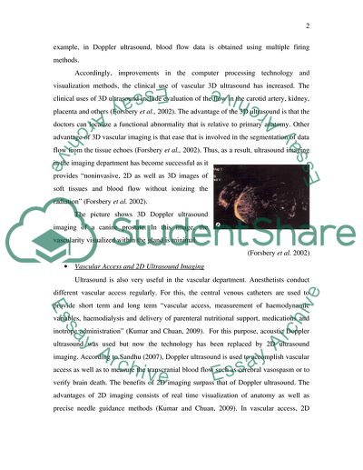
The clinical uses of 3D ultrasound include evaluation of the flow in the carotid artery, kidney, placenta and others (Forsbery et al., 2002). The advantage of the 3D ultrasound is that the doctors can localize a functional abnormality that is relative to primary anatomy. Other advantage of 3D vascular imaging is that ease that is involved in the segmentation of data flow from the tissue echoes (Forsbery et al., 2002). Thus, as a result, ultrasound imaging in the imaging department has become successful as it provides “noninvasive, 2D as well as 3D images of soft tissues and blood flow without ionizing the radiation” (Forsbery et al. 2002). Ultrasound is also

...Download file to see next pages Read More
Cite this document
  • APA
  • MLA
  • CHICAGO
(“The main uses of Ultrasound in an imaging department and a vascular Essay”, n.d.)
The main uses of Ultrasound in an imaging department and a vascular Essay. Retrieved from https://studentshare.org/miscellaneous/1558437-the-main-uses-of-ultrasound-in-an-imaging-department-and-a-vascular-department
(The Main Uses of Ultrasound in an Imaging Department and a Vascular Essay)
The Main Uses of Ultrasound in an Imaging Department and a Vascular Essay. https://studentshare.org/miscellaneous/1558437-the-main-uses-of-ultrasound-in-an-imaging-department-and-a-vascular-department.
“The Main Uses of Ultrasound in an Imaging Department and a Vascular Essay”, n.d. https://studentshare.org/miscellaneous/1558437-the-main-uses-of-ultrasound-in-an-imaging-department-and-a-vascular-department.
  • Cited: 0 times

CHECK THESE SAMPLES OF The main uses of Ultrasound in an imaging department and a vascular department

Diagnostic Imaging and Herniated Nucleus Pulposus

the main goal of the hospital is to provide a quality experience to its patients (Reiser and Andrea, 2008).... Diagnostic imaging and Herniated Nucleus Pulposus Name Institution Date Diagnostic imaging and Herniated Nucleus Pulposus Introduction Diagnostic imaging is the technologies used by doctors to look inside a human body in order to look any medical condition clues.... Types of diagnostic imaging include CT scans, X-rays, MRI scans, Ultrasound and nuclear medicine scans....
8 Pages (2000 words) Research Paper

Professional Behavior and Evidence-Based Practice

02) presents that promoting professional behaviour is currently the main concern across the continuum of medical education.... This can be confirmed by an ultrasound crucial for determination of the exact location of the lamp, the nature of the lamp whether inflammation or blood clots.... - ultrasound is quite crucial, especially when abdominal aortic aneurysm among other conditions is suspected because it aids in measuring the size and other important aspects of the condition to help determine the risk for rapture....
4 Pages (1000 words) Essay

Medical Terminology

This report also briefly mentions the company's products and their uses.... This paper "Medical Terminology" discusses medical terminology by the company that is specializing in the manufacture and marketing of endoscope products of day to day use in multi-specialty hospitals.... hellip; Endoscopy an examination technique since the mid-1950s with the use of mirrors usually to view vocal cords directly has now been developed as a process to inspect the body parts through an optical instrument inserted onto the tubes or cavities to reach the body parts intended to be examined....
12 Pages (3000 words) Assignment

Determine the Best Diagnostic Image Modality in Peripheral Arterial Disease

These include digital pulse volume recordings; Doppler flow velocity waveform analysis; duplex ultrasonography, which combines B-mode imaging and pulse-wave Doppler examination; segmental pressure measurements; transcutaneous oximetry; stress testing, usually using a treadmill; and tests of reactive hyperemia (Mohler, III, 2003, 2306-2314)....
24 Pages (6000 words) Essay

The Main Uses of Ultrasound in Actual Departments or Centres

The paper "the main uses of ultrasound in Actual Departments or Centres" highlights that the introduction of ultrasound into the medical profession was a great revolutionary act.... vascular sonographies are therefore important in helping doctors to view the flow of the blood and any blockages or constrictions in the vessels carrying it.... The Doppler technique helps in imaging the rate and direction of the flow of blood.... he type of sonography known as imaging is a noninvasive test method that enables medical practitioners to examine the internal organs of the body without necessarily cutting the patient open....
8 Pages (2000 words) Coursework

Bleeding Esophageal Varices in Patient With Liver Cirrhosis

nbsp;… The aims and objectives of this research are to establish and develop a Doppler ultrasound-based investigative modality with defined parameters to quantitate the extent of portal hypertension, hence the severity of the disease in patients with cirrhosis of the liver, where associated measurements of flow dynamics through the portal vein and esophageal varices may provide a predictive assessment of changes of variceal bleeding....
10 Pages (2500 words) Research Proposal

3D Doppler Ultrasound

This work called "3D Doppler Ultrasound" describes the developments of a number of 3D Doppler ultrasound imaging systems using free-hand, mechanical, and 2D array scanning techniques.... hellip; A technological breakthrough in this field has led to the advancement of ultrasonic imaging with the coming of the 3D ultrasound.... This novel approach in ultrasound imaging is speedily achieving extensive use with numerous applications.... 3D surmounts the limitations of 2D ultrasound imaging as this study will show....
10 Pages (2500 words) Research Paper

Design of a New Imaging and Cancer Care Facility

A functional design of an imaging and cancer facility is also required to be in line with the specific government regulations and codes that govern such facilities' construction in the locality while promoting convenience, comfort, energy conservation, and cost reduction.... The radiology department's available section will help approximately 60 patients per day about the number of persons expected to be served at the facility....             On the other hand, the nuclear medicine department, general/cardiac section will serve 15 patients per day while the Positron Emission Tomography (PET)/ Computed Tomography (CT) section will handle 50 patients per day....
7 Pages (1750 words) Coursework
sponsored ads
We use cookies to create the best experience for you. Keep on browsing if you are OK with that, or find out how to manage cookies.
Contact Us