StudentShare
Contact Us
Sign In / Sign Up for FREE
Search
Go to advanced search...
Free

The ABO System of Blood Groups - Term Paper Example

Cite this document
Summary
The paper 'The ABO System of Blood Groups' presents the ABO system of blood groups which was developed by Karl Landsteiner in 1900 – he discovered three blood groups, viz. ‘A’, ‘B’ and ‘O’, that are responsible for acute hemolytic reaction seen in mismatched blood transfusion. …
Download full paper File format: .doc, available for editing
GRAB THE BEST PAPER93.2% of users find it useful
The ABO System of Blood Groups
Read Text Preview

Extract of sample "The ABO System of Blood Groups"

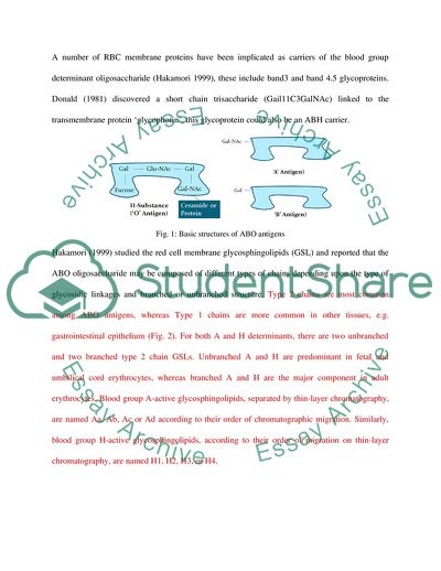
Introduction The ABO system of blood groups was developed by Karl Landsteiner in 1900 – he discovered three blood groups, viz. ‘A’, ‘B’ and ‘O’, that are responsible for acute hemolytic reaction seen in mismatched blood transfusion. His students discovered the forth group i.e. ‘AB’ in 1902. Today, the blood group matching of the donor and the recipient is essential foundation of the pre-transfusion testing because transfusion of even 10-15 ml of incompatible blood can trigger a life-threatening hemolytic reaction. ABO System of Blood Groups The ABO system of blood groups is based on the presence or absence of three antigens, viz., ‘A’, ‘B’ and ‘H’ on the red blood cell membrane. Structure of ABO antigens: The three antigens are oligosaccharide units, attached to either a sphingolipid (ceramide) or a protein, thus forming a glycolipid or a glycoprotein, respectively. Morgan and Watkins (1969), and Kabat et al. (1973) established the antigenic structures responsible for the specificity of blood group A, B and H. The three oligosaccharides have very subtle but immunopotent differences (Fig.1). The basic oligosaccharide unit is a tetra-saccharide which, after taking up a fucose, makes the ‘H-substance’ and is responsible for the ‘O’ blood group. Attachment of an N-acetylgalactosamine to the terminal galactose in H-substance transforms it into the ‘A’ antigen; whereas substitution with a galactose makes the ‘B’ antigen (Fig. 1). A number of RBC membrane proteins have been implicated as carriers of the blood group determinant oligosaccharide (Hakamori 1999), these include band3 and band 4.5 glycoproteins. Donald (1981) discovered a short chain trisaccharide (Gail11C3GalNAc) linked to the transmembrane protein ‘glycophorin’, this glycoprotein could also be an ABH carrier. Fig. 1: Basic structures of ABO antigens Hakamori (1999) studied the red cell membrane glycosphingolipids (GSL) and reported that the ABO oligosaccharide may be composed of different types of chains depending upon the type of glycosidic linkages and branched or unbranched structure. Type 2 chains are most common among ABO antigens, whereas Type 1 chains are more common in other tissues, e.g. gastrointestinal epithelium (Fig. 2). For both A and H determinants, there are two unbranched and two branched type 2 chain GSLs. Unbranched A and H are predominant in fetal and umbilical cord erythrocytes, whereas branched A and H are the major component in adult erythrocytes. Blood group A-active glycosphingolipids, separated by thin-layer chromatography, are named Aa, Ab, Ac or Ad according to their order of chromatographic migration. Similarly, blood group H-active glycosphingolipids, according to their order of migration on thin-layer chromatography, are named H1, H2, H3, or H4. Fig. 2: Types of blood group antigens (Hakamori 1999) Synthesis of ABO antigens: The antigen specificity of an individual develops during 5-6 weeks of the embryonic life when ABO antigen can be detected on the surface of the red cells. Watkins (1980) and Kobata et al [1970] clarified the enzymatic basis for synthesis of the blood group antigenic determinants. The H-substance is expressed on the red cell membranes during the earlier stages of development and is modified to A or B antigens with the help of two specific transferase enzymes. The N-acetylgalactosaminyltransferase enzyme, known as ‘A-enzyme’, coverts the H-substance into A-antigen by transferring an N-acetylgalactosamine residue from UDP-N-acetylgalactosamine to its terminal galactose. The blood group of the individual thus becomes ‘A’. Similarly, the ‘B-enzyme’ (a galactosyl transferase) transfers a galactose from a molecule of UDP-galactose to the H-substance galactose thus transforming the blood group specificity to ‘B’. Absence of both the transferases leaves the H-substance galactose open and confers blood group ‘O’ specificity. The development of the blood group specificity is, thus, dependent upon the expression of the either (or none) of the two transferases. Genes and inheritance of ABO antigens The molecular basis of the antigenic diversity of ABO system was elucidated by Yamamato et al. (1990). The gene for the glycosyltransferase enzymes resides on chromosome 9 on a position known as ABO locus. The genes for H-substance are present on chromosome 19. AB alleles – There are mainly three alleles responsible for A and B antigens, viz. A1, A2 and B. A1 and A2 – code for the enzyme N-acetylgalactosamine transferase. B codes for the enzyme galactosyl transferase. It must be remembered that presence of a H-substance is must for the formation of A or B antigens. The addition of GalNAc or Gal units takes place only if the H-substance has a fucose unit attached to the terminal galactose. H/h alleles – The H gene on chromosome 19 shows two allele, viz. H and h. The H allele codes for the enzyme L-fucosyltransferase that adds fucose residues to the basic unbranched H-substance. On the other hand, h gene is an amorph and does not code for the enzyme. Presence of at least one H gene is essential to produce mature H-substance and hence O antigen. Inheritance of ‘hh’ genotype results in a condition in which the individuals lack H-antigen; this phenotype is known as Bombay phenotype (see box). Rh System The Rh blood group system was first described in year 1940 by Landsteiner and Wiener, when they found that serum from rabbits and guinea pigs immunized with red blood cells of Macacus rhesus could agglutinate 85% of the blood from New York blood donors. The heteroantibody responsible for the agglutination was renamed anti-LW (Landsteiner and Wiener) and the human alloantibody was called anti-D (Levine et al. 1963). The system is based on three pairs of common types of Rh antigens called as Rh factor. These antigens are designated D-d, C-d and E-e. The D antigen is much more prevalent in the population and is highly antigenic, presence of ‘D’ makes the individual positive whereas absence will make Rh negative. There is no ‘d’ antigen and the letter denotes absence of D antigen. Although Rh system is recognized by DdEeCc antigens, there are at least 45 antigens discovered so far that make the Rh system. The agglutination / hemolytic reactions due to other antigens are milder and hence are not important clinically. Rh positive individuals are much more (85%) common than Rh negative (15%) in the world. The Rh system appears to have originated from Africa since all Africans blacks are Rh positive. Structure of Rh Antigens The Rh blood group system is the most polymorphic of the human blood groups. The Rh antigen complex on the surface of red cells is formed of two major components, viz. Rh antigens and Rh-associated glycoproteins (RhAG). Presence of RhAG is must for the expression of Rh antigens on the membrane. The proteins Rh and RhAG have ~40% structural homology and collectively termed as Rh protein family. Rh and RhAG proteins have 12 transmembrane spans (Huang 1998), with both the N-terminus and C-terminus oriented to the cytoplasm (Figure 3). Figure 3. Model of topology for RhAG, RhCE, and RhD (Avent and Reid 2000). Rh protein family The size of Rh complex has been estimated to be ~170 kDa and consists of a tetramer of two RhAG proteins with two Rh antigen proteins (RhCcEe or RhD). Characteristics of the RhD protein and of the RhCcEe protein are depicted in Figure 3 and summarized in Table 1. Together, the association of the Rh protein family and the Rh accessory proteins is called the ‘Rh complex.’ Table 1. Proteins in the Rh Complex in Normal RBC Membranes (Avent and Reid 2000) Protein Antigens Gene Location Mol.Wt. (kDa) Rh protein family RhD D 1p36.13-p34.3 30-32 RhCcEe Ce, CE, ce, cE 1p36.13-p34.3 32-34 RhAG Carries MB2D10 epitope 8p21.1-p11 45-100 Rh accessary proteins LW LW 19p13.3 37-47 IAP - 3q13 47-52 GPB N, S, s, U 4q28-q31 20-25 Band 3 Diego 17q12-q21 90-100 Comparison of ABO and Rh systems ABO and Rh are the two most important systems used in transfusion medicine; the ABO as well as the Rh of the donor and recipient of the blood transfusion must be matched. ABO system is also important for the tissue transplants since these antigens are also present on the other tissues cells. The Rh system, on the other hand is more important in materno-fetal situations as discussed below. Structures of antigens in two systems ABO antigens, as discussed above, are oligosaccharides attached to sphingolipids or glycoproteins. The Rh antigens on the other hand are complex aggregates of transmembrane proteins. Inheritance of ABO and Rh blood groups The inheritance pattern of the two types of blood group system (Table 2) is well established and is regularly used by the clinicians and epidemiologists. Table 2: The possible blood types of an offspring based on ABO and Rh systems. Blood Type Mothers's Type O A B AB Fathers' Type O O O, A O, B A, B A O, A O, A O, A, B, AB A, B, AB B O, B O, A, B, AB O, B A, B, AB AB A, B A, B, AB A, B, AB A, B, AB   Rh Factor Mother's Type Rh + Rh - Father's Type Rh + Rh +, Rh - Rh +, Rh - Rh - Rh +, Rh - Rh - Immune response against ABO antigens ABO oligosaccharide antigens are highly immune-potent and hence trigger the generation of antibodies. The ABO antibodies found in different blood groups are natural antibodies, i.e. they are presence in the blood even without a recognised exposure to the incompatible antigen. The ABO antibodies develop in the first 3 to 6 months after birth, continue to rise till the age of 5-6 years when their concentration becomes stable for the life, except in the old age when their levels decline (Spalter et al. 1999). An individual with A blood group will have anti-B antibodies and vice versa; and O group individuals have antibodies that react with A as well as B antigens. Since these antibodies are being formed due to continuous stimuli of non-self antigens without any exposure to RBCs, they are called ‘non-red cell stimulated antibodies’. Majority of these antibodies are IgM immunoglobulins, very small proportion is IgG. Transfusion Reaction If one blood type is transfused into a recipient who has an incompatible blood type, a transfusion reaction is likely to occur in which the red blood cells of the donor blood are agglutinated. It is rare that the transfused blood causes agglutination of the recipient’s cells, because the plasma, and hence antibodies, of the donor blood get diluted by the plasma of the recipient. But the recipient’s agglutinins do agglutinate and hemolyse the mismatched donor cells. The red cells are phagocytosed and destroyed. The hemoglobin released from RBCs is converted into bilirubin in liver and is excreted into the intestines. Jaundice usually does not appear in an incompatible tranfusion unless > 400 milliliters of blood is hemolysed in a day. The antibodies can also stimulate the complement system and release of cytotoxins that can trigger a potentially fatal anaphylactic reaction. Immune response against Rh system So-called "natural" antibodies to Rh do not exist in humans, as they do for the AB antigens. When red blood cells containing Rh factor are mixed with / injected into an Rh-negative person, anti-Rh agglutinins develop slowly, reaching maximum concentration about 2 to 4 months later. With multiple exposures to the Rh factor, an Rh-negative person eventually becomes strongly “sensitized” to Rh factor. If a negative person has never before been exposed to Rh+ blood, transfusion of Rh-positive blood will cause no immediate reaction. However, anti-Rh antibodies can develop in sufficient quantities during the next 2 to 4 weeks to cause agglutination of the transfused cells. These cells are then hemolyzed by the tissue macrophage system. Thus, a delayed transfusion reaction occurs, although it is usually mild. On subsequent transfusion of Rh-positive blood the transfusion reaction is greatly enhanced and can be immediate and as severe as a transfusion reaction caused by mismatched type A or B blood. Rh-incompatibility and RhoGam Therapy The Rh factor assumes a special importance in maternal-fetal interactions. If an Rh- mother gets pregnant with a Rh+ fetus (if the father is Rh+), since there are no natural anti-Rh antibodies, this poses no special risk for the first pregnancy. At the time of delivery, a significant amount of fetal blood can get mixed with the maternal circulation; and may stimulate a strong anti-Rh response in the mother. If the same mother then bears a second Rh+ child, the existing anti-Rh antibodies can cross the placenta during the pregnancy and cause agglutination and phagocytosis of the fetal red blood cells and can cause severe damage to the vital organs. The condition is known as Erythroblastosis Fetalis (also known as Hemolytic Disease of the Newborn, or HDN). The production of anti-Rh antibodies in an Rh- mother can be prevented by injecting a large dose of anti-Rh antibody (Anti-D or RhoGam) into the mother within 72 hours of the birth of her Rh+ baby. These antibodies result in rapid removal of the infant's red blood cells from the maternal circulation and can greatly reduce the likelihood of sensitization of the mother's immune system. The RhoGam injections have to be given to the mother during her every Rh+ pregnancy. Organ Transplant The ABO antigens are present on cells of most of the organs of our body, and each bodily tissue has its own additional complement of antigens. Consequently, foreign cells transplanted anywhere into the body of a recipient can produce immune reactions (Warner and Nester 2006). However, unlike the A and B antigens, the Rh antigens are present only on red blood cells. Therefore, while they are important for blood transfusion, they do not normally play a role in organ transplantation, and Rh typing of organ donors and recipients therefore not a significant consideration. ABO Antigens and disease The ABO blood groups also show variation in theie susceptibility to certain diseases. The incidence of malaria is lesser in people with O blood group (Cserti and Dzik 2007). Similarly, incidence of tumor occurrence in blood group A individuals compared to O individuals was higher for gastric cancer and ovarian cancer, clearly higher for vulvar cancer and much higher for salivary gland cancer (Anstee 2010). The O blood group people are more susceptible to H.pilori infection. Conclusions The ABO and Rh are the two most important system of blood groups in human being. ABO antigens are oligosaccharide in nature and are attached to glycosphingolipids or glycoproteins, whereas the Rh antigens are transmembrane proteins complexes that interact with other membrane proteins. Antibodies to ABO antigens are naturally occurring and are important for blood transfusion as well as organ transplants. The Rh antibodies are provoked only after red cell exposure and are important for materno-fetal system incompatibilities. References Anstee, D.A., 2010. The relationship between blood groups and disease. Blood, 115, 4635-4643. Morgan, W.T.J., Watkins, W.M., 1969. Genetic and biochemical aspects of human blood group A-, B-, H-, Lea- and Leb specificity, Br. Med. Bull. 25, 30-34. Kabat, E.A., 1973. Immunochemical studies on the carbohydrate moiety of water soluble blood group A, B, H, Lea and Leb substances and their precursor I antigens. In: H. Isbell (Ed.), Carbohydrates in solution (Adv Chemistry Series 117), American Chemical Society, Washington, DC, 334-361. Watkins, W.M., 1980. Biochemistry and genetics of the ABO, Lewis and P blood group systems, In: H. Harris, K. Hirschhorn (Eds.), Advances in human genetics, Vol. 10, Plenum Press, New York, 1-136. Kobata, A., Grollman, E.F., Torain, B., Ginsburg, V., 1970. Genes, glycosyltransferases and blood types, In: D. Amino (Ed.), Blood and tissue antigens, Academic Press, New York, 497-506. Kobata, A., Grollman, E.F., Ginsburg, V., 1968. An enzymic basis for blood type A in humans, Arch. Biochem. Biophys., 124, 609-612. Hartel-Schenk, S., Agre, P., 1992. Mammalian red cell membrane Rh polypeptides are selectively palmitoylated subunits of a macromolecular complex. J Biol Chem., 267, 5569-5574. Yamamoto, F., Clausen, H., White, T., et al., 1990. Molecular genetic basis of the histo-blood group ABO system. Nature (Lond),345,229-233. Donald, A.S.R., 1981. A-active trisaccharides isolated from A1 and A2 blood-group-specific glycoproteins. Eur. J. Biochem., 120, 243-249. Levine, P., Celano, M.J., Wallace, J., Sanger, R., 1963. A human ‘D-like’ antibody. Nature, 198, 596-597. Huang C-H., 1998. The human Rh50 glycoprotein gene—structural organization and associated splicing defect resulting in Rhnull disease. J Biol Chem., 273, 2207-2213. Avent N.D. and Reid M.E., 2000. The Rh blood group system: a review. Blood, 95(2), 375-387. Spalter S.H., Srini V. Kaveri, S.V., Bonnin, E., Mani, Jean-Claude, Cartron Jean-Pierre and Kazatchkine M.D., 1999. Normal Human Serum Contains Natural Antibodies Reactive With Autologous ABO Blood Group Antigens. Blood, 93, 4418-4424. Warner,P.R. and Nester, T.A., 2006. ABO-Incompatible Solid-Organ Transplantation. Am J Clin Pathol,125(Suppl 1), S87-S94. Cserti, C.M. and Dzik, W.H., 2007. The ABO blood group system and Plasmodium falciparum malaria. Blood, 110, 2250-2258. Read More
Cite this document
  • APA
  • MLA
  • CHICAGO
(The ABO System of Blood Groups Term Paper Example | Topics and Well Written Essays - 2792 words, n.d.)
The ABO System of Blood Groups Term Paper Example | Topics and Well Written Essays - 2792 words. Retrieved from https://studentshare.org/health-sciences-medicine/1746072-compare-the-inheritance-of-the-abo-system-with-that-of-the-rh-system-highlighting-antigen-and-antibody-formation-and-structure-as-well-as-clinical-significance
(The ABO System of Blood Groups Term Paper Example | Topics and Well Written Essays - 2792 Words)
The ABO System of Blood Groups Term Paper Example | Topics and Well Written Essays - 2792 Words. https://studentshare.org/health-sciences-medicine/1746072-compare-the-inheritance-of-the-abo-system-with-that-of-the-rh-system-highlighting-antigen-and-antibody-formation-and-structure-as-well-as-clinical-significance.
“The ABO System of Blood Groups Term Paper Example | Topics and Well Written Essays - 2792 Words”, n.d. https://studentshare.org/health-sciences-medicine/1746072-compare-the-inheritance-of-the-abo-system-with-that-of-the-rh-system-highlighting-antigen-and-antibody-formation-and-structure-as-well-as-clinical-significance.
  • Cited: 0 times

CHECK THESE SAMPLES OF The ABO System of Blood Groups

The ABO Blood Grouping

Blood Staining A thin blood smear was prepared using a drop of blood on a glass slide and allowed to dry.... BLOOD GROUPING, BLOOD STAINING, AND HEMATOCRIT Laboratory Report 06 March 2013 Introduction the abo blood grouping entails the categorisation of human blood depending on heritable traits of red blood cells as established by the existence or absence of antigens (Rogers, 2011).... hellip; blood grouping is also an essential procedure to determine the presence of the Rhesus factor (Rh)....
5 Pages (1250 words) Assignment

Multiple alleles and sex chromosomes

hellip; The author analyzes the concept of blood transfusion from a risky and fatal system to a safe and secure process with accurate projections of possible outcomes.... This paper “Multiple alleles and sex chromosomes” explains the role of multiple alleles in the inheritance of the abo blood group system that is determined genetically.... DNA consists of nucleotide substances that are further comprised of deoxyribose, phosphate groups and the four bases....
5 Pages (1250 words) Assignment

Hemolytic Disease of the Newborn

Hemolytic disease of the newborn (HDN) occurs due to maternal IgG antibodies crossing the placenta thereby producing hemolysis mainly due to Rh, ABO and Kell groups.... When the baby's Rh factor is positive, like the father's, problems can develop if the baby's red blood cells cross to the Rh negative mother.... hellip; However, it may also happen anytime blood cells of the two circulations mix, such as during a miscarriage or abortion, with a fall, or during an invasive prenatal testing procedure (i....
7 Pages (1750 words) Essay

Blood Groups and Rh Factor Incompatibility

People with antigen A present on the surface of… When both A & B antigens are found on the surface of blood cells the blood group thus described is AB and the absence of any blood groups blood groups The four blood groups i.... The terms universal donors and universal recipients are described for the two blood groups i.... When both A & B antigens are found on the surface of blood cells the blood group thus described is AB and the absence of any antigen defines the blood group O....
1 Pages (250 words) Essay

Blood and cardivascular

blood groups are determined due to presence of certain type of proteins present in the blood.... Different blood groups are due to the different combination of antigens and antibodies.... Platelets are binding agents in the blood stream.... When a person gets an injury in shape of external cut even a small one, the platelets in blood make a temporary bond at that place to stop the blood loss (Day, 2014).... It represents coordination between the platelets and clotting proteins to cease the blood loss in case of any blood vessel injury....
4 Pages (1000 words) Assignment

The Semai Culture

heir cultural and social system is isolated from the rest of the world and they are left with practicing and reinventing the mentality, beliefs and customs of their forefathers.... This essay "The Semai Culture" focuses on Semai culture that has been shaped over centuries and the people have learned to survive within their means, without the aid of the foreign world....
7 Pages (1750 words) Essay

Multiple Alleles, the Inheritance of Sex Chromosomes and Sex-Linked Characteristics

This paper explains the role of multiple alleles in the inheritance of the abo blood group system.... NA consists of nucleotide substances that are further comprised of deoxyribose, phosphate groups and the four bases.... A person's blood type is determined genetically.... It is imperative that a thorough understanding of the DNA is ascertained for better grasping of the multiple alleles' direct contribution to the inheritance of ABO blood systems....
5 Pages (1250 words) Assignment

Analysis of the Patients ABO RHD Blood Group Results

… Analysis of the patient's ABO RhD blood group resultsThe patient's ABO RhD blood group as interpreted from the DiaMed CAT reaction results is AB Rh-ve.... This means that the patient's blood group is AB and the individual does not have the Rh antigens Analysis of the patient's ABO RhD blood group resultsThe patient's ABO RhD blood group as interpreted from the DiaMed CAT reaction results is AB Rh-ve....
6 Pages (1500 words) Essay
sponsored ads
We use cookies to create the best experience for you. Keep on browsing if you are OK with that, or find out how to manage cookies.
Contact Us