StudentShare
Contact Us
Sign In / Sign Up for FREE
Search
Go to advanced search...
Free

The basic principles of MR image production - Essay Example

Cite this document
Summary
This essay briefly explores the basic principles of MR image production. In 1946, Bloch and Purcell discovered nuclear magnetic resonance (NMR).  Later, in 1973, Lauterbur produced the first nuclear magnetic resonance image.  …
Download full paper File format: .doc, available for editing
GRAB THE BEST PAPER97.7% of users find it useful
The basic principles of MR image production
Read Text Preview

Extract of sample "The basic principles of MR image production"

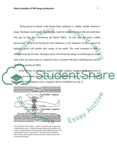
Introduction In 1946, Bloch and Purcell discovered nuclear magnetic resonance (NMR). Later, in 1973, Lauterbur produced the first nuclear magnetic resonance image. Over the years, magnetic resonance imaging (MRI) has constantly evolved and proved to be a very useful and accurate clinical diagnostic imaging tool. This essay briefly explores the basic principles of MR image production. Basic principles Initially, MRI was known as Nuclear Magnetic Resonance (NMR). However the basic principles have remained the same (Faulkner, n.d). When compared to computed tomography (CT), it can provide more anatomical details, with lesser risk of ionizing radiation and with much lesser patient discomfort (Armstrong and Keevil 2003). MR imaging involves a combination of a strong magnetic field and radiofrequency (RF) energy in order to study the distribution and behavior of hydrogen protons in fat and water (Weir and Murray 2004). (From Armstrong and Keevil 2003. Magnetic resonance imaging-1: basic principles of image production. BMJ; 303:35-40.) Being present in plenty in the human body, hydrogen is a highly suitable element to image. Hydrogen nuclei can be considered as small bar magnets having north and south poles that spin on their axes (Armstrong and Keevil 2003). As they spin, the nuclei wobble (precession). Radiowaves having the same frequency as the frequency of precession of the hydrogen nuclei will transfer their energy to the nuclei. The word resonance in MRI is obtained from the fact that “hydrogen nuclei will absorb the energy of radiofrequency pulses only if they are precessing at a frequency that is resonant with those radiofrequency pulses” (Armstrong and Keevil 2003). In the absence of an external magnetic field, the magnetic moments of all the protons in the body are randomly arranged. However, with the patient lying in a ‘tunnel’ formed by the magnet and radiofrequency coils, a magnetic field is established (see fig. 2). Fig 2. The patient lies within a “tunnel” formed by the magnet and radiofrequency coils. A high field magnet along with gradient coils creates a predictable but varied magnetic field. Radiofrequency coils transmit and receive energy pulses. The signals from within the selected slice of the patient can be measured, localized and used to create the final image (From Armstrong and Keevil 2003. Magnetic resonance imaging-1: basic principles of image production. BMJ; 303:35-40.) RF energy is used to rotate or ‘flip’ the protons in the static magnetic field. When the RF field is switched off, the protons experience only the effects of the static magnetic field, and flip back to their original position (Weir and Murray 2004). During this return to equilibrium, a process called relaxation, protons emit a small RF signal. This energy is detected by the antenna in the MRI machine, which is in turn digitized, amplified and finally, spatially encoded. The resulting images are displayed on the operator’s console and can be recorded on hard copy (for viewing) or transferred to optical disk (for storage) (Weir and Murray 2004). MR imaging systems are graded according to the strength of the magnetic field they produce. High-field systems are those capable of producing a magnetic field strength of 1-2 Tesla (T), mid-field systems produce a magnetic field strength of 0.35-0.5 T, and low-field systems produce field strength of less than 0.2 T (Weir and Murray 2004). MR images may be obtained in any plane. There is a wide range of pulse sequences, each of which provides a different image contrast (Weir and Murray 2004.) Four factors determine the size of the signals. These include: proton density, i.e, the number of hydrogen nuclei per unit volume, spin-lattice (T1), spin-spin (T2) relaxation times and the motion of protons (Armstrong and Keevil 2003). By changing the pulse sequence parameters, the MRI contrast can be manipulated. The specific number, strength, and timing of the RF and gradient pulses are set by a pulse sequence (Hesselink, n.d). The repetition time (TR) and the echo time (TE) are the two most important parameters. “The TR is the time between consecutive 90 degree RF pulses. The TE is the time between the initial 90 degree RF pulse and the echo” (Hesselink, n.d). “Echo time (TE) is the time from the application of an RF pulse to the measurement of the MR signal; TE determines how much decay of the transverse magnetization is allowed to occur before the signal is read” (Hesselink, n.d). Based on their specific signal characteristics, pathologic lesions can be separated into five major groups: T1-weighted, T2-weighted, and proton density-weighted (PD)/FLAIR (Fluid Attenuated Inversion Recovery) (Hesselink, n.d). Fig 3. (a) (b) (From Armstrong and Keevil 2003. Magnetic resonance imaging-1: basic principles of image production. BMJ; 303:35-40.) As far as brain pathology is concerned, T2-weighted images are the most sensitive. T2-weighted spin-echo and FLAIR images are used for patients with suspected intracranial disease (Hesselink, n.d). Fig 4. (From Armstrong and Keevil 2003. Magnetic resonance imaging-1: basic principles of image production. BMJ; 303:35-40.) T1 and T2 relaxation times The physicochemical environment of the hydrogen protons will eventually determine the TI and T2 relaxation times. TI and T2 relaxation times are a depiction of the rate at which energy is lost by the excited protons (Armstrong and Keevil 2003). Although every MR image has information about both TI and T2 relaxation times, it is possible to weight the image (on one or other of these relaxation times or to represent mainly proton density) by the correct choice of timing and length of the radiofrequency pulses (Armstrong and Keevil 2003). While TI represents the time taken for 63% of the magnetization due to the excited protons to realign with the field, T2 represents the time it takes for 37% of the magnetization due to the spinning protons to decay due to dephasing. The final variable is motion. (Armstrong and Keevil 2003). Contrast agents in Magnetic Resonance Imaging Contrast mediums used in MRI are given either orally or by an injection. The first contrast agent to be used in MRI was Gadolinium diethylenetriaminepenta-acetic acid (DTPA). By acting as a Ti shortening agent at standard tissue concentrations, DTPA makes the tissues containing gadolinium to appear bright on TI weighted images (Armstrong and Keevil 2003). Some uses for contrast agent in MRI include: detection of very small tumors, differentiating tumor tissue from surrounding edema or differentiating active inflammation from established scar tissue (Armstrong and Keevil 2003). Advances in MRI Fast magnetic resonance imaging The time taken for conventional spin echo images ranges from several minutes to as long as 10-15 minutes to acquire. Although patients can keep their head and body still for several minutes, it is not possible to do so for the chest or abdomen. Therefore, motion artifacts can result and degrade the quality of these images (Armstrong and Keevil 2003). In order to negate motion artifacts, several approaches have been adopted. For images of the heart and larger arteries, cardiac gating of the data collection is widely used and is successful. (Armstrong and Keevil 2003). Spin echo and inversion recovery are two most commonly used imaging sequences. For clinical imaging, the major MR imaging technique used is multiecho spin-echo imaging. One of the main reasons for the development of multislice imaging techniques is to reduce the patient examination time. In addition, in order to minimize the gaps between adjacent tissue slices, various techniques have been developed; one such technique is an optimization approach. Another technique is the acquisition of two multislices, which minimize the gaps that is obtained with the first (Scherzinger and Hendee 2004.) There are fundamental limitations in the improvement of the spatial resolution of conventional MRI into high-resolution microscopy. At present, therefore, the highest resolution MRI microscopes are limited to voxel volumes >40 μm3. There have been some developments in order to address this issue (Degen et al 2008.) Magnetic resonance force microscopy (MRFM) is a recent breakthrough in magnetic resonance detection, which has increased the sensitivity of images. This technique has resulted in great progress in single spin detection for electrons and nuclear spin detection. Magnetic resonance force microscopy is based on mechanical measurement of ultrasmall magnetic forces between nuclear spins in a sample and a nearby magnetic tip (Degen et al 2008.) Functional MRI (fMRI) is another technique, which, like conventional MRI, uses similar imaging techniques and the same equipment. Any change in neuronal activity, like after a particular stimulus or task, causes blood oxygen level–dependent (BOLD) changes in the MRI signal. Functional MRI detects these changes. FMRI is useful in clinical (mapping of sensory and motor functions before neurosurgery or radiation therapy) and more basic neuroscience (studies of patients recovering from stroke, understanding Alzheimer disease, and various cognitive deficits etc (Gore 2003). Conclusion Basically, an MRI system consists of a magnet and coils to generate the magnetic field, a radiofrequency (RF) coil to transmit a radio signal into the specific body part that is being imaged, a receiver coil that detects the incoming radio signals, gradient coils for spatial localization of the signals, and finally, a computer that deciphers the signals and constructs the image. Four factors determine the signal intensity of the MR image size: proton density, T1 & T2 relaxation times and the motion of protons. The contrast agent used in MRI is Gadolinium diethylenetriaminepenta-acetic acid (DTPA). Contrast agents are used in MRI to detect very small tumors, differentiating tumor tissue or active inflammation from surrounding edema or scar tissue respectively etc. Multiecho spin-echo imaging is the major MR imaging technique used for clinical imaging. Multislice imaging techniques has helped to reduce the patient examination time. Some of the advances in MRI include: fast magnetic resonance imaging, Magnetic resonance force microscopy (MRFM) and Functional MRI (fMRI) Magnetic Resonance Imaging is a versatile and highly useful technology that has allowed the clinician to study any body part with great accuracy and come to a definitive conclusion to aid diagnosis. This technique has evolved greatly over the years, and it is being further refined to increase the sensitivity. References Armstrong, P and SF. Keevil. 2003. Magnetic resonance imaging-1: basic principles of image production. BMJ; 303:35-40. Degen, C. L, Poggio, M, Mamin, H. J, Rettner, C. T and D. Rugar, 2008. Nanoscale magnetic resonance imaging. PNAS 106 (5); 1313-1317. Faulkner, WM. n.d. Basic principles of MRI. http://www.e-radiography.net/mrict/Basic_MR.pdf. Gore, JC. 2003. Principles and practice of functional MRI of the human brain. J. Clin. Invest. 112(1): 4-9. Hesselink, JR. n.d. Basic principles of MR imaging. http://spinwarp.ucsd.edu/NeuroWeb/Text/br-100.htm#anchor51907. Scherzinger, AL and WR. Hendee. 2004. Basic principles of magnetic resonance imaging-An update, In High-tech medicine. West J Med. 143:782-792. Weir, J, and AD. Murray. 2004. Mosby’s atlas and text of clinical imaging. Mosby-Wolfe. Read More
Cite this document
  • APA
  • MLA
  • CHICAGO
(The basic principles of MR image production Essay - 1, n.d.)
The basic principles of MR image production Essay - 1. https://studentshare.org/health-sciences-medicine/1726842-explain-the-basic-principles-of-mr-image-production
(The Basic Principles of MR Image Production Essay - 1)
The Basic Principles of MR Image Production Essay - 1. https://studentshare.org/health-sciences-medicine/1726842-explain-the-basic-principles-of-mr-image-production.
“The Basic Principles of MR Image Production Essay - 1”. https://studentshare.org/health-sciences-medicine/1726842-explain-the-basic-principles-of-mr-image-production.
  • Cited: 0 times

CHECK THESE SAMPLES OF The basic principles of MR image production

Identifying Stakeholders and Developing Charter

the basic aim of this research is to describe how various organizational structures impact the PM process.... The company's culture determines a company's managerial actions, performance, image, and policies, also having impact on the operational programs of the PM practice....
6 Pages (1500 words) Essay

Technologies and Predictions

Moreover, they do not need backlights to form the image, which means that their size is the thinnest in the market and so flexible that one can even roll it like a T-shirt.... It took centuries for the first human to discover about communication, clothing, and food and adopt other basic societal reforms....
7 Pages (1750 words) Essay

Computer Hardware, Systems Software & Networking

An easy way to think of a raster graphic is to picture an image in a comic book that is made up of many small dots of color.... The raster graphic arrangement can also be referred to as an arrangement of an array of pixels that combine to display an image.... Direct memory access facilitates the printers to fetch data directly from main memory without the intervention of CPU by stealing CPU cycles....
3 Pages (750 words) Essay

Magnetic Resonance Imaging

Reference: Brown, Mark A and Semelika, Richard C (2010) MRI: basic principles and Application, 4th Edn.... References: Brown, Mark A and Semelika, Richard C (2010) MRI: basic principles and Application, 4th Edn.... Reference: Brown, Mark A and Semelika, Richard C (2010) MRI: basic principles and Application, 4th Edn.... eferencesBrown, Mark A and Semelika, Richard C (2010) MRI: basic principles and Application, 4th Edn.... When the x or y component is plotted as a function of the number of phase encoding steps and time, it varies sinusoidally, slower at the centre of the image, and faster along the right and left edges....
5 Pages (1250 words) Essay

Historical Poetics in The Devils Backbone

The director Guillermo del… In order to bring out the story in a way that captures the audience, del Toro utilizes recurring symbols and images throughout the film. According to The method of production itself includes the principles that determine how the project was composed, the effects, functions and uses of the project and the basic rules that govern cinematography and how they have been applied in the film.... The film then presents a disturbing image of a deformed baby inside a jar suspended in fluid....
6 Pages (1500 words) Essay

Reflected Light Imaging

There has been an improvement over time in the image formation system.... In all improvements, the image formation in the eyes still has great similarity to all the devices.... Light properties form the central part in the ability of any device to form an image with principle remaining the same for all image-forming devices.... Cameras form the central part in understanding the image formation processes.... The principle of using reflected light from the object still forms an integral part of image formation....
8 Pages (2000 words) Essay

Principles of Human Resources

This paper "principles of Human Resources" focuses on the fact that the workforce is one of the vital resources for an organisation; the companies pay extra attention to the selection of its human resource.... nbsp;  Apart from these costs, until the organisation recruit a new substitute for the vacant position, production process suffers and results in loss of production.... Hence, this loss in production also adds up with the cost of employee turnover....
10 Pages (2500 words) Essay

Harnessing Nuclear Power in Saudi Arabia

uclear energy is used in the production of hydrogen needed in transportation.... hellip; Its scope is only limited to the very basic challenges that every country can face.... This work called "Harnessing Nuclear Power in Saudi Arabia" describes nuclear power as one of the non-fossil technologies of generating electricity....
7 Pages (1750 words) Essay
sponsored ads
We use cookies to create the best experience for you. Keep on browsing if you are OK with that, or find out how to manage cookies.
Contact Us