StudentShare
Contact Us
Sign In / Sign Up for FREE
Search
Go to advanced search...
Free

Respiratory, Endocrine, and Cardiovascular Systems - Assignment Example

Cite this document
Summary
This paper "Respiratory, Endocrine, and Cardiovascular Systems" discusses the following issues: the intrinsic and extrinsic factors that control the cardiac output, starling's law of the heart states, how do adrenaline and noradrenaline differ in this respect, etc…
Download full paper File format: .doc, available for editing
GRAB THE BEST PAPER97.7% of users find it useful
Respiratory, Endocrine, and Cardiovascular Systems
Read Text Preview

Extract of sample "Respiratory, Endocrine, and Cardiovascular Systems"

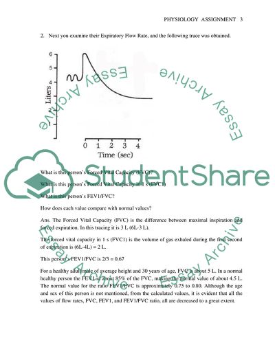
Respiratory, Endocrine, and Cardiovascular Systems You are working in a pulmonary function laboratory, and a person comes in for testing due to shortness of breath and reduced ability to work. The person has been a heavy smoker most of his life. You examine his lung volumes with a spirometer, and the following trace is obtained. Assuming this trace is accurate and the volume scale is correct. What are the following volumes and how do they differ from normal? Tidal volume 0.5 L 0.1 L less than normal of 0.6 L Inspiratory reserve volume 1.25 L 1.25 L less than the normal 2.5 L Expiratory reserve volume 2.0 L 1.0 L more than the normal 1.0 L Vital capacity 3.75 L 1.05 L less than the normal of 4.8 L Functional residual capacity 5.0 L 2.8 L more than the normal of 2.2 L Residual Volume 3.0 L 1.8 L more than the normal of 1.2 L Total Lung Capacity 6.75 L 0.75 L more than the normal of 6 L 2. Next you examine their Expiratory Flow Rate, and the following trace was obtained. What is this person’s Forced Vital Capacity (FVC)? What is this person’s Forced Vital Capacity in 1 s (FVC1) What is this person’s FEV1/FVC? How does each value compare with normal values? Ans. The Forced Vital Capacity (FVC) is the difference between maximal inspiration and forced expiration. In this tracing it is 3 L (6L-3 L). The forced vital capacity in 1 s (FVC1) is the volume of gas exhaled during the first second of expiration is (6L-4L) = 2 L. This person’s FEV1/FVC is 2/3 = 0.67 For a healthy adult male of average height and 30 years of age, FVC is about 5 L. In a normal healthy person the FEV1 id about 85% of the FVC, making the normal value of about 4.5 L. The normal value for the ratio FEV1/FVC is approximately 0.75 to 0.80. Although the age and sex of this person is not mentioned, from the calculated values, it is evident that all the values of flow rates, FVC, FEV1, and FEV1/FVC ratio, all are decreased to a great extent. 3. From the data you have obtained, what do you think is wrong with this persons lung? Give the reasons for your answer. The person is smoker all his life. The person has shortness of breath and reduced ability to work. All the parameters measured with spirometer indicate reduced pulmonary function, and expiratory flow rates are also significantly decreased. This limitation in airflow is a hallmark of emphysema. Emphysema combined with shortness of breath and reduced ability to work indicates hypoxia which might have been caused by perhaps small airway disease and associated chronic bronchitis. Everything taken together, this person is suffering from chronic obstructive pulmonary disease (Phillipson and Slutsky, 2000, 2139-2152). 4. Explain why this person has reduced ability to work? In this disease, there is emphysema that occurs due to destruction and enlargement of the lung alveoli. Along with this there is narrowing of small bronchioles. Airflow limitation or obstruction is evident from persistent reduction of forced expiratory flow rates. The lungs remain hyperinflated. With decrease in FEV1, the PaO2 will gradually decrease and PaCO2 will increase. Although other factors such as right ventricular failure, ventilation-perfusion mismatch, pulmonary hypertension may be implicated in hypoxia, the final common pathway, in this case remains reduction in PaO2 due to airflow limitation. This hypoxia will lead to reduced ability to work (Senior and Shapiro, 1998, 659-681). 5. Describe how the ventilation-perfusion ratio changes throughout the lungs. Gravitational effects on blood flow throughout the different regions of the lungs and in different postures of the body lead to an uneven distribution of blood in the lungs, and even in the normal lungs, this causes changes in the ventilation-perfusion ratio. In an erect human being, the gravitational pull is maximally effective downward. The pulmonary vessels are actually belong to a high-flow, low resistance, and low-pressure exchange system. Since the vessels are compliant, gravity causes the blood volume and hence flow is to be greater at the base of the lungs than in the apices. This causes a difference in pulmonary arterial pressure of about 30 cm of H2O. Moreover, the apical region being situated about 15 cm higher than the heart and base being about 15 cm lower than the heart indicated by levels of pulmonary artery, the arterial pressure is 11 mmHg less in the apices and 11 mmHg more in the bases. Comparatively lower arterial pressure in the apices would result in lower blood flow in the sheet of capillary network surrounding the apical alveoli. In contrast the basal capillaries would be distended, and basal blood flow would be augmented. Thus in an upright person, the capillary perfusion will be altered in such a manner that from apex to base, the pulmonary blood flow will increase from apex to base and hence the ratio of ventilation-perfusion ratio in an inverse manner. 6. Describe 4 reasons why someone might be hypoxic and briefly describe how you might differentiate between the 4 different conditions. Regional Hypoventilation: About 90% cases of hypoxia are due to regional hypoventilation. This occurs due to a regional ventilation-perfusion mismatch due to a partial airway obstruction. Thus a fraction of the blood flowing through the lung does not get oxygenated totally. This results in an increased venous admixture, whereas in order to lower a systemic arterial Po2 as predicted by the oxyhaemoglobin denaturation curve, only a small venous admixture is necessary. Therefore, these people with abnormally low ventilation-perfusion ratio would have a high alveolar-arterial O2 gradient, leading to reduction in partial pressure of O2 and low O2 content. Measurement of A-aO2 gradient, low PO2, and low O2 content would be helpful to identify these patients, but in these cases PaCO2 would not be altered much. Clinically large pulmonary embolism leads to such a clinical picture. Shunts: Either right-to-left shunt due to anatomic anomalies or an intrapulmonary shunt also cases hypoxia. Intrapulmonary shunts are seen with total obstruction of airway. The blood gas pictures are almost same, but ventilation-perfusion test results are characteristic. Shunt hypoxia also presents with high A-aO2 gradient, low PO2, and low O2 content and a normal or slightly high PaCO2. The distinction between shunt hypoxia and regional hypoventilation can be made with a specific test, when the patient is made to breathe 100% O2 for 15 minutes. If the PaO2 is greater than 150 mmHg is cause is low ventilation-perfusion ratio. If, however it is less than 150 mmHg, the probable cause is a shunt. Generalized Hypoventilation: This occurs with depressed ventilation. This form of hypoventilation is encountered in patients with chronic obstructive pulmonary disease or depressed respiratory drive due to head injury or drug overdose. With depressed ventilation, PCO2 increases with increase in arterial PH. The ventilation is insufficient to maintain systemic arterial PO2 and PCO2. This condition can be distinguished from shunt or ventilation perfusion mismatch by normal A-aO2 gradient since both arterial and alveolar PO2 are lowered in an equal extent. Thus a combination of high PCO2, low PaO2, and normal A-aO2 distinguishes this condition. Moreover administering oxygen would not be able to correct the hypercapnia. Diffusion Block: Most frequently a block in the diffusion occurs with pulmonary edema. Physiologically, the diffusion distance between the alveolar capillary membrane is increased die to thickened alveolar wall leading to decreased gas exchange, which may also result from compromised permeability of the alveolar-capillary membrane. This is distinguished by a low PaO2, high A-aO2 gradient, and a high PaCO2 due to CO2 retention and impaired exchange. 7. Describe how the body responds to a decrease in plasma calcium. The plasma calcium is maintained within narrow limits through minute-to-minute adjustments by the combined actions of three different hormones, namely, active vitamin D and parathyroid hormone (PTH) acting on bone, kidney, and intestine. The primary function of PTH is to maintain a constant concentration of Ca2+. As the concentration of Ca2+ diminishes, PTH secretion increases. PTH secretion are all stimulated by low concentrations of Ca2+ and suppressed by high concentrations. There are calcium sensing receptors on PTH cells. These sense minute changes in plasma calcium, and acting on bone, increases bone resorption through promotion of osteoclastic activity. Calcium is released into the blood from the bone stores. At the same time PTH stimulation also release active vitamin D from the kidneys, which increase absorption of calcium from the intestine. Moreover, acting on the kidney, it increases renal tubular reabsorption of calcium, conserving about 150 mg of calcium per day which is reflected through proportionate increase in plasma calcium. This process is facilitated by inhibition of tubular reabsorption of phosphate, and stimulation of conversion of vitamin D to its biologically active form, calcitriol. As a result, filtered Ca2+ is avidly retained, and its concentration increases in plasma, whereas phosphate is excreted, and its plasma concentration falls. Newly synthesized calcitriol interacts with specific high-affinity receptors in the intestine to increase the efficiency of intestinal Ca2+ absorption, thereby also increasing the plasma Ca2+ concentration. PTH increases tubular reabsorption of Ca2+ with concomitant decreases in urinary Ca2+ excretion (Bilezikian and Potts, 2002, N57). 8. Draw a cross section through the adrenal gland and explain what hormones are secreted from which zone. The inner part of adrenal gland is known as medulla. The medulla has chromaffin, neuroendocrine cells, which secrete hormones epinephrine and norepinephrine. Both zona fasciculata and zona reticularis of the adrenal cortex secrete glucocorticoids cortisol and corticosterone. Zona glomerulosa secrete mineralocorticoid, aldosterone. Adrenal androgens are secreted by both zona fasciculata and zona reticularis of the adrenal cortex. 9. a. How are the actions of catecholamines (adrenalin and noradrenaline) mediated? Adrenaline and noradrenaline bind to adrenergic receptors, α1, α2, β1, and β2. These mediate the cellular effects of these hormones. 9. b. How do adrenaline and noradrenaline differ in this respect? Both adrenaline and noradrenaline raise the systolic blood pressure by stimulating heart rate and contractility, thereby increasing cardiac output. However, epinephrine reduces diastolic pressure as a result of causing vasodilatation of certain vessels, particularly those of skeletal muscle, while norepinephrine raises diastolic pressure by causing a more generalized vasoconstriction. Both catecholamines cause piloerection and dilatation of the pupils. Adrenaline also acts as a bronchodilator and reduces the motility of the gut (Vander et al., 2001, 710-716). 10. Starlings Law of the Heart states The greater the ventricular volume the greater the force of ventricular contraction. Explain what is meant by this statement and how this is achieved? The relation between the initial length of the muscle fibers and the developed force is of prime importance for the function of heart muscle. This forms the basis of the Starlings law of the heart, which states that, within limits, the force of ventricular contraction is a function of the end-diastolic length of the cardiac muscle; in the intact heart the latter is closely related to the ventricular end-diastolic volume. In cardiac muscle and in fact, in any muscle, the force of contraction depends on initial muscle length. The sarcomere length associated with the most forceful contraction is approximately 2.2 microm. At this length, the two sets of overlapping myofilaments of the sarcomere are configured so as to provide the greatest area for their interaction. The length of the sarcomere also regulates the extent of activation of the contractile system, its sensitivity to Ca2+. According to this concept, termed length-dependent activation, at the optimal sarcomere length of 2.2 microm, the myofilament sensitivity to Ca2+ is also maximal. In diastole, blood begins to fill the ventricle. The ventricular pressure rises with stretching of the myocardial fibers which are placed under a degree of tension known as preload. An increase in filling pressure leads to both an increase in end-diastolic volume and an increase in the subsequent stroke volume, enabling the cardiac muscle to respond to increased stretch with a more forceful contraction, thus heart can automatically adjust its cardiac output with that of the venous return (Starling, 2002, 19-35). 11. A patient has been admitted to hospital suffering from a cardiac problem that results from loss of SA node function. Although her heart rate is slow, her ventricles appear to be operating normally, but with a lightly reduced cardiac output. How this is possible? The excitation of heart normally begins at SA node because of the pace making potential of this specialized tissue. This reaches the threshold before the pacemaker potential of the AV node. Although the pacemaker rate of SA node is 60 to 100 beats per minute, in this case there is a loss of SA node function. However, the AV node has also intrinsic pacemaker rate of 40 to 50 beats per minute. In the absence of SA node function, this activity continues, so the contraction happens at a slower rate and since atrial contraction is absent, the preload is less on the ventricles. The rates become slower, and the presystolic stretch on the ventricles is less. Therefore, the force of contraction of the ventricles is less following Starlings Law of Heart leading to lesser cardiac output. 12. Describe the intrinsic and extrinsic factors that control the cardiac output. The extrinsic factors that control cardiac output are factors that influence cardiac contractility from outside. These include sympathetic stimulation via noradrenalin and circulating adrenaline acting on beta-1 receptors. Numerous physiological and psychological conditions may be responsible for this. Many drugs such as positive and negative inotropic drugs such as digitalis and general anesthetics or toxins respectively may influence this. The intrinsic factors that may influence cardiac output will be factors determining stroke volume among others. Those working through force of contraction are end-diastolic fiber length determined by preload, determined in turn by end-diastolic pressure and ventricular diastolic complaince play important roles. Intrinsic changes in contractility in response to changes in heart rate and afterload also play roles. Diseases involving coronary arteries and myocardium may also influence this. Ventricular hypertrophy is an important intrinsic factor. Different patterns of electrical excitation and abnormalities from them are also known intrinsic factors along with conditions that influence the afterload also influence cardiac output. These occur due to changes in ventricular systolic pressure, due to changes in ventricular radius. Reference List Bilezikian JP, Potts JT Jr., (2002). Asymptomatic primary hyperparathyroidism: New issues and new questions — bridging the past with the future. J Bone Min Res. 17(Suppl 2):N57 Phillipson EA, Slutsky AS, (2000). Hypoventilation and hyperventilation syndromes, in Textbook of Respiratory Medicine, 3d ed, JF Murray, JA Nadel (eds). Philadelphia, Saunders,pp 2139-2152. Senior RM, Shapiro SD, (1998). Chronic obstructive pulmonary disease: Epidemiology, pathophysiology, and pathogenesis, in Fishmans Pulmonary Diseases and Disorders, 3d ed, AD Fishman et al (eds). New York, McGraw-Hill, 1998, pp 659-681 Starling, MR, (2002). Physiology of myocardial contraction, in WS Colucci and E Braunwald (eds). Atlas of Heart Failure: Cardiac Function and Dysfunction, 3d ed, Philadelphia: Current Medicine, pp 19-35. Vander et al., (2001), Human Physiology: The Mechanism of Body Function, Eighth Edition. The McGraw−Hill. Companies, 710-716. Read More
Cite this document
  • APA
  • MLA
  • CHICAGO
(Respiratory, Endocrine, and Cardiovascular Systems Assignment, n.d.)
Respiratory, Endocrine, and Cardiovascular Systems Assignment. Retrieved from https://studentshare.org/health-sciences-medicine/1717232-human-physiology
(Respiratory, Endocrine, and Cardiovascular Systems Assignment)
Respiratory, Endocrine, and Cardiovascular Systems Assignment. https://studentshare.org/health-sciences-medicine/1717232-human-physiology.
“Respiratory, Endocrine, and Cardiovascular Systems Assignment”. https://studentshare.org/health-sciences-medicine/1717232-human-physiology.
  • Cited: 0 times

CHECK THESE SAMPLES OF Respiratory, Endocrine, and Cardiovascular Systems

Dehydration and Medical Physiology

It mainly targets the kidney and cardiovascular system.... High levels of phosphorus in the body results to increase risk of cardiovascular diseases.... It becomes produced in the anterior pituitary gland and gets involved in the regulation of endocrine functions of the thyroid gland.... Thyroid disease- the thyroid is an endocrine gland.... alkalemia can be caused by respiratory or metabolic alkalosis.... Acidosis gets caused by respiratory acidosis or metabolic acidosis....
7 Pages (1750 words) Essay

Human Body Systems

It is common fact that the respiratory system and cardiovascular system synchronize in a proper manner.... Respiratory system provides… In the similar way digestive system provides the food to the cells that is delivered by the blood circulatory system. Functions as to control the body functions and systems.... All the systems are connected to the nervous system through nerves and send and Brain, nerves & spinal cord.... The oxygenated blood is transported by the cardiovascular system and the heart pumps the blood as faster as the oxygen is required....
7 Pages (1750 words) Essay

2014 BC Metabolism Meets 2014 AD Lifestyle

Developing countries has recorded high cases of cardiovascular diseases.... Atherosclerosis, metabolic syndrome and diabetes are evidences of variation in lifestyle and genetic makeup.... As opposed to the 2014BC, 2014AD have been depicted with a host of… Biotechnological methods has taken over the traditional methods....
4 Pages (1000 words) Essay

Exploring Endocrine and Respiratory Systems

Blood glucose can go down during starvation or intense exercises thus through glycogenesis, The endocrine pancreas plays a very important role in the metabolism of glucose.... While many tissues are capable of utilizing other energy sources such as fat and protein, brain and the red blood cells can only use glucose....
2 Pages (500 words) Essay

The Hands-on Experience of Working with Patients

ll systems reviewed & negative except as marked.... cardiovascular: Regular rate and rhythm, normal heart sounds, normal capillary refill, BP & pulses = bilaterally.... oral; Heart Rate 83; BP 140/65; respiratory Rate 18: O2 saturation 100% RA; Height 5'2; Weight 172lbs....
5 Pages (1250 words) Essay

Yoga and Anatomy

According to Kurz, the aim of a good warm-up is to give "an increased awareness, improved coordination, improved elasticity and contractibility of muscles, and a greater efficiency of the respiratory and cardiovascular systems.... Therefore Yoga is nothing but the union of all the organ systems of the body in correlation to the consciousness of the mind....
5 Pages (1250 words) Essay

A Nurse and Patient

The paper contains the questionnaire of the female patient who states that she has been feeling ill for two days and that she “has a cold.... rdquo; Symptoms include “exhaustion, runny nose, stuffy head, blurred vision.... rdquo; The patient also mentioned that it “could be some kind of flu” … The patient states that she has been feeling ill for two days....
8 Pages (2000 words) Assignment

Harry Houdini - A Body under Control

The paper "Harry Houdini - A Body under Control" discusses the phenomenon of Harry Houdini, who was known for the control of his breathing and training his muscles, besides an amazing showman.... The paper highlights the main contributions of this person to the world and community.... hellip; Harry Houdini was born in Budapest, Hungary, in 1874, moving to Appleton, Wisconsin in 1878 with his mother and siblings....
9 Pages (2250 words) Essay
sponsored ads
We use cookies to create the best experience for you. Keep on browsing if you are OK with that, or find out how to manage cookies.
Contact Us