StudentShare
Contact Us
Sign In / Sign Up for FREE
Search
Go to advanced search...
Free

The Common Image Artifacts Associated with Breast MRI, and Strategies Used to Minimize Them - Essay Example

Cite this document
Summary
The paper "The Common Image Artifacts Associated with Breast MRI, and Strategies Used to Minimize Them" discusses that generally, malignancies and other associated pathological processes are often characterized by changes in tissue mechanical properties…
Download full paper File format: .doc, available for editing
GRAB THE BEST PAPER95.4% of users find it useful
The Common Image Artifacts Associated with Breast MRI, and Strategies Used to Minimize Them
Read Text Preview

Extract of sample "The Common Image Artifacts Associated with Breast MRI, and Strategies Used to Minimize Them"

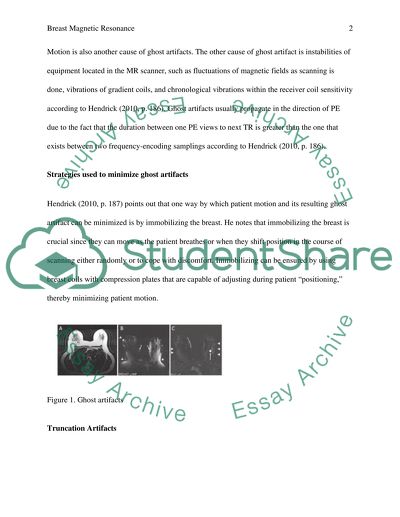
?Discuss the common image artifacts associated with Breast MRI, and strategies used to minimize them The application of Magnetic Resonance Imaging (MRI) has grown in recent years as a tool used in medical imaging diagnosis (Bernstein, Huston & Ward, 2006, p. 735–737). This has particularly been witnessed in creasing of breast as a way of treating cancer and other related malignant tumors. However, technicians and radiologists working on these systems quite often encounter image artifacts related to radio waves with strong magnets in the scanner. There are numerous artifacts associated with MRI scanning. Therefore, understanding their causes may be the surest way of minimizing, correcting or avoiding either at the source before any data can be put in or at the reconstruction stage after the acquisition of data (Morris & Liberman, 2005, p. 14–19). The artifacts commonly related to breast MIR include ghost artifacts, truncation, aliasing, RF transmission, chemical shift, reconstruction, and metallic artifacts such as biopsy marker clip artifacts and other randomly occurring image artifacts. Ghost Artifacts Hendrick (2010, p. 187) notes that ghost artifacts that feature in images are more that often induced by patients themselves as a result of signal-producing tissues when data is being acquired. He notes that the motion produces a blurred image and ghosts of bright moving objects (Fingure 1). These artifacts manifest as patterns of noise propagating in the phase encoding direction, irrespective of the route moved by bright tissues. The main cause of ghost artifacts is miss-registration of images data of altering signal values of rigid structures obtained from diverse phase encoding views according to Bernstein, Huston and Ward (2006, p. 735–737). Motion is also another cause of ghost artifacts. The other cause of ghost artifact is instabilities of equipment located in the MR scanner, such as fluctuations of magnetic fields as scanning is done, vibrations of gradient coils, and chronological vibrations within the receiver coil sensitivity according to Hendrick (2010, p. 186). Ghost artifacts usually propagate in the direction of PE due to the fact that the duration between one PE views to next TR is greater than the one that exists between two frequency-encoding samplings according to Hendrick (2010, p. 186). Strategies used to minimize ghost artifacts Hendrick (2010, p. 187) points out that one way by which patient motion and its resulting ghost artifact can be minimized is by immobilizing the breast. He notes that immobilizing the breast is crucial since they can move as the patient breathes or when they shift position in the course of scanning either randomly or to cope with discomfort. Immobilizing can be ensured by using breast coils with compression plates that are capable of adjusting during patient “positioning,” thereby minimizing patient motion. Figure 1. Ghost artifacts Truncation Artifacts Truncation artifacts, or “Gibbs”, “ringing”, or “edge” artifacts, are those that occur due to finite sampling taking place in MRI. These artifacts more than often occur near high-contrast, sharp artifacts as shown in Fig. 2. Hendrick (2010, p. 193) notes that reconstruction of images with a discrete number of samples tends to overshoot the actual signal changes across sharp interfaces because of the finite sampling of the image in each of the in-plane direction, thereby producing a ringing artifact beyond the image interface. The overshooting in this case is what is usually referred to as the Gibbs phenomenon. The phenomenon augments contrast across the interface and it is associated with the ringing which causes the dark and light banding that takes place near sharp interfaces and reduces with the increase in the distance from the interface. Strategies used to minimize truncation artifacts Hendrick (2010, p. 193) notes that truncation artifact can be minimized using a higher matrix. He adds that as this is done, it is imperative that the scan time be increased to ensure that the size of matrix is increased in the PE direction. Figure 2. Truncation artifacts Aliasing Artifacts Aliasing artifacts, “image wrap” or “wraparound” is the type of artifact that occurs when tissue-producing signals exceeds the prescribed field-of-view in either PE or FE direction as noted by Hendrick (2010, p. 190). Hedrick adds that aliasing occurs when MRI needs the gathering of a distinct number of signals in every course to produce an image. Having a number of discrete pixels here implies that 3D or 2D reconstruction cannot differentiate between the corresponding scan field-of-view and well assigned signal-producing tissues with signal-producing tissues outside the scan FOV (Hendrick, 2010, p. 190). This makes signals originating from tissues exterior to the approved FOV add up to signals from pixels within the FOV found at the opposite end of the image (Dietrich, Reiser & Schoenberg, 2008, p. 30–33). Usually aliasing artifacts tend to add structured noise capable of obscuring details in the breast as well as occasionally simulating pathology. It is noted that aliasing artifacts usually take place in the PE direction as opposed to the FE direction. The reason is that MR manufacturers apply measures that suppress signals originating from tissues located behind the chosen FOV and automatically oversampling the quantity of data points in the FE direction. Strategies used to minimize aliasing artifacts Hendrick (2010, p. 190) points out that the surest way to do away with an aliasing artifact in the FE direction is by using a combination of low-pass frequency filters capable of suppressing signals just beyond the selected FOV and frequency oversampling. This is because it ensures that the image wrap from signal-producing tissues within one FOV on both sides of the prescribed FOV are suppressed, thereby eliminating the majority of these artifacts (Hendrick, 2010, p. 190). Chemical Shift Artifacts Hendrick (2010, p. 1940) notes that chemical shift artifacts are caused by diverse resonance frequencies of hydrogen in water and in lipid, where miss-registration of fat and water protons result in a signal void between areas of water and fat as shown in Figures 3A and B. Radiol (2009, p. 264) notes that these events take place in high field strength where chemical shift is not significant and compensation is usually unnecessary. Strategies used to minimize Chemical shift artifacts One way of minimizing chemical shift artifact is by broadening the smallest possible FOV and the receiver bandwidth. Radiol notes that a wider bandwidth also reduces ANR. Therefore, by reducing the bandwidth, the best SNR can be obtained using chemical saturation (Dietrich, Reiser & Schoenberg, 2008, p. 30–33). Figure 3A Figure 3B Radiofrequency Transmission Artifacts Radiofrequency (RF) transmission artifacts occur because of the incomplete radio frequency shielding of the MRI exam room. Hendrick (2010, p. 198) notes that RF shielding is usually done by a Faraday cage consisting a shell of wire mesh covering the doors, the inside wall, and windows of the MRI scan room. In case the door remains partially closed or when a break occurs in the RF shield, this may result in the appearance of radio transmission as a dominant line in the image near the Larmor frequency (Figure 4). RF feedthrough originating from transmission coils to receiving coils can also cause line artifacts .The sources include patient monitoring equipment and faulty lighting fixtures in the MRI scanner room. Hendrick (2010, p. 198) cites that these normally produce a broad spectrum of RF interferences manifesting themselves as broad lines criss-crossing the image in the PE direction. Strategies for minimizing Radiofrequency transmission artifacts The artifacts are secluded by obtaining phantom images with lights on. This is followed by their removal from the scan room until the artifacts completely disappear (Hendrick, 2010, p. 198). Figure 4. Radiofrequency transmission artifacts Reconstruction Artifacts These artifacts normally appear as recurring patterns of dots or lines at fixed periodicity in planar images (Fig. 5a & 5b). The reconstruction artifacts are caused by corrupted data measured as signal is being acquisition, or due to alteration prior to 2 or DFT image reconstruction (Dietrich, Reiser & Schoenberg, 2008, p. 30–33). Strategies for minimizing reconstruction artifacts This type of artifact can be corrected by checking the k-space depiction of image data as it reveals the corrupted data (Dietrich, Reiser & Schoenberg, 2008, p. 30–33). Figure 5 Discuss the clinical usefulness of spectroscopy in Breast MRI Stanwell et al. (2010, p. 13) note that breast cancer has emerged as the deadliest health problem for women over the past few decades. As such, since research still shows that cancer cannot be cured, its early detection is very important. Breast magnetic resonance imaging has been found to provide both physiologic and physical tissue features that are useful for suppressing malignant from benign lesions. The most important is the recently development of the use of the breast MRI spectroscope in the treatment of cancer and other tumors related to it. Vivo MR Spectroscopy (MRS), for instance, has been found to be a valuable method for obtaining biochemical status of normal and disease tissues (Warrem, 2000, p. 123–125). Researchers argue that the vivo magnetic resonance spectroscopy, when performed alongside MRI, can be so helpful in obtaining information relating to the chemical content of breast lesions. The information obtained from the vivo can be used for a number of clinical applications, including improving the lesion diagnosis accuracy and examining the response to cancer therapies (Bolan et al., 2005, p. 150–152). Earlier MRS researches of breast cancer have been promising and have seen many research groups factoring in the technique in their breast MRI protocols. It is argued that malignant tissues usually contain high concentration of choline containing compounds, which can act as a biochemical marker. As a result, magnetic resonance spectroscopes have been found to be helpful in adjusting the specificity of MRI in lesions that are larger than 1 cm, as they monitor the tumor response as well. Tozaki and Maruyama (2007, p. 42) note that MRS can be useful in assessing the response to neoadjuvant chemotherapy. He notes that contrast-enhancement patters may provide inaccurate results and false-negative findings associated with the effects of chemotherapeutic agents. Contrary to this, MRS and diffusion-weighted imaging are showing great promise in evaluating the direct effects of chemotherapeutic agents (Yeh, 2003). Yeh argues that the existence of the Cho peak in breast cancer as shown may show the rise in cell proliferation, with a fall in the peak after treatment which reflects a lower viability of the tumor. Physicians have also been finding it hard diagnosing certain hidden tumors on the breast, which are associated with cancer. This has mainly been witnessed when using the mammography technique to screen women at risk of cancer. However, it is argued that with the introduction of MRS, this will be a thing of the past (UnitedHealthcare, 2012, p. 4). The reason is that MRS techniques are very sensitive and are proficient of detecting hidden tumors that might otherwise escape unnoticed if examined using other techniques. Tozaki and Maruyama (2007, p. 42) reveal that MRS has been providing promising results with regard to differentiation between benign and malignant breast lesions. They note that in malignant lesion, MR achieved a sensitivity level of more than 80%. In addition, MRS proved to be very sensitive with regard to detection of invasive carcinoma from sarcoma and ductal carcinoma in situ (UnitedHealthcare, 2012, p. 5). Fig 6: MRS and MRI measurement in a patient with mixed invasive ductal and lobular carcinoma. Courtesy of http://www.tswj.com/2012/508295/fig2/ What is Magnetic Resonance Elastography (MRE)? What information would it add to a Breast MRI examination? Proulx (2011, p. 58) defines magnetic resonance elastography (MRE) as a non-invasive imaging technique used for measuring mechanical properties of biochemical waves quantitatively. This technique incorporates both MRI imaging with sound waves to develop an elastogram or visual map showing how stiff or elastic the body tissues are. The technique has mainly been applied in detecting the hardening of the liver caused by different types of liver ailments. Fig 7: MRE in application Ehman (2001, p. 1) notes that malignancies and other associated pathological processes are often characterized by changes in tissue mechanical properties. He argues that these accounts for the efficacy of palpation as a clinical device for detecting cancer in body regions which are accessible. This technique has assisted in detection of a number of tissue ailments such as the breast, thyroid, and the prostate. Research reveals that MRE is vital in detection of breast ailments. In this regard, MRE is helpful in assessing the viscoelastioc sheer properties of lesions through direct MRI visualization of acoustic waves. It also quantifies the decreased elasticity of malignant tumors. Ehman (2001, p. 6) argues that the quantification of the differential elasticity or stiffness between the breast lesion and the adipose tissue in the background as well as the fibroglandular tissue is achieved by assessing through the propagation of the mechanical waves created by the electromechanical driver, via the breast using gradient echo phase contrast sequence. He notes that the stiffness of the tissue map (or electrogram) is based on a linear scale, attuned into kilopascals and represented as a color map. Research also reveals that elastographic technique is showing promising result with regard to detecting carcinoma and other suspicious breast lesions (Ehman 2001, p. 2) It is also well; known that is becoming the most deadliest life-threatening malignancy among women in America and the world at large causing a several premature deaths if not diagnosed early enough. Despite the fact that physicians have for a long time relied on screening mammography, research show that it has several limitations. The limitations on mammography have been seen with regard to its specificity and sensitivity, which decreases considerably with radiographically dense breasts as noted by Proulx (2011, p. 59). It is here that elastographic screening has been found to be very useful. The reason being, it is highly sensitive with high specificity ratio which is able to detect even small tumors in radiographically dense breast (Proulx 2011, p. 60). Electrographic technique does this since it is able to quantitatively portray the elasticity of breast tissues in vivo, which other MRI techniques have not been able to. Furthermore, elastographic technique is able to portray high shear elasticity in recognized breast tumors. In conclusion, adopting this technology will indeed add value to breast MRI examination which in turn improve early diagnose of breast cancers, which other techniques have not been able to reveal early enough. This will see a reduction in death rates caused by breast cancers and other ailments associated with it due to early diagnosis. References Bernstein, M., Huston J. & Ward, H. (2006). Imaging artifacts at 3.0T. Journal of Magn Reson Imaging. 24 (4) p. 735–746. Bolan, P., Nelson, M., Yee, D. & Garwood, M. (2005). Imaging in breast cancer: magnetic resonance spectroscopy. Breast Cancer Res. 7 (4), p. 149-152. Dietrich, O., Reiser, M. & Schoenberg, S. (2008). Artifacts in 3-T MRI: physical background and reduction strategies. Eur. J. Radiol. 65(1), p. 29–35. Ehman, R. (2001). Magenetic Resonace Elastography: An Emerging Tool for Cellular Mechanobiology. Mayo Clinic, Rochester, MN: USA. Hendrick, R. (2010). Breast MRI: Fundamentals and Technical Aspects. New York, NY: Sringer. Morris, E. & Liberman, L. (2005). Breast MRI: Diagnosis and Intervention. New York, NY: Springer. Proulx, T. (2011). Mechanics of Biological Systems and Materials, Volume 2: Proceedings of the 2011 Annual Conference on Experimental and Applied Mechanics. Upper Saddle River, NJ: Springer. Radiol, I. (2009). MRI artifacts: Letter to the editor. 6(4): 263-265. Stanwell, P., Mountford, C., Baltzer, P., Dietzel, M., Maylycha, P. & Kaiser, W. (2010). proton magnetic resonance spectroscopy of the breast: clinical women’s health. Magnetom Flash. p. 13–42. Tozaki, M. & Maruyama, K. (2007). HRM spectroscopy of the breast: women’s health clinic. Magnetom Flash. p. 41–43. UnitedHealthcare. (2012). Breast imaging for screening and diagnosis cancer. Medical Policy, No. 2012T03751, May 1. p. 1–13. Warrem, R. (2000). Localization of breast lesions shown only on MRI-a review for the UK study of MRI screening for breast cancer. The British Journal of Radiology. 73 p. 123–132. Yeh, E. (2003). Characterization of breast lesions with proton MR spectroscopy. American Roentegen Ray Society, May. Read More
Cite this document
  • APA
  • MLA
  • CHICAGO
(“Breast Magnetic Resonance Essay Example | Topics and Well Written Essays - 2000 words - 1”, n.d.)
Breast Magnetic Resonance Essay Example | Topics and Well Written Essays - 2000 words - 1. Retrieved from https://studentshare.org/health-sciences-medicine/1456784-breast-magnetic-resonance
(Breast Magnetic Resonance Essay Example | Topics and Well Written Essays - 2000 Words - 1)
Breast Magnetic Resonance Essay Example | Topics and Well Written Essays - 2000 Words - 1. https://studentshare.org/health-sciences-medicine/1456784-breast-magnetic-resonance.
“Breast Magnetic Resonance Essay Example | Topics and Well Written Essays - 2000 Words - 1”, n.d. https://studentshare.org/health-sciences-medicine/1456784-breast-magnetic-resonance.
  • Cited: 0 times

CHECK THESE SAMPLES OF The Common Image Artifacts Associated with Breast MRI, and Strategies Used to Minimize Them

Breast Magnetic Resonance

Name: Tutor: Course: Date: University: breast mri #1 Breast magnetic resonance is a powerful tool that has gained immense prominence in breast cancer, research and treatment over the past decades (Hendrick 2007, p.... Potential applications of breast mri include the examination of the extent of damage of breast cancer in affected women, examination of contralateral breast cancer in the newly diagnosed breast cancer patients, screening of breast cancer in women at high risk of developing the condition, and assessment of the efficacy of various cancer treatments....
9 Pages (2250 words) Essay

Non-Invasive Multislice CT Coronary Angiography

A properly planned and executed procedure of a 64-slice MDCT, imaging and contrast protocol, and intelligent image reconstruction can reduce the risk of radiation exposure, thus establishing this as the future clinical imaging modality of choice in the patients at risk of coronary artery disease and adverse cardiac events.... From this anomalous ostium, two vessels arose, one a small anomalous left anterior descending artery and a common trunk that eventually trifurcated into a large anomalous circumflex artery, right coronary artery, and a small septal branch....
14 Pages (3500 words) Essay

Artifacts Produced on the Images During CT Imaging

In this article, various types of CT artifacts and strategies to avoid or minimize them will be discussed.... The author of this paper states that artifacts are defined as 'as any structure that is seen on an image but is not representative of the actual anatomy' (Goldman, 2007, 222).... CT imaging is more prone to artifacts when compared to conventional radiography because "the image is reconstructed from something on the order of a million independent detector measurements....
9 Pages (2250 words) Term Paper

Computed Tomography, Patient-Based Artifacts

The methods used to reduce artifacts due to beam hardening include filtration of low energy particles, calibration correction, and use of appropriate software algorithms to correct the hardening (Barrett & Keat, 2004; Huang, n.... Computed Tomography (CT) is extensively used in the field of imaging to study the geometry, composition and mass density of interior organs by taking two-dimensional x-ray projections of the organs from which 3D images are reconstructed (Yazdi & Beaulieu, 2008; Barrett & Keat,....
5 Pages (1250 words) Essay

Artifacts in CT Images

Other strategies to minimize metallic artifacts are based on replacing the missing projections from metal regions by several other techniques like multi-resolution analysis of the wavelets, linear prediction methods (Yazdi and Beaulieu, 2008, 136), visual sinogram concepts (Abdoli, n.... While some experts have classified artifacts based on appearances, like ring artifacts, shading artifacts, and streak artifacts (Goldman, 2007, 222), others have classified them based on the causes (Yazdi and Beaulieu, 2008, 135)....
5 Pages (1250 words) Article

The Artifacts Produced on the Images during CT Scans

This work called "The artifacts Produced on the Images during CT Scans" describes artifacts in CT and the methods to reduce or remove these artifacts.... The author focuses on the classification of artifacts, different methods to reduce or prevent artifacts in CT imaging, the role of the image quality of a CT scan.... artifacts compromise the quality of the CT image, and therefore, impair the diagnostic capability of the physician....
6 Pages (1500 words) Case Study

Breast Magnetic Resonance

The paper "Breast Magnetic Resonance" examines the common image artifacts associated with breast mri, and strategies used to minimize them.... The author of this paper focuses on the clinical usefulness of spectroscopy in Breast MRI, strategies used to minimize truncation artifacts, aliasing artifacts.... The artifacts commonly related to breast MIR include ghost artifacts, truncation, aliasing, RF transmission, chemical shift, reconstruction, and metallic artifacts such as biopsy marker clip artifacts and other randomly occurring image artifacts....
9 Pages (2250 words) Report

Common Image Artifacts Associated With Breast MRI

common image artifacts associated with breast mri and the Strategies used to Minimize them:Over the past years, breast cancer has been the killer in the United States.... common image artifacts associated with breast mri and the Strategies used to Minimize them:Over the past years, breast cancer has been the killer in the United States.... breast mri is increasingly becoming an adjunctive diagnostic imaging technique Q1.... breast mri is increasingly becoming an adjunctive diagnostic imaging technique for diagnosing breast cancer....
10 Pages (2500 words) Essay
sponsored ads
We use cookies to create the best experience for you. Keep on browsing if you are OK with that, or find out how to manage cookies.
Contact Us