StudentShare
Contact Us
Sign In / Sign Up for FREE
Search
Go to advanced search...
Free

Surface Analysis - Assignment Example

Cite this document
Summary
This assignment "Surface Analysis" shows that  Surface Analysis is a non-destructive method of testing used to analyze the chemical and molecular composition of different surfaces depending upon its use. This method uses the simple principle of pounding a sample specimen with electrons…
Download full paper File format: .doc, available for editing
GRAB THE BEST PAPER98.1% of users find it useful
Surface Analysis
Read Text Preview

Extract of sample "Surface Analysis"

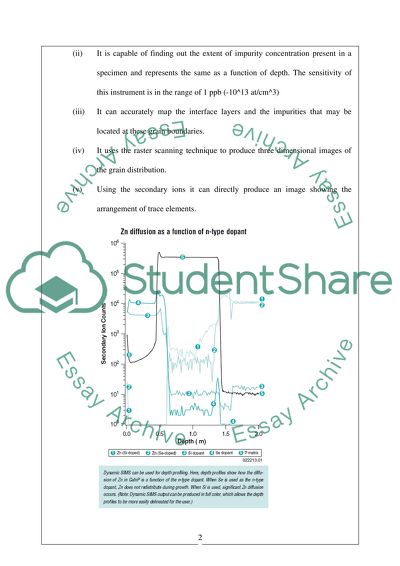
?Surface Analysis 508581) Introduction Surface Analysis is a non destructive method of testing used to analyse the chemical and molecular composition of different surfaces depending upon its use. This method uses the simple principle of pounding a sample specimen with electrons, x rays or photons in a vacuum atmosphere which thereby generates a map of the sample complete with grain boundaries, intermolecular spaces and location of any impurities. (Surface Science and Analysis, n.d) Surface Analysis in Material Science There are a number of sophisticated techniques that provide a comprehensive study of the surface of material specimen. Some of these are 1. Dynamic Secondary Ion Mass Spectrometry (Dynamic SIMS) In this process the surface of the sample is subjected to a steady stream of focussed primary ions. These ions on collision dislodge atoms from the surface creating secondary ions. Using a combination of electrostatic and magnetic fields which distinguishes the ions on the mass to charge ratio, these secondary ions are drawn into a mass spectrometer. By varying the strength of the magnetic field one can assess ions of different mass to charge ratios. (Surface Analysis, 2000) Some of the applications of SIMS include (i) Identifying the isotopes in a material that might have same atomic masses as it is capable of producing mass resolutions to tune of 7000. (ii) It is capable of finding out the extent of impurity concentration present in a specimen and represents the same as a function of depth. The sensitivity of this instrument is in the range of 1 ppb (-10^13 at/cm^3) (iii) It can accurately map the interface layers and the impurities that may be located at these grain boundaries. (iv) It uses the raster scanning technique to produce three dimensional images of the grain distribution. (v) Using the secondary ions it can directly produce an image showing the arrangement of trace elements. Source: Surface Analysis, 2000 2. Auger Electron Spectroscopy (AES) (Surface Analysis, 2000) In this method the specimen is pounded with a stream of electrons which releases on auger electrons from the material on collision. These auger electrons have specific kinetic energies which depend on the electrons emitted. This method therefore creates element maps capable of identifying chemical composition. Some of the applications of AES include (i) This is used to identify compositions of elements in solid materials with high output sensitivities in the range of 0.5 atomic percent for lithium to uranium. (ii) This is useful in carrying out the volumetric analysis of a specimen. (iii) This is capable of producing magnified images to the tune of 20000x and is used extensively in microelectronics. 3. X-Ray Photoelectron Spectroscopy (XPS) (Surface Analysis, 2000) This advanced technique uses x rays in dislodging electrons from specimens and these have distinct kinetic energies depending on the nature of emitted electrons. It is particularly useful in providing information regarding the type of bonds that exist between electrons. This is because any change in binding energy is reflected in the XPS spectrum chart which shows crests and troughs. The crests obviously reflect the high chemical boding energy that existed and the troughs vice versa. Its applications include (i) Determining molecular composition of surfaces along with information regarding the atomic bonding. (ii) It can identify very accurately to the tune of 0.5 atomic % the presence of lithium and uranium. (iii) This can create depth profiles for materials in the range of 1µm thickness. (iv) This method of analysis is particularly useful to study functional groups in polymers. 4. Scanning Probe Microscopy (SPM) This is also referred to as Atomic Force Microscopy (AFM). This is capable of generating a profile that shows the topographical nature of the specimen surface. Schematic of atomic force microscope operation Source: Surface Analysis, 2000 This essentially consists of a tip mounted on a cantilever. The specimen is placed below this cantilever and dragged back and forth. The laser which is focussed on the cantilever bounces off the surface onto the photodiodes. As the cantilever tip moves up and down due the surface irregularities of the specimen, the surface of the sample is plotted showing irregularities. Uniform movement of the sample is achieved by using a piezoelectric crystal which generates a voltage on applying a pressure. This helps in plotting out the surface profile. This differs from Scanning Electron Microscopy in that it does not require a vaccum atmosphere for the sample to be tested. These have a wide variety of applications and are extensively used in the study of super conducting materials and in the analysis of insulating coatings, wear resistant films and cutting edges. 5. Scanning Electron Microsope (SEM) This system compared to the SPM uses a vacuum atmosphere to direct electrons onto the specimen. The vacuum prevents the scattering of electron beams and restricts dispersion. To achieve this two classes of pump are used. A low vacuum pump brings down the air pressure from atmosphere to 10-3 Torr and a high vacuum pump bring it further down from 10-3 Torr to 10-6 Torrthe interaction of the electron beam produced from the electron gun and the specimen generates different type of signals which can be detected. The electrons on specimens are typically accelerated in voltage ranges of 2 to 40kV. This interaction in a zone called the ‘interaction volume’ zone produces Secondary electrons (SE) and back scattered electrons (BSE). When primary beams are directed on specimens at angles less than 90? secondary electrons are produced but manage to escape from the surface layers. Its escape energy is limited to 50eV. Back scattered electrons escaping from the surface have an energy of >50eV. The higher the potential energy and smaller the atomic number of the specimen the BSE production is of a higher quantity. This results in greater resolution. (Scanning Electron Microscopy, n.d) Different detectors that are sensitive to emissions of different energy levels are used to detect emissions from sample. The nature of specimen topography forms the basis of most SEM’s. This information is provided by the secondary electrons. A 200 volt applied to the secondary electron detector attracts these secondary electrons and is further accelerated by a voltage of 10kV on to the scintillator. The secondary electrons hitting the scintillator release photons which travel through the light pipe onto the photomultiplier (PM). This amplifies the original signal. Every striking photon generates number of electrons and hence the amplification produced is high. The X rays released from the specimens are characterised to the nature of the material from which it is released. Background X rays on the other hand lose this characteristic after losing its energy on striking other particle. An EDS X ray detector collects the entire spectrum of x rays from 0eV to 30eV and is generally used when dealing with large concentration of x rays. However when wavelengths of smaller wavelengths need to be detected a WDS detector is used. This offers greater accuracy in dealing with more sensitive detection. The signal processing system processes the signal that has been generated and offers additional electronic modification of the image to enhance its quality. The amplifier in the detector amplifies the signals and is converts it into an image to be viewed on the cathode ray tube (CRT). The brightness control, contrast all form part of this processing system. The display and recoding system allows for the recording of the end results using the photographic medium. The well defined image on the CRT is transferred to a film by making certain adjustments in brightness and contrast. (Dunlap Michael & Adaskaveg, 1997) Surface Analysis in Medical Science-Biocompatibility It has been described in extensive medical researches that most interactions in the biological realm occur at interfaces. This interface as in the medical or biological space covers the entire gamut of the cell surfaces, cellular matrix, hydrated tissues and proteins. This is because the degree of resistance is least across surfaces or interfaces. There are three areas of great interest as far as medical science is concerned. These are 1. The study of chromatographic separations 2. Study of compatibility between different blood groups. 3. Cell nurturing and study of bone marrow specifics A scanning electron micrograph of a myoblast cell interacting with synthetic surface Source: Castner David & Ratner Buddy, 2001 The TOF-SIMS technique is predominantly used in field. The specimen that is to be analysed is bombarded with ions of short pulses. The time of movement is subsequently measured. The secondary ions have the same kinetic energy; the time of movement thus depending on the square root of mass. This information is then calibrated to give information regarding different cells undergoing biological reactions. Static TOF SIMS can generate an entire mass spectrum using 1-2nm of the sample from the outer periphery. This gives precise information regarding the structure of these biological materials. For liquid sources spatial resolutions that are close to 0.1µm are obtained. Analysis of static TOF SIMS involves three major steps. (Castner David & Ratner Buddy, 2001) (i) Firstly the raw information is assessed and boxcar filtering methods are used to de-noise images i.e disturbances are reduced so that the weak signals are identified in the first stage, (ii) Secondly, the chemical constituents in the cell specimen are identified. It also provides information as to which peaks shown in the static TOF SIMS spectrum have been produced from the same chemical part. (iii) The third part involves generating the image after successful component identification. Source: IonTOF, n.d Surface analysis assumes importance in the study of materials that are termed bio-compatible. Bio compatible materials are defined as those which do not contaminate or give out harmful toxins when in permanent contact with the human body. In a normal wound it has been found out that the ‘macrophage’ normally heals the wounded site by cleaning the affected area and giving out cytokine molecules which thereby activate fibroplast, keratinocyte that are required for healing. In the case of a biomaterial this process is hampered. The macrophages do not recognize this foreign body and tries to consume and connect with the entire mass failing in creating a seamless adherence with the parent body. (Castner David & Ratner Buddy, 2001) To study how this biocompatible material reacts with different proteins the TOF SIMS becomes a handy instrument. Proteins are generally adsorbed into the biocompatible material very quickly and the composition of this protein layer depends on the fluid phase composition. Since the TOF SIMS is a highly sensitive device, sampling depths of 1-2nm can be studied to ascertain the degree, nature and composition of proteins that has been adsorbed with the biocompatible structure. The protein surrounding this biocompatible or ‘foreign body’ have a tendency to realign itself with nature of this surface and spread to other areas with new amino acid compositions. Using TOF SIMS helps in assessing this protein spread, calculate protein concentration and source out different variants of proteins. This information proves vital for the development of biomaterials. (Castner David & Ratner Buddy, 2001) Conclusion Apart from the application of surface analysis tools in identifying material structures and also in the study of biocompatible materials in medical science, it has number of varied other applications in Forensics where surface analysis of the human tooth can establish the identity of human faces rendered unrecognizable by fires. (New tooth enamel dating technique could help disaster victims, 2005,) Other applications in the field of archaeology where surface analysis of rocks or early tools found at sites can be used to find the exact dates during which they were supposedly used. Reference Lists 1. Castner David & Ratner Buddy, 2001, Biomedical surface science: Foundation to frontiers, Surface Science 500. 2. Dunlap Michael & Adaskaveg, 1997, Introduction to the Scanning Electron Microscope, Facility for Advanced Instrumentation 3. New tooth enamel dating technique could help disaster victims, 2005, Available at:http://www.physorg.com [Accessed 10th March 2011] 4. Surface Science and Analysis, n.d, XV-Analysis-A-Surfaces 5. Surface Analysis, 2000, Measurements and Characterization, National Renewable Energy Laboratory 6. Scanning Electron Microscopy, n.d, Technical Faculty of Christian Albrechts University Read More
Cite this document
  • APA
  • MLA
  • CHICAGO
(“Surface analysis Assignment Example | Topics and Well Written Essays - 1750 words”, n.d.)
Retrieved from https://studentshare.org/family-consumer-science/1410939-surface-analysis
(Surface Analysis Assignment Example | Topics and Well Written Essays - 1750 Words)
https://studentshare.org/family-consumer-science/1410939-surface-analysis.
“Surface Analysis Assignment Example | Topics and Well Written Essays - 1750 Words”, n.d. https://studentshare.org/family-consumer-science/1410939-surface-analysis.
  • Cited: 0 times

CHECK THESE SAMPLES OF Surface Analysis

Surface Analysis and Materials Science

This essay "Surface Analysis and Materials Science" is about the primary objective of materials science is to develop materials that possess specific mechanical, electrical, magnetic, optical, thermal, and chemical properties.... The combination of probes and corresponding responses provides 36 basic classes of experimental techniques which may be utilized for Surface Analysis.... AnalysisAuger electron spectroscopy or AES is considered a key chemical Surface Analysis tool for conducting material samples....
7 Pages (1750 words) Essay

Bell Helicopter Textron

This paper "Bell Helicopter Textron" presents one of the best helicopter company in the world, the Bell helicopter company which is a wholly-owned subsidiary of Textron Incorporated.... The company has over 50 year's experience of testing and flight innovation (Bell helicopter, 2006).... hellip; The company makes commercial and military helicopters and tiltrotor aircraft....
7 Pages (1750 words) Case Study

Forensic Material Identification Techniques, Spectroscopy

The current instrumental methods in forensic identification and analysis not only relies on technologies but also on sound theories on the application of forensic techniques in the analysis, identification and characterisation of toxic and nontoxic biological and chemical substances (Skoog, 2007)....
8 Pages (2000 words) Coursework

Atomic force microscopy

ifferent imaging techniques are used to picture the surface or for different Surface Analysis, they include the contact mode, non-contact mode and tapping mode.... This tip is attached to the free end of a The material to be scanned is placed below the tip, as the tip moves across the surface it is attracted or repelled by different forces, the atomic deflection from rest is recorded and an imaging software is used to process the results.... The results normally appears as a topographical image of the surface imaged2....
5 Pages (1250 words) Lab Report

Interaction Framework

On the same note, failure to adhere to network and Surface Analysis means the novice cannot distinguish mapping operations and superseding receptions.... Understanding of interaction framework is integral when viewed from the perspective of its Evaluation Action Cycle because of a few reasons worth noting....
2 Pages (500 words) Essay

The Impact of Geospatial Science on the 21st Century Geography

This essay "The Impact of Geospatial Science on the 21st Century Geography" specifically analyzes the role of the geographic information system and the global positioning system in the study of 21st Geography.... nbsp;Geospatial science involves the study and application of information technology....
5 Pages (1250 words) Essay

Failure Criteria of Aluminium

nbsp;In failure assessment, fracture Surface Analysis is hardly used in seclusion.... In this paper, an analysis of the failure criteria for aluminum is given.... In other instances, a wide array of methods and in-depth analysis is needed.... It is pertinent to have a deep understanding of the elements that impact the topography and analysis of pictures of fracture surfaces (Hull, 1999).... It also involves the review of the magnitude of the application of the component in conformity to terms, stress assessment, non-destructive assessment of the botched parts, chemical review, and fractographic analysis....
8 Pages (2000 words) Essay

Electropolymerized Conducting Polymers

The redox reactions only occur at the surface of the electrode with a moderate rate of transfer of electrons, and no absorption or adsorption.... Electrochemical oxidation of polyaniline, polypyrrole, or polythiophene molecules frequently results in the formation of an organic polymer film that is electrically active when used at the surface of an electrode.... The current produced from the reaction is dependent on the rate of diffusion from the bulk solution to the surface of the electrode....
7 Pages (1750 words) Lab Report
sponsored ads
We use cookies to create the best experience for you. Keep on browsing if you are OK with that, or find out how to manage cookies.
Contact Us