StudentShare
Contact Us
Sign In / Sign Up for FREE
Search
Go to advanced search...
Free

Elbow as the Joint of the Upper Limb - Essay Example

Cite this document
Summary
This paper 'Elbow as the Joint of the Upper Limb" portrays the elbow located between the distal end of the humerus and the proximal end of the forearm. It connects the humerus with the two forearm bones, Radius, and Ulna. The connection these bones form is known as the elbow joint…
Download full paper File format: .doc, available for editing
GRAB THE BEST PAPER93.9% of users find it useful
Elbow as the Joint of the Upper Limb
Read Text Preview

Extract of sample "Elbow as the Joint of the Upper Limb"

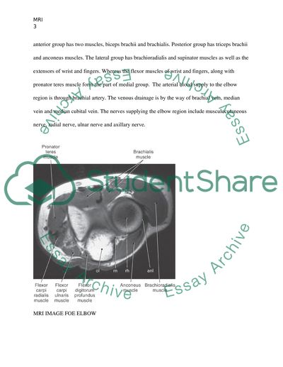
? MRI Institute MRI Answer no Elbow is the joint of the upper limb. It is located between the distal end of humerus and the proximal end of forearm. It connects the humerus with the two forearm bones, Radius and Ulna. The connection these bones form is known as the elbow joint. It is a complex type of joint of hinge- pivot variety. A number of bony surfaces are involved in the formation of the elbow joint. The distal end of humerus has two condyles, one on its medial side and another on the lateral side. These condyles, along with their epicondyles provide the site for the attachment of the ligaments and tendons. Apart from these condyles, which are bony prominences, the distal end of humerus has two depressions as well. The bony depression on the anterior side is known as coronoid fossa, while the posterior depression is known as olecranon fossa. The areas where the bones connect to each other are referred to as articular surfaces. The elbow joint is articulated at Radioulner, Radiohumeral and Ulnohumeral joints. These articular surfaces are covered by the hyaline cartilage. A joint capsule, which is lined by Synovial membrane, encloses the entire joint in such a way that all three joint cavities remain in communication with each other. The elbow joint is stabilized by ligaments. These include Radial and Ulnar Collateral ligaments and the annular ligament of the head of radius. The medial side of the joint is strengthened by the Ulnar collateral ligament whereas the radial collateral ligament stabilizes the lateral side. The head of radius is connected to the radial notch of ulna by the annular ligament which runs around the head of radius. The movements at the elbow joint are facilitated by the muscles attached in this region. These muscles are organized into four different compartments according to their position, namely anterior, posterior, lateral and medial. The anterior group has two muscles, biceps brachii and brachialis. Posterior group has triceps brachii and anconeus muscles. The lateral group has brachioradialis and supinator muscles as well as the extensors of wrist and fingers. Whereas the flexor muscles of wrist and fingers, along with pronator teres muscle form the part of medial group. The arterial blood supply to the elbow region is through brachial artery. The venous drainage is by the way of brachial vein, median vein and median cubital vein. The nerves supplying the elbow region include musculocutaneous nerve, radial nerve, ulnar nerve and axillary nerve. MRI IMAGE FOE ELBOW The daily activities performed by the hands, grasping and the coordinated movements at the forearm and wrist are possible because of the complex synovial joint at the elbow which is referred to as the elbow joint (Lee et al 2003). It connects the distal end of the upper arm bone humerus to the proximal ends of the two forearm bones radius and ulna. The bones are articulated at trochlea and capitulum of humerus above and trochlear notch of ulna and the head of the radius below (Snell 2004). The three bones are articulated at four joints, together known as cubital articulations, i.e, humeroulner, humeroradial, superior radioulner and inferior radioulner (Stroyan et al 1993). The articular surfaces are covered by hyaline cartilage and the joint is enclosed by the capsule, lined by synovial membrane. The capsule serves to stabilize the joint along with the two ligaments, i.e. lateral collateral ligament and the ulnar collateral ligament. Anteriorly, the capsule attaches to the radial and coronoid fossae and the medial and lateral epicondyles of humerus above and to the coronoid process of ulna and annular ligament of radius head below. The posterior attachments of capsule are to the olecranon processes of humerus above and ulna below, and the annular ligament of radius head below (Snell 2004). The fan shaped lateral collateral ligament attaches lateral epicondyle of humerus to the annular ligament of radius. The triangular ulnar collateral ligament forms three strong bands: anterior band between the medial epicondyle and coronoid process, posterior band running from medial epicondyle to the olecranon process and the third transverse band traversing the path between ulnar attachments of the anterior and posterior bands. These ligaments stabilize the joint and allow flexion and extension movements and axial rotation. Flexion is brought about by Brachialis, Biceps Brachii, Brachioradialis and Pronator teres muscles. Anconeus and triceps are responsible for extension. Carrying angle, which is 170 degrees in male and 167 degrees in female, is the normal angle between the long axis of extended arm and forearm (Chaurasia 2004). Answer no.2. 1- First Position: The common position for obtaining the MRI of the elbow is with the patient lying supine position. The elbow is placed by the side of the patient. Arm is kept in prone position with the palm resting on the thigh. (Reiser et al 2000). If the range of movement at elbow and shoulder joint is not compromised, the MRI can be obtained in the body coil position with the patients arm elevated above the head level. 2- Second Position: Another position to obtain the MRI elbow is commonly referred to as FABS position. FABS stands for flexed abducted and supinated position. In this position, the arm is flexed at the elbow and abducted at shoulder with the forearm in supination (Steinbach et al 2010). Comparison: Patients are generally more comfortable in supine position. It reduces the chances of motion artifacts and is preferred for examinations extending over longer time duration. Patient lying oblique in body coil position brings him close to the isocenter of the magnetic field and thus the fat suppression is more homogenous in the MRI film. The FABS position allows a better view of the ligament complexes due to the slight flexion. Answer no.3. The common artifacts encountered during the MRI elbow are motion artifacts and the metal artifacts. The most suitable patient position to reduce motion artifacts is patient lying supine with arms in pronation alongside body. Overhead position increases the likelihood of motion artifacts so the limb should be immobilized (Reiser et al 2000). Metal artifacts can be because of the metal type and surface complexity, or due to the screws, bullet fragments and prosthesis. The consequences range from moderate to severe. Titanium does not cause much artifact (Steinbach et al 2010). Furthermore artifacts can be avoided by reducing fat suppression, positioning the patient along the axis of metallic equipment, tilting the view angle, avoiding gradient echo sequences and upgrading the read-out bandwidth (Vandevenne et al 2007). Answer no.4 (a) A close clinical evaluation and a detailed history is necessary in case of calcifying tendonitis, as it mostly affects the athletes and volleyball or baseball players which require repeated abduction and external rotation of the shoulder (Yochum et al 1996).  People from occupations like typists and assembly workers are also more prone to developing this condition due to prolonged slight abduction and the resultant hypovascularity in the tendon Supraspinatus muscle (Kulkarni 2008). The condition can present in its acute resorptive phase with acute painful symptoms or as a chronic (sub-acute) shoulder pain (Iannoti et al 2007). The condition is asymptomatic and painless in initial stages. Pain develops in a chronic manner as the condition slowly progresses through formative phase and may present with constant severe clicking pain, restricted movement, low grade fever and malaise (Micheo 2011). Acute resorptive phase presents with shoulder pain radiating towards the insertion of deltoid muscle in humerus. The severe pain is because of the influx of inflammatory cells, edema and increased intratendinous pressure because of the proliferation of the vessels (Ianotti et al 2007). There is tenderness at greater tuberosity of humerus and there may be subacromial impingement. The disease may take chronic course, which is characterized by diffuse pain even at rest, intermittent worsening of symptoms and difficulty with overhead movement (Wilk et al 2009).  Answer no.4 (b) Calcifying tendonitis is a slowly evolving chronic disease associated with deposition of calcium in the tendons. It mostly affects the tendons of rotator cuff muscles, as shoulder joint is the most used joint in the body. There are several factors involved in the pathogenesis of the condition which include increased intraosseous pressure, prolonged usage leading to increased Oxygen demand and wear and tear at the glomerulohumeral joint.  The course of calcium deposition in the tendons evolves through four stages of disease progression, namely precalcific, formative, resorptive and the postcalcific stage. The precalcific stage sets in with the growth of fibrocartilaginous material in the tendon. With continued growth, these deposits form crystals of hydroxyapatite, which is the hallmark of formative phase. These foci of crystals coalesce to form areas of chalky white calcification. During this stage, there is pain on movement. The further course of the disease is variable in terms of time. After a certain time period, resorptive phase sets in spontaneously. It is characterized by resorption of hydroxyapatite crystals, which is associated with severe excruciating pain. As a result, the range of movement of the joint is severely compromised. The release of thick paste like deposit induces local irritation and inflammation (Kulkarni 2008). Answer no.4 (c) Calcifying tendonitis can be accurately diagnosed by taking into consideration the clinical presentation along with the radiological appearance. Plain X rays as well as the MRI are helpful. Plain X rays provide baseline information about the general variations in normal anatomy for management of the condition. Bony structures are visualized in an excellent way. The draw back with plain radiographs is that the soft tissues cannot be visualized. This is where the magnetic resonance imaging has revolutionized the radiologic diagnosis of the calcifying tendonitis. Plain radiographs of true anteroposterior view help visualize the glomerulohumeral joint and shoulder in external and internal rotation to see the tendon insertions and tuberosities. Calcifications are most frequently found in the tendon of supraspinatus muscle. They can be seen in the anteroposterior view of X- ray with the humerus in neutral position which brings the greater tuberosity in profile. MRI is helpful as it allows the visualization of soft tissue pathology as well. The calcifications are seen on the MRI as intermediate or decreased signal intensity on T1 weighted images. Whereas on T2 weighted images, there is increased uptake around the calcifications due to the edema during the resorptive phase (Ianotti et al 2007). Depending upon the chronic level of the condition, the T2 weighted images may show intermediate or even show decrease in signal intensity (Yochum et al 1996). Answer no.5 (a) The wrist region is formed by eight small carpal bones. These bones are arranged in a proximal and a distal row. The proximal row bones include scaphoid, lunate, triquetrum and pisiform. While the distal row is formed by the bones trapezium, trapezoid, capitate and hamate. These bones connect the forearm to the bones of hand and are responsible for the versatile range of motion at the wrist joint. Cystic lesions containing fluid can develop around the tendons of the bones. These lesions are known as ganglions or ganglion cysts. Intraosseous ganglions can arise near the ends of long bones by extension from the soft tissue ganglia or synovial rests (Meyers 2008). They are often asymptomatic, but chronic ones are associated with inflammation and intermittent pain. They can be single or multiple cystic lesions, covered by a layer of connective tissue. Unlike the synovial cysts, ganglion cysts are not lined by synovium. They usually range in size from 1 to 4 cm and are filled with viscous gelatinous material composed of polysaccharides. They can be identified radiologically as lytic lesions with well defined margins below the subchondral plate. On the MRI, intraosseous ganglia appear in shape from round to oval serpiginous configurations. Ganglia give low signals on T1W1. Signal intensity on PDW1 appears as low intermediate while on T2W1, it is high (Zhang et al 2010).  Answer no. 5(b) Carpal tunnel syndrome is the most frequent surgically treated condition in hand (Luchetti et al 2007). The Median nerve is compressed at the carpal tunnel. The compression can be caused by any intrinsic space occupying lesion in the region, like tumors, ganglia, cysts, muscular hypertrophy, edema or deposition of fat or amyloid. Extrinsic compression can be because of a dislocated fractured fragment of a bone or callus (Vahlensieck et al 2000). The diagnosis rests on clinical symptoms, nerve conduction studies and the radiologic imaging. As the nerve is affected, the muscles innervated by it undergo atrophy.MRI is of particular importance as it identifies the soft tissue as well as bony pathology. It helps identify muscle atrophy pattern consistent with denervation of a particular nerve. T1 and T2 weighted images are obtained on axial scale. It helps identify any obvious pathologic masses in the region responsible for compression as well as variations in size and shape of the nerve. The compression of the Median nerve results in ischemia, which is evident as increased signal intensity on T2 weighted images. The acute and chronic compression can be differentiated on the MRI, as increased signal intensity on T1 images in chronic compression whereas acute compression gives increased signal intensity on T2 due to edema and swelling (Khanna 2010) References CHUNG, C. B., & STEINBACH, L. S. (2010). MRI of the upper extremity shoulder, elbow, wrist and hand. Philadelphia, Wolters Kluwer Health/Lippincott Williams & Wilkins. FORNALSKI, S., GUPTA, R., & LEE, T. Q. (2003). Anatomy and Biomechanics of the Elbow Joint. SPORTS MEDICINE AND ARTHROSCOPY REVIEW. 11, 1-9. IANNOTTI, J. P., & WILLIAMS, G. R. (2007). Disorders of the shoulder: diagnosis & management. Philadelphia, Lippincott Williams & Wilkins. KHANNA, A. J. (2010). MRI for orthopaedic surgeons. New York, Thieme. KULKARNI, G. S. (2008). Textbook of orthopedics and trauma. New Delhi, Jaypee Brothers. LUCHETTI, R., & AMADIO, P. C. (2007). Carpal tunnel syndrome. Berlin, Springer.  MCCARTHY, E. F., ZHANG, P. J., & KHURANA, J. S. (2010). Essentials in bone and soft tissue pathology. New York, Springer. http://dx.doi.org/10.1007/978-0-387-89845-2. MEYERS, S. P. (2008). MRI of bone and soft tissue tumors and tumorlike lesions: differential diagnosis and atlas. Stuttgart, Thieme.     MICHEO, W. (2011). Musculoskeletal, sports, and occupational medicine. New York, Demos Medical.  STROYAN M, & WILK KE. (1993). The functional anatomy of the elbow complex. The Journal of Orthopaedic and Sports Physical Therapy. 17, 279-88.  VAHLENSIECK, M., GENANT, H. K., & REISER, M. (2000).MRI of the musculoskeletal system. Stuttgart, Thieme  VANDEVENNE JE, VANHOENACKER FM, PARIZEL PM, BUTTS PAULY K, & LANG RK. (2007). Reduction of metal artefacts in musculoskeletal MR imaging. JBR-BTR : Organe De La Socie?te? Royale Belge De Radiologie (SRBR) = Orgaan Van De Koninklijke Belgische Vereniging Voor Radiologie (KBVR). 90.)  WILK, K. E., REINOLD, M. M., & ANDREWS, J. R. (2009). The athlete's shoulder. Philadelphia, PA, Churchill Livingstone/Elsevier. YOCHUM, T. R., & ROWE, L. J. (1996). Essentials of skeletal radiology. Baltimore, Williams & Wilkins. Read More


Read More
Cite this document
  • APA
  • MLA
  • CHICAGO
(“MRI Essay Example | Topics and Well Written Essays - 1500 words”, n.d.)
Retrieved from https://studentshare.org/environmental-studies/1413012-mri
(MRI Essay Example | Topics and Well Written Essays - 1500 Words)
https://studentshare.org/environmental-studies/1413012-mri.
“MRI Essay Example | Topics and Well Written Essays - 1500 Words”, n.d. https://studentshare.org/environmental-studies/1413012-mri.
  • Cited: 0 times

CHECK THESE SAMPLES OF Elbow as the Joint of the Upper Limb

What is Work Related Upper Limb Disorder

There has been a lot of interest in the group of disorders termed 'work-related upper limb disorders' or WRULD because it is thought to be increasingly prevalent in the workplace.... Palmer and Cooper (2005) also report that the Health and Safety Executive estimated that in 2004-/5 Work-Related Musculoskeletal Disorders that included 450,000 people with back pain and 375,000 with neck and upper limb disorders cost British society about 5....
22 Pages (5500 words) Research Paper

Amputation: Causes and Results

As you see most amputations are stipulated by diseases (like diabetes etc) (Jeffcoate et al, 2003) "In general, amputations through the lower limb account for 85% of all amputations at the present time.... (Girdhar et al, 2001) The loss of the limb leads to a permanent disability that can impact a patient's self-image, self-care, and mobility (movement) negatively.... Amputation rehabilitation programs usually include pain management for both post-operative and phantom pain (a sensation of pain that occurs below the level of the amputation)Nutritional counselingPatient and family education (Pandian, Kowalske, 1999; Gibson, Certcouns, 2001)Biomechanics of transfemoral amputation:1,6For rehabilitation to pass more successfully, it is desirable to have a more distal level of limb amputation....
13 Pages (3250 words) Essay

I will attach the document

lexion denotes to the pose made possible by the joint, which decrease twist in anatomical terms.... ronation denotes the turning motion of the forearm via the radioulnar joint, or on the bottom of the leg at the subtalar and talocalcaneonavicular lower limb joints in the field of anatomy.... During the normal standing posture, pronation-turning motion shifts the palm of the right upper appendage anti-clockwise and clockwise for the left forearm from a superiorly vision....
4 Pages (1000 words) Essay

Whether Incorrect Manual Handling Procedures Cause Work Related Upper Limb Disorders in Radiographers

This paper "Whether Incorrect Manual Handling Procedures Cause Work Related upper limb Disorders in Radiographers" focuses on the fact that there has been a lot of interest in the group of disorders termed 'work-related upper limb disorders' or WRULD because it is prevalent in the workplace.... Palmer and Cooper (2005) also report that the Health and Safety Executive estimated that in 2004-/5 Work-Related Musculoskeletal Disorders that included 450,000 people with back pain and 375,000 with neck and upper limb disorders cost British society about 5....
23 Pages (5750 words) Case Study

Nerve Lesions of Lower Limb (Muscle Testing of Lower Limb)

For a proper understanding of the nerves, their functions and the dangers that they may suffer in the event of a lesion, the lower limb is divided into the nerves of the thighs and nerves of the legs.... The conclusion from this study states that maintaining a healthy neural system and muscles of the hind limb is the secret to walking strong for long.... Neurological supply of the lower limb is composed of spinal nerves that emanate from the vertebral column....
19 Pages (4750 words) Book Report/Review

Innovations in Prosthetic Athletic Limbs

upper limb amputations are of less common but where it is found to affect all ages.... People with upper limb mutations have better choices rather than lower limbed ones.... A person with limb amputated experiences a staggering emotional and financial lifestyle changes.... limb prosthesis includes both upper and lower extremities.... 'Throughout the world, the major objective of prosthetics is to restore the functional capacity as well as the best cosmetic result afforded to, held by a limb deficient person' (Strait, 2006)....
8 Pages (2000 words) Research Paper

Elbow, Forearm and Wrist

The occurrence of medial epicondylitis in golf players is enhanced by poor golf swings and overuse of upper limb muscles.... In golf, the muscles used range from those in the upper limbs, the trunk, and the lower limbs.... The author of the paper "elbow, Forearm, and Wrist" will begin with the statement that golf is a recreational activity undertaken in various parts of the world.... Medial epicondylitis is characterized by the inflammation and tenderness that originates from the inner elbow....
6 Pages (1500 words) Essay

Selecting Materials for Prosthesis

A prosthesis is an artificially manufactured limb for substituting a natural limb that has been lost through a congenital defect, illness, accident, or wartime injury.... These are:- Below the knee (BK), transtibial – A prosthetic lower leg that is usually attached to an intact upper part of the leg.... Above the knee (AK), transfemoral – A prosthetic upper and lower leg, includes a prosthetic knee.... Above the elbow (AE), transhumeral – A prosthetic upper and lower arm, includes a prosthetic elbow....
11 Pages (2750 words) Report
sponsored ads
We use cookies to create the best experience for you. Keep on browsing if you are OK with that, or find out how to manage cookies.
Contact Us