StudentShare
Contact Us
Sign In / Sign Up for FREE
Search
Go to advanced search...
Free

Pathogens and Disease - Coursework Example

Cite this document
Summary
This work called "Pathogens and Disease" focuses on a biological agent, which causes an illness or disease in its host. From this work, it is clear about the examples of pathogens include bacteria, viruses, fungus, and protozoa. The author outlines the transmission and development of infection in V. Cholerae…
Download full paper File format: .doc, available for editing
GRAB THE BEST PAPER91.8% of users find it useful
Pathogens and Disease
Read Text Preview

Extract of sample "Pathogens and Disease"

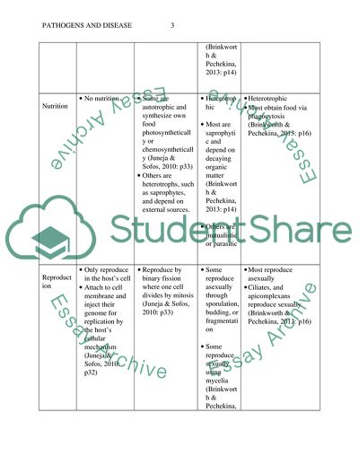
Pathogens and Disease TAQ 326 words) a) A pathogen is a biological agent, which causes an illness or disease in its host (Austin, 2011: p41). Examples of pathogens include bacteria, virus, fungus, and protozoa. b) Features Virus Bacteria Fungus protozoa Structure Have a single strand of RNA or DNA surrounded by the capsid or protein coating (Juneja & Sofos, 2010: p32) Their nucleoproteins and genome together form the nucleocapsid (Crumpton, 2012: p44) Some have lipid envelope Have cell walls Do not have a nucleus Have chromosomal and plasmid DNA (Juneja & Sofos, 2010: p33) Some have a flagellum for locomotion Pathogenic bacteria have fimbriae, pili, and glycocalyx for attachment to surfaces, which is critical to their survival (Crumpton, 2012: p57) Have hyphae (Brinkworth & Pechekina, 2013: p14) Cell walls made of chitin Multi-nucleated Main body of the fungus is made up of a vast number of hyphae threads that combine to form a web of mycelium (Markovics, 2014: p32) Unicellular with one or more nuclei Have a pellicle, which is a tough outer membrane (Brinkworth & Pechekina, 2013: p16) Some have more complex organelles for excretion, digestion, and respiration Locomotion is enabled by cilia, flagella, and amoeboid movement, depending on the type of protozoan (Markovics, 2014: p48) Size 20-100x10-9M (Juneja & Sofos, 2010: p32) 0.1-1.0 x 10⁻⁶M (Juneja & Sofos, 2010: p33) Microscopic or macroscopic (Brinkworth & Pechekina, 2013: p14) 1-100x10-6M (Brinkworth & Pechekina, 2013: p16) Nutrition No nutrition Some are autotrophic and synthesize own food photosynthetically or chemosynthetically (Juneja & Sofos, 2010: p33) Others are heterotrophs, such as saprophytes, and depend on external sources. Heterotrophic Most are saprophytic and depend on decaying organic matter (Brinkworth & Pechekina, 2013: p14) Others are mutualistic or parasitic Heterotrophic Most obtain food via phagocytosis (Brinkworth & Pechekina, 2013: p16) Reproduction Only reproduce in the host’s cell Attach to cell membrane and inject their genome for replication by the host’s cellular mechanism (Juneja & Sofos, 2010: p32) Reproduce by binary fission where one cell divides by mitosis (Juneja & Sofos, 2010: p33) Some reproduce asexually through sporulation, budding, or fragmentation Some reproduce sexually using mycelia (Brinkworth & Pechekina, 2013: p14) Some reproduce both sexually and asexually Most reproduce asexually Ciliates, and apicomplexans reproduce sexually (Brinkworth & Pechekina, 2013: p16) References Austin, B. (2011). Pathogens in the environment. Oxford: Blackwell. Brinkworth, J. F. & Pechenkina, E. A. (2013). Primates, pathogens, and evolution. New York: Springer. Crumpton, M. (2012). Bacteria and viruses. Logan: Perfection Learning. Juneja, V. K. & Sofos, J. N. (2010). Pathogens and toxins in foods: Challenges and interventions. Washington, DC: ASM Press. Markovics, J. L. (2014). Tiny invaders! Deadly microorganisms. Mankato, MN: Capstone Press. TAQ 2 (800 words) Transmission and Development of Infection in V. Cholerae Transmission Sources of transmission for Vibrio Cholerae include eating raw fruits and vegetables contaminated by irrigation water, fertilizers, and manure, drinking contaminated well and surface water, eating contaminated sea-food, and eating grains contaminated by the bacterium after cooking and keeping at room temperature, which provides a perfect medium for V. Cholerae growth (Michael & Spear, 2010: p52). Transmission of the bacteria occurs between humans via the faecal-oral route. Development of Infection The bacteria has two distinct life cycles as in Figure 1, one of which is the environment where they attach onto the surface of copepod crustaceans, acquiring strength in numbers by using the formation of bio-films on the copepod’s surface as a protective barrack to survive unfavourable conditions. In addition, their aggregation on the copepod results in an effective vehicle for the bacteria’s transmission to humans (Ochsendorf, 2012: p30). The other stage of its lifecycle is in humans, where they have an incubation period of ~1–5 days. Once in the intestines, the bacteria reach the walls of the intestines and produce membrane pili for attachment to the wall, while simultaneously producing potent enterotoxin that binds onto the outer membrane of mucosa cells lining the intestines. This toxin leads to overproduction of cAMP, which activates continuous pumping of Cl- into the small intestines, altering the ionic gradient and causing Na+ and water to be expelled into the small intestines (Michael & Spear, 2010: p51). It is this process that causes production of painless, odourless, watery, and copious amounts of watery diarrhoea. Life-Cycle of V. Cholerae Figure 1: Lifecycle of Cholera Bacterium, (Sack et al, 2004) Transmission and Development of Infection in Trichophyton Transmission The mode of transmission for the Trichophyton fungus is via direct and indirect contact with lesions of the skin from infected fomites such as shower stalls and floors, animals, and people contaminated with desquamated epithelium. Additionally, transmission may also occur through broken skin if the host has suppressed cell-mediated immunity. Walking on one’s bare feet increases the chance that one will be infected by the disease (Khan, 2011: p22). Once they are transmitted to the host, the fungi have an incubation period of between a few days to a few weeks, which is dependent on the host’s susceptibility. Development of Infection Outside the host, it can survive in various media and surfaces and can survive for up to two years at room temperature on skin scales (Campbell et al., 2013: p44). The fungi attach to the skin of the host, colonizing the keratinized surface and using keratinase and elastase to invade the host’s epidermis. However, the fungi remain in the skin’s stratum corneum, which is not vascularised, evading the host’s immune. In the absence of nutrients necessary for Trichophyton metabolism, it enters a log phase, during which the fungi degrade keratin to form required proteins for reproduction and growth. After gaining sufficient nutrition, they enter the stationary phase and degradation of keratin slows down with production of spores as in figure 2. The metabolic products of this process are what result in an inflammatory eczematous and allergic response characteristic of athlete’s foot in the host (Khan, 2011: p22). Life-Cycle of Trichophyton Fungi Figure 2: Lifecycle of Trichophyton Fungi, (Bear & Rintoul, 2014) Transmission and Development of Infection in Malaria Transmission Malaria is a disease that is caused by protozoa belonging to the Plasmodium genus, which is typically transmitted to the host through the Anopheles mosquito, while contact with contaminated blood may also transmit the pathogen, albeit rarely (Khan, 2011: p41). Because the pathogen is found in the erythrocytes, it is also possible for them to be transmitted through organ transplant, blood transfusion, or shared syringes and needles that are contaminated with infected blood. Development of Infection Normally, however, infection begins when the host is bitten by an infected mosquito, injecting malaria sporozoites into its blood. These sporozoites are carried to the liver by blood as in figure 3, where they grow and divide into merozoites, which eventually leave the liver and infect erythrocytes in the host’s blood and reproduce asexually to produce thousands of infected cells that lead to malaria complications and illness. Infected cells undergo apoptosis, releasing more pathogens into the blood to infect other erythrocytes. A mosquito that draws blood from the host ingests the infected erythrocytes that travel to the mosquito’s stomach and invade its salivary glands. The lifecycle of the sporozoites is completed and repeated when the mosquito bites another host and infects it with the pathogen through its saliva (Khan, 2011: p42). Life-Cycle of Malaria Figure 3: Lifecycle of Malaria Protozoan, (nih.gov, 2012) Transmission and Development of Infection in Influenza Transmission Influenza is caused by the influenza virus that is mainly spread from one host to another through droplets expelled by an infected host through sneezing or coughing, which are then deposited on the nose or mouth of nearby hosts or on surfaces that other host then touch, becoming infected (Ackerman et al., 2013: p44). The virus then enters into the lungs and alveolar sacs, where it makes its way into the host’s cells. Development of Infection On binding to the surface of endothelial cells in the respiratory tract, the virus undergoes endocytosis and is delivered to cellular endosomes, fusing with the endosomal membrane and releasing viral ribonucleoprotein complexes into the cytoplasm. These complexes travel to the nucleus to begin synthesis of viral mRNA that is exported to the cytoplasm for translation of viral proteins, which are then transported to the nucleus for synthesis of viral ribonucleoprotein complexes. At the same time, membrane viral proteins are synthesized by the RER and processed in the Golgi complex followed by transport to the plasma membrane where they meet viral ribonucleoprotein complexes from the nucleus, forming multiple progeny virions (Ackerman et al., 2013: p44). These virions are released from the cells by budding as in figure 4, after which the epithelial cells die. Lifecycle of Influenza Virus Figure 4: Lifecycle of Influenza Virus, (Izstein, 2007) References Ackermann, H.-W., Berthiaume, L. & Tremblay, M. (2013). Viral pathogenesis in diagrams. Boca Raton, Fla: CRC Press. Bear, R., & Rintoul, D. (2014). Kingdom Fungi . Retrieved October 13, 2014, from OpenStax College : http://cnx.org/contents/b95751e9-54aa-43b7-9bb0-406671635905@5/Kingdom_Fungi Campbell, C., Johnson, E. & Warnock, D. W. (2013). Identification of pathogenic fungi. Chicester: Wiley. Itzstein, M. (2007, December ). FIGURE 2 | Life cycle of the influenza virus and targets for therapeutic intervention. Retrieved october 13, 2014, from Nature Reviews: http://www.nature.com/nrd/journal/v6/n12/fig_tab/nrd2400_F2.html Khan, N. A. (2011). Microbial pathogens and human diseases. Enfield: Science Publishers. Michael, E. & Spear, R. C. (2010). Modelling parasite transmission and control. New York, N.Y: Springer Science+Business Media. nih.gov. (2012, April 03). Malaria . Retrieved October 13, 2014, from National Institute of Allergy and Infectious Diseases: http://www.niaid.nih.gov/topics/malaria/pages/lifecycle.aspx (nih.gov, 2012) Ochsendorf, F. (2012). Cholera and the ecology of vibrio cholerae. New York: Springer. Sack, D. A., Sack, B., & Nair, B. (2004, January 17). Cholera. Retrieved October 13, 2014, from The Lancet: http://education.thelancet.com/journals/lancet/article/PIIS0140673603153287/images?imageId=gr4§ionType=green&hasDownloadImagesLink=true TAQ 3 (See Attached PowerPoint Presentation) TAQ 4 (900 words) The body has to defend itself from these pathogens to remain healthy, which it does through specific and non-specific defence systems. The first part of the human defence system acts as a barrier to stop entry of all pathogens, thus is considered the non-specific defence system (Thames, 2013: p62). One component of the non-specific system involves physical barriers. The skin is part of the system and prevents entry of pathogens by creating a physical barrier, while sweat and sebaceous glands produce bacteria-killing chemicals. In addition, lysozymes in the tear glands and saliva are also used to kill bacteria pathogens. If the skin breaks, blood clots form to prevent pathogen entry. Mucous membranes secrete mucus that traps pathogens, preventing entry, while nasal hairs trap air-borne pathogens entering the nose. HCL in the stomach also kills pathogens, while lactic acid produced in the vagina prevents pathogen growth (Thames, 2013: p62). Where the pathogens are able to evade the physical barriers of the non-specific defence system, a second line of defence takes over defence responsibility. One component of this second line of defence is phagocytes, which are leukocytes that engulf, ingest and digest pathogens (Thames, 2013: p63). Some forms of phagocytes are especially effective against parasitic worms in the larvae and egg stages. Macrophages, which are larger than phagocytes and longer living move around the body scavenging for pathogens, while others are fixed in location, for example in the lymph system and lymph nodes. Natural killer cells are also present in the lymph and blood and act to kill all types of cells infected by viruses. Complement defence proteins are also produced when pathogens and their products are present in the body, triggering, as well as taking part, in complement reactions (Thames, 2013: p63). This involves enzymatic reactions that lead to engulfing or lysis of the pathogen. The non-specific defence system also consists of interferons, which refer to defence proteins that are produced by cells in the body after they become infected by all types of viruses (Thames, 2013: p65). Following their production, interferons then travel cells in their proximity to prevent the virus from spreading. Finally, inflammation is also part of the non-specific system. When the pathogens manage to infect the host’s cells, these cells produce chemicals referred to as histamines that cause blood vessels to dilate and increase in porosity. This results in the area becoming red, swelling, warming up, and becoming painful. As a result, leukocytes are recruited to the infected area in order to counter the infection. As the inflammation lasts for longer, the host develops a fever, which is another way for the body to combat pathogens since increased temperature tends to inhibit bacteria and viruses from reproducing (Thames, 2013: p66). When these non-specific defence systems fail, the specific system becomes active and is targeted at specific pathogen invaders either through engulfing of specific pathogens by leukocytes or the production of antibodies (Derkins, 2011: p22). Monocytes and lymphocytes are produced in the bone marrow and transported to the thymus gland, spleen, lymph nodes, and lymph vessels by the blood. Monocytes are large leukocytes that engulf pathogens and present part of the pathogen on their cell surface as an antigen. Lymphocytes, on the other hand, attack the cells and monocytes that present antigen on their surface. In addition, other lymphocytes also produce antibodies that are targeted at specific antigens. These antibodies, which are as a result of the antigenic material from the pathogens, are proteins from the immunoglobulin group. In this case, each antigen only stimulates production of a particular antibody that fits onto its receptor surface (Derkins, 2011: p22). The most abundant lymphocytes are T-cells and B-cells, which are both produced in the bone marrow, although T-cells leave to mature in the thymus. The T-lymphocytes take part in cell-mediated immunity, compared to B-lymphocytes that produce antibodies. Types of T-lymphocytes include helper T-cells that release cytokines to stimulate B-cells to produce antibodies, while also stimulating and activating macrophages, suppressor T-cells, and cytotoxic T-cells (Sompayrac, 2012: p33). Cytotoxic T-cells release chemicals for lysis of invading pathogens, while T-cells suppress immune response to avoid destruction of normal cells. Finally, memory T-cells remain to aid the defence system respond faster to future invasion by the specific pathogen. B-cells, on the other hand, are activated by helper T-cells when exposed to pathogens, resulting in the production of antibodies. After the pathogen is neutralised, memory B-cells remain, allowing the defence system to produce antibodies faster in subsequent pathogen encounters (Sompayrac, 2012: p35). Some antibodies cause the identified pathogens to congregate and clump together for phagocytes to engulf and ingest them, while others bind onto the antigen on the pathogen’s surface and prevent the pathogen from accessing cells of the host (Sompayrac, 2012: p35). In addition, other antibodies act by activating the complement system that results in the eventual lysis of the pathogen. Protection by antibodies remains active in the host’s body and, if the same pathogen is encountered again, lymphocytes present from the initial invasion quickly release antibodies to prevent infection by the pathogen. The specific defence system can also be induced artificially, resulting in artificial active immunity. This type of defence requires inoculation of the potential host using a part of the pathogen that does not cause disease, while dead pathogens may also be used, which carry the antigen that triggers antibody production without harming the host (Sompayrac, 2012: p36). References Derkins, S. (2011). The immune system. New York: Rosen Pub. Group. Sompayrac, L. (2012). How the immune system works. Chichester, West Sussex: Wiley-Blackwell. Thames, S. (2013). Our immune system. Vero Beach, FL: Rourke Pub. TAQ 5 (200 words) a) Immunity Natural Acquired Similarities Both involve the action of antibodies Both are the result of previous contact with antigens, whether, partial, dead or living (Janeway, 2014: p38) Differences Does not change with repeated infection, i.e. has no memory (Janeway, 2014: p38) Improves with continuous and repeated infection, i.e. has memory (Janeway, 2014: p38) Mediated by Natural Killer cells and macrophages Mediated by T and B cells Acts within minutes of exposure to pathogen Requires several days after pathogen exposure to become effective Complement system activated through lectin and alternative pathways Activated via the classical pathway Encounter with an antigen does not require the intervention of medical practice or therapy (Parham, 2013: p29) Encounter with the antigen requires intervention of medical practice or therapy (Parham, 2013: p29) b) Immunity Active Passive Similarities Both can result in specific immunity (Abbas et al., 2012: p52) Both modes of immunity can result from artificial or natural processes Differences Provides relief after a long period Provides immediate relief Does not produce side effects Could cause a reaction Lasts for a long time Does not last for a long time Developed as a result of host cells producing antibodies as a response to vaccines or infection (Abbas et al., 2012: p52) Developed as a result of injecting antibodies from other organisms to counteract antigen (Abbas et al., 2012: p52) Involves the introduction of antigens into the body via vaccines (Alt, 2012: p43) Involves the introduction of pre-formed antibodies via injection (Alt, 2012: p43) References Abbas, A. K., Lichtman, A. H. & Pillai, S. (2012). Basic immunology: Functions and disorders of the immune system. Alt, F. W. (2012). Advances in Immunology. Burlington: Elsevier Science. Janeway, C. (2014). Immunobiology: The immune system in health and disease. New York: Garland Science. Parham, P. & Janeway, C. (2013). The immune system. London: Garland Science. Read More
Cite this document
  • APA
  • MLA
  • CHICAGO
(Pathogens and Disease Coursework Example | Topics and Well Written Essays - 2500 words, n.d.)
Pathogens and Disease Coursework Example | Topics and Well Written Essays - 2500 words. https://studentshare.org/biology/1840990-pathogens-and-disease
(Pathogens and Disease Coursework Example | Topics and Well Written Essays - 2500 Words)
Pathogens and Disease Coursework Example | Topics and Well Written Essays - 2500 Words. https://studentshare.org/biology/1840990-pathogens-and-disease.
“Pathogens and Disease Coursework Example | Topics and Well Written Essays - 2500 Words”. https://studentshare.org/biology/1840990-pathogens-and-disease.
  • Cited: 0 times

CHECK THESE SAMPLES OF Pathogens and Disease

The Role of Hydrochloric Acid in Protecting the Body from Disease

It not only plays a crucial role in digestion but also helps in the protection of the body against some organism that may cause disease.... HCL plays a vital role in the protection of the body from pathogens.... It does so by maintaining a sterile environment in the stomach, this is through protection against pathogens that are ingested during food intake.... pathogens cannot achieve overgrowth in the small intestines due to the action of this acid hence the gastrointestinal tract remains free of pathogens....
4 Pages (1000 words) Essay

Host resistance and immunity

Microbes invade host organisms and lead to disease infections.... Microbes invade host organisms and lead to disease infections.... here are however three characteristics that ought to worry any individual exposed to a disease.... An environment that favours the growth of the agent in the host body and its ability to damage the host determines the resultant outcome of the association between disease causing agent and the host body....
2 Pages (500 words) Essay

Scientific Writing

The… It has the mechanisms of recognizing pathogens and external materials as non-self and consequently mounts an immune response against them.... The immune system is the body system that protects the body from attacks of disease causing pathogens and the antigen that are harmful to the body.... It has the mechanisms of recognizing pathogens and external materials as non-self and consequently mounts an immune response against them.... It works by recognizing disease causing pathogens and triggering mechanisms that destroy them....
2 Pages (500 words) Essay

Human Eukaryotic Pathogenic Disease: Dracunculiasis

The other common name for the condition is guinea worm disease.... Moreover, the disease is mostly Additionally, the condition has a vast negative impact in the society as it often leads to temporary disability.... Consequently, there has been progress in controlling the spread of the disease.... The causative agent for the infection Drancunculus medinensis (Centers for disease Control and Prevention (CDC) 1).... The other name for the condition is the Guinea worm disease (GWD)....
6 Pages (1500 words) Essay

Entamoeba histolytica

The parasitic The Entamoeba histolytica is the protozoan responsible for a disease called amoebiasis1.... However, though there are numerous species of protozoa in the genus Entamoeba, not all of them are disease causing organisms.... Even though some members of the Entamoeba genus are not associated with disease, they are important since they often may be confused with Entamoeba histolytica in diagnostics....
8 Pages (2000 words) Essay

Identification of Possible Pathogens in an Office Environment

The disease-causing pathogens lead to various illnesses that may lower the efficiency of personnel in the offices.... However, they can be opportunistic disease-causing organisms to the immune-compromised beings.... Effective Cross-Border Monitoring Systems for Waterborne Microbial pathogens: A Plan for Action....
1 Pages (250 words) Research Paper

The Primary Rights of a Civilian Residing in Sydney

The essay “The Primary Rights of a Civilian Residing in Sydney” looks at the city of Sydney, which is ranked as one of the most modern cities of the world in the present context.... The recent survey by the National Medical Journal reported some problems regarding the quality of the general water supply....
5 Pages (1250 words) Article
sponsored ads
We use cookies to create the best experience for you. Keep on browsing if you are OK with that, or find out how to manage cookies.
Contact Us