StudentShare
Contact Us
Sign In / Sign Up for FREE
Search
Go to advanced search...
Free

Basic Principles of Image Production of Magnetic Resonance Imaging - Case Study Example

Cite this document
Summary
This paper "Basic Principles of Image Production of Magnetic Resonance Imaging" focuses on the fact that MRI is one of the most preferred diagnostic tools in healthcare. Most hospitals are equipped with this technology because of its advantages in the efficient delivery of health care services. …
Download full paper File format: .doc, available for editing
GRAB THE BEST PAPER95.9% of users find it useful
Basic Principles of Image Production of Magnetic Resonance Imaging
Read Text Preview

Extract of sample "Basic Principles of Image Production of Magnetic Resonance Imaging"

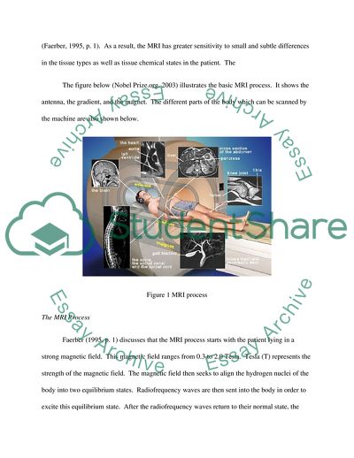
Basic Principles of Magnetic Resonance Imaging (MRI) Image Production Introduction Magnetic Resonance Imaging (MRI) is presently one of the most preferred diagnostic tools in healthcare. Most hospitals are equipped with this technology because of its advantages and potentials in the efficient delivery of health care services. This paper shall discuss what an MRI is and the principles which surround its use. Magnetic Resonance Imaging defined The MRI is defined in Medicine.net (Shiel, 2004) as a “special radiology technology designed to image internal structures of the body using magnetism, radio waves, and a computer to produce the images of body structures”. A combination of the above tools makes this technology possible. Meriles, et.al., (2006, p. 106) discuss that MRI is “based on the use of inhomogeneous or ‘gradient’ fields supplementing on an otherwise uniform invariant magnetic field”. The relation between the magnetic resonance and the location makes it possible to reconstruct the image being scanned by the imaging process. MRI principles The CT scan and the MRI are one of the major diagnostic examinations available in the healthcare industry. While the CT scan relies on X-ray attenuation properties of tissues, the MRI, on the other hand, “depends on the magnetic spin properties of hydrogen nuclei in tissues and how these nuclei recover after excitation with radio frequency electromagnetic waves” (Faerber, 1995, p. 1). As a result, the MRI has greater sensitivity to small and subtle differences in the tissue types as well as tissue chemical states in the patient. The The figure below (Nobel Prize.org, 2003) illustrates the basic MRI process. It shows the antenna, the gradient, and the magnet. The different parts of the body which can be scanned by the machine are also shown below. Figure 1 MRI process The MRI Process Faerber (1995, p. 1) discusses that the MRI process starts with the patient lying in a strong magnetic field. This magnetic field ranges from 0.3 to 2.0 Tesla. Tesla (T) represents the strength of the magnetic field. The magnetic field then seeks to align the hydrogen nuclei of the body into two equilibrium states. Radiofrequency waves are then sent into the body in order to excite this equilibrium state. After the radiofrequency waves return to their normal state, the excitation energy is released into the body as radiofrequency energy. The energy is detected by special energy coils making it possible to detect the course from excitation to relaxation. Relaxation times are usually measured in the T1 and T2 parameters. These two parameters are largely dependent on the tissue involved and on other elements like water, swelling, tumour, and such similar characteristics in body tissues. Consequently, “images can be produced with proper spatial localization of these signals, and diagnostic decisions made” (Faerber, 1995, p. 1). Figure 2: Schematic Diagram of MR imaging (Oxford Centre for Functional MRI of the Brain, n.d) MRI Parameters (T1, T2, PD weighted) Jacobson (2008) expands on Faerber’s explanation by mentioning that the impact and rate of energy release which manifest as protons resume the T1 relaxation alignment; as they wobble or precess during such process, they are recorded by a coil as spatially localized signal intensities. The computer then analyzes these signals in order to project the anatomic images. The brightness of the tissues in the MRI image is determined by radiofrequency pulse and gradient wavelengths which obtain images, tissue characteristics, and tissue density. When the radiofrequency pulse and waveforms are controlled, the computer program creates specific pulses which later resolve into images. These images can be weighted or projected based on T1, T2, or proton density. Body tissues manifest in different ways. Jacobson (2008) discusses that fat appears bright with a high signal intensity when the T1 weight is used, but they appear dark on T2 images. Water and other body fluids appear dark when the T1 weight is used, and appear bright on the T2 weight. The T1 weight would normally project soft tissue and fat, therefore, they are used to confirm fat tissues with masses. The T2 weight is used in order to show fluid and abnormalities. Both T1 and T2 weighted images are actually used to provide complementary information; hence they are both important elements in helping detect abnormalities in the body (Jacobson, 2008). The figures (Wang, n.d, p. 46) below compare the T1, T2, and PD images. Figure 3 a. PD-weighted b. T2 weighted c. T1 weighted Figure 4 T1 weighted Figure 5 T2 weighted (Wang, n.d, p. 49) Figure 6 PD weighted (Kumar, 2005, p., 670) Parts of the MRI Topol & Califf (2007, p. 898) explain that the MRI scanner has five major parts. These are: the magnet, the transmitter, the antenna, the receiver, and the computer. The MRI magnet works best with a magnet that has high field strength which will help in achieving optimum signal-to-noise ratio. The transmitter functions by sending out radiofrequency transmissions to an antenna, which then sends out RF to the patient. The antenna then receives returning transmissions. The coil is usually placed directly on the patient’s surface depending on the part of the body from where information is being gathered. The receiver would then amplify the signal picked up from the coils. Information gathered from the coils shall then be processed by the computer (Topol & Califf, 2007, p. 898). Types of magnets in MRI systems Three types of magnets are used in the magnetic resonance systems. They are the permanent, the resistive, and the superconductive magnets. The magnetic field is actually measured in the unit known as Tesla or ‘T’. This magnetic field is also important in establishing a net magnetization of hydrogen protons which are needed for the MRI (Consiglio, 2006, p. 5). The static magnetic force – torque and translational force – all act simultaneously on fixed magnetic objects and these forces can be dangerous for patients with implanted devices or magnetic foreign bodies. These forces can cause projectile effects and then cause the object to gain speed and kinetic energy on its path to the magnet. This projectile effect can cause serious injury and can also be fatal to people within the trajectory (Consiglo, 2006, p. 5). Basis of MRI Magnetic Resonance Imaging is also based on Nuclear Magnetic Resonance where there is an “absorption and emission of energy by atomic nuclei in the presence of an externally applied magnetic field” (McKie & Brittenden, 2005, pp. 13-14). The NMR is described as a non-invasive means of acquiring clinical images and of studying tissue metabolism in vivo (Easy Measure, 2002). The hydrogen nuclei functions to generate the image seen later in the computer screens. The hydrogen proton nuclei are most concentrated in the body’s water and fat molecules. When the patient is entered in the machine, energy is released in the form of radiofrequency pulses which are then matched to the frequency of hydrogen atoms – atoms that absorb energy pulses. When radiofrequencies are applied, hydrogen atoms then re-release this energy as magnetic resonance; this resonance then stimulates a small voltage in a receiver coil near the patient (McKie & Brittenden, 2005, pp. 13-14). Van Geuns, et.al. (2003, p. 149) discuss that quantum mechanics plays a large role in the imaging process. Nuclei spins around their axes and act like small magnets. Aside from hydrogen, other nuclei like carbon-13, sodium and phosphorus also play a role in clinical imaging. These tiny magnets are distributed in space and normally cancel each other out. However, when the patient is also submitted to a strong external magnetic field, the nuclei would either adapt an orientation parallel or anti-parallel to the external field (Van Geuns, et.al., 2003, p. 149). The parallel orientation is often the preferred alignment because it is at the lower energy state. In order to achieve successful imaging, high resolution images, large field of views, and insensitivity to field in homogeneities and motion, parallel orientation is preferred. These are the elements which make MR imaging a challenging feat to undertake. Motion is a challenge because it actually creates artifacts from other sources of movement like the cardiac wall, blood flow through blood vessels, respiratory movements, and even peristalsis. However, advancements have recently been made in order to address these problems and in order to ensure successful image acquisition (Constable, 2003, p. 1). MRI Variations Jacobson (2008) discusses that the MRI has different variations. The diffusion-weighted MRI is seen when the intensity of the signal is related to the diffusion of water in the molecules or in the tissues. This is used to detect ischemia and infarctions. The echo planar imaging is a very fast technique and is used in the functional imaging of the brain and the heart. The gradient echo imaging is conducted through pulse sequence, and is used to obtain images of moving blood and CSF. The perfusion MRI is used to assess relative cerebral blood flow and to detect ischemia during imaging for stroke (Jacobson, 2008). For quantitative MRI, biophysical importance is estimated from “a collection of MR signals that are related to one another through a function of one or more experimentally controlled variables “ (Koay, et.al., 2009, p. 108). There is a higher sensitivity achieved through MRI because of improved spatial and temporal resolution. Functional MRI (fMRI) The functional MRI is blood-oxygenation-level dependent (BOLD), and is the most “commonly employed method for noninvasive localization of activity in the brain in cognitive psychology investigations” (Sutton, et.al., 2008, p. 33). Functional MRIs (fMRI) are used to measure the ‘function’ and the activity of the brain. Through the BOLD, the fMRI now evaluates differences in magnetic susceptibility between oxygenated and deoxygenated haemoglobin in microcirculation through the brain pathways. More importantly, the functional MRI “detects changes of brain activity rather than brain activity per se” (Frahm, et.al., 2004, p. 3). Depending on changes in metabolism, perfusion and blood velocity affects neuronal function; and such images can be detected through fMRI based on blood oxygenation and magnetic susceptibility. However, oxygenation and magnetic susceptibility are not directly related to each other. The intensity of the signal and the spatial focus in the MRI relies on a uniform magnetic field which exists in the sample being tested. Protons present in the body’s water will align with the strong electromagnetic field, and such field can be manipulated in order to identify the location of activity in the brain (Sutton, et.al., 2008, p. 33). The fMRI was started in 1990 when Owaga and colleagues attempted to demonstrate that images from an MRI can also reflect information dependent on blood oxygenation level (BOLD). BOLD is said to measure haemodynamic changes that react during neuronal activation. The BOLD approach has also been used in some instances to measure pharmacological MRI and to check the effect of stimulants on brain haemodynamics (Marzola, et.al., 2003, p. 165). Contrast Agents In some instances, contrast agents are used in order to “highlight vascular structures and to help characterize inflammation and tumors” (Jacobson, 2008). Most commonly, agents used are gadolinium derivatives. And these derivatives have magnetic properties that affect proton relaxation joints and thereby enabling resonance imaging. Water diffusion and magnetic field gradients Magnetic Resonance Imaging is sensitive to water self-diffusion. As a result, magnetic field gradients undergo phase shifts based on loss of coherence and on decrease in the intensity of signal. Through DW-MRI, there is now a “unique form of MR contrast that enables the diffusional motion of water molecules to be measured and, as a consequence of the interactions between tissue water and cellular structures, provides information about size, shape, orientation and geometry of brain structures” (Bozzali & Cherubini, 2007, p. 970). This sensitivity to water now allows assessments on neurodegeneration in dementia as exhibited by altered diffusion coefficient. Such sensitivity can also be used to explain pathways in the brain based on the assumption that such same sensitivity and algorithms are relevant to segment specific tracts (Bozzali & Cherubini, 2007, p. 970). Consiglio (2006, p. 6) discusses that the gradient fields of the MRI function by localizing tissue signals. Three gradients are found in the system, and each field functions through the flow of electrical current through separate loops of wire mounted in one gradient coil. During an MRI, the gradient fields send out electrical currents in the nerves and muscles, and these currents later act as conductors in the patient’s body. Because of these gradients, a patient is bound to feel mild cutaneous sensations, muscle contractions, and even cardiac arrhythmias. The noises heard during MRI are a result of vibrations and forces in the gradient system. As electrical currents are changed, forces and vibration are then produced. Vibrations then generate the acoustic noise heard during MRI (Consiglio, 2006, p. 5). Conclusion Magnetic resonance imaging functions through a special radiology technology which allows structures of the body to be projected into images through radiofrequency waves. The principles behind the MRI process rely on the magnetic spin properties of hydrogen nuclei in the tissues. The MRI has 5 major parts which work together. The magnet realigns the proton nuclei of the body tissues. The transmitter sends radiofrequency transmissions to the antenna which in turn sends frequencies into the patient. And the antenna also returns the transmissions. The coil helps to gather the information needed, and the receiver amplifies the signals gathered. Lastly, the computer processes the information gathered from the 4 previous elements. And this complex process then allows for the transmission and projection of accurate information about the tissues of the body. Works Cited Bozzali, M. and Cherubini, A. 11 January 2007. Diffusion tensor MRI to investigate dementias: a brief review. Magnetic Resonance Imaging 25: 969–977 September 2009 Chapter 2 Principles of Magnetic Resonance Imaging. (n.d) Oxford Centre for Functional MRI of the Brain. http://users.fmrib.ox.ac.uk/~stuart/thesis/chapter_2/section2_6.html Accessed 18 September 2009 Consiglio, N. 2006. MRI and Patient Safety. The Canadian Journal of Medical Radiation Technology Constable, R. 2003. Magnetic Resonance Imaging. Radiologic Clinics of North America, 41: 1- 15 Faerber, E. 1995. CNS Magnetic Resonance Imaging in Infants and Children. London: Mac Keith Press Frahm, J., Dechent, P., Baukewig, K., and Merboldt, D. 13 October 2003. Advances in functional MRI of the human brain. Progress in Nuclear Magnetic Resonance Spectroscopy 44: 1–32 Jacobson, J. July 2008. Magnetic Resonance Imaging. Merck.com. http://www.merck.com/mmpe/sec22/ch329/ch329d.html. Accessed 18 September 2009 Koay, C. Ozarslan, E. and Basser, P. 22 July 2008. A signal transformational framework for breaking the noise floor and its applications in MRI. Journal of Magnetic Resonance 197: 108-119 Kumar, Y. 2005. Comparison of Fusion techniques applied to preclinical images: fast discrete curvelet transform using wrapping technique and wavelength transform. Journal of Theoretical and Applied Information Technology, 5 (6): 668-673 Marzola, P., Osculati, F., and Sbarbati, A. 5 August 2003. High field MRI in preclinical research. European Journal of Radiology, 48: 165-170 McKie, S. and Brittenden, J. 2005. Basic science: magnetic resonance imaging. Current Orthopaedics, 19: 13-19. Meriles, C., Sakellariou, D. and Trabesinger, A. 2006. Theory of MRI in the presence of zero to low magnetic fields and tensor imaging field gradients. Journal of Magnetic Resonance, 182: 106-114 Puddephat, M. 21 December 2002. Principles of Magnetic Resonance Imaging. Easy Measure. http://www.easymeasure.co.uk/principlesmri.aspx Accessed 18 September 2009 Shiel, W. 2004. Magnetic Resonance Imaging. Medicine.net. http://www.medicinenet.com/mri_scan/article.htm Accessed 18 September 2009 Sutton, B. Ouyang, C., Karampinos, D., and Miller, G. 26 September 2008. Current trends and challenges in MRI acquisitions to investigate brain function. International Journal of Psychophysiology, 73: 33-42 The Nobel Prize in Physiology or Medicine. 2003. Nobel Prize.org. http://nobelprize.org/nobel_prizes/medicine/laureates/2003/illpres/ Accessed 18 September 2009 Topol, E. and Califf, R. 2007. Textbook of cardiovascular medicine, Volume 355. Pennsylvania: Lippincott Williams and Wilkinson Van Geuns, R., Wielopolski, P., and Bruin, H., Rensing, B., Ooijen, P., Hulsoff, M., Oudkerk, M., and de Feyter, P. 2003. Basic Principles of Magnetic Resonance Imaging. Progress in Cardiovascular Diseases, 42 (2): 149-156 Wang, Y. (n.d) Physics of MRI. Polytechnic University Department of Electrical and Chemical Engineering. http://eeweb.poly.edu/~yao/EL5823/MRI_physics_ch12.pdf Accessed 18 September 2009 Read More
Cite this document
  • APA
  • MLA
  • CHICAGO
(Basic Principles of Image Production of Magnetic Resonance Imaging Case Study - 1, n.d.)
Basic Principles of Image Production of Magnetic Resonance Imaging Case Study - 1. https://studentshare.org/technology/1727268-explain-the-basic-principles-of-magnetic-resonance-imaging-mri-image-production
(Basic Principles of Image Production of Magnetic Resonance Imaging Case Study - 1)
Basic Principles of Image Production of Magnetic Resonance Imaging Case Study - 1. https://studentshare.org/technology/1727268-explain-the-basic-principles-of-magnetic-resonance-imaging-mri-image-production.
“Basic Principles of Image Production of Magnetic Resonance Imaging Case Study - 1”. https://studentshare.org/technology/1727268-explain-the-basic-principles-of-magnetic-resonance-imaging-mri-image-production.
  • Cited: 0 times

CHECK THESE SAMPLES OF Basic Principles of Image Production of Magnetic Resonance Imaging

Basic Principles Of MR Imaging

The paper "Basic Principles Of MR Imaging" discusses the features of the process of magnetic resonance therapy.... basic principles of MR ImagingQuestion 1Magnetic resonance is the technology that allows an individual to study an image of the anatomic section and the disease procedure under observation.... basic principles of MRI.... basic principles of MR Imaging.... magnetic resonance is the technology that allows an individual to study an image of the anatomic section and the disease procedure under observation....
4 Pages (1000 words) Essay

Magnetic Resonance Instrumentation

The causes of undesirable sounds produced by magnetic resonance imaging at runtime are due to gradient coil experiencing magnetic forces and torques when being pulsed.... The main cause of nerve stimulation due to magnetic resonance imaging is the slew rate push and the maximum gradient strengths that induce field in the patient, thereby stimulating the nerves.... The two types of image quality reduction caused by zonal impurities in the static magnetic field are: Artifacts; Homogeneity....
3 Pages (750 words) Essay

Improvements in the Use of Magnetic Resonance Imaging

Improvements in the Use of magnetic resonance imaging Student ID Number & Code Date Total Number of Words: 1,000 1.... Aside from problems related to setting appointments for magnetic resonance imaging (MRI) procedures, communication problems can also occur between the MRI department and other radiology departments and pharmacy.... The use of old MRI machines can affect the quality service given to our patients in the sense that the use of new MRI machines can offer better and higher imaging resolution as compared to the old machines....
4 Pages (1000 words) Assignment

Magnetic Resonance Imaging Goes on the Road

magnetic resonance imaging Goes on the Road Nuclear Magnetic Resonance (NMR) has been used by chemists to analyze chemical properties for over 50 years, but due to the complexity of the equipment it has seldom been outside the laboratory.... Nuclear magnetic resonance (NMR) has been used by chemists to analyze chemical properties for over 50 years, but due to the complexity of the equipment it has seldom been outside the laboratory.... According to an article titled "Nuclear magnetic resonance, On the Go", Vasilki Demas is about to change all that....
2 Pages (500 words) Essay

Use of magnetic resonance imaging as lie detector

magnetic resonance imaging is a tried and tested technique to record the activities of brain.... MRI is used to diagnose brain tumors using the functional magnetic resonance imaging (fMRI) scans (Reese, 2005).... This technique uses a radio wave antenna and takes images of brain using the principle of nuclear magnetic resonance.... 'Brain imaging with MRI Could Replace Lie Detector'.... Exposing the soft tissues to such magnetic rays is quite safe as compared to some other forms of diagnostic techniques like X-ray and CAT scans....
2 Pages (500 words) Essay

The Vectors of the Magnetic Moment

The probability of the net magnetic moment approaching zero is when there are many nuclei aligned in different directions If RF pulse is applied at the resonance, then the protons can absorb that energy at the quantum level, a single proton jumps to higher energy state.... Both of these entities play a significant role in producing a MRI active nuclei having active magnetic moment property.... n electromagnetic laws a nucleus spining around its own axis with an electric charge will cause a small magnetic moment (composed of spin and charge) (Donald W et al....
3 Pages (750 words) Essay

Magnetic Resonance Imaging

To rebuild the distribution of… When there are no other position encoding components present, the FT of the signal that results is simply a one-dimensional projection profile of the magnetic resonance imaging by of the school/ of the Describe how MRI sequences spatially en nuclei in the read and phase direction.... Reference: Brown, Mark A and Semelika, Richard C (2010) MRI: basic principles and Application, 4th Edn.... References: Brown, Mark A and Semelika, Richard C (2010) MRI: basic principles and Application, 4th Edn....
5 Pages (1250 words) Essay

Maintenance

hpParts Function Fault or failure PWM amplifier It is designed to drive the magnetic resonance imaging systems and other electromagnetic functions A clogged filter assembly leading to accumulation of dust.... ata monitoring All the compressors are normally equipped with some kind of monitoring equipment in order to protect the compressor and prevent production downtime.... Atlas CopcoCompressed Air Manual (CENTRIFUGAL COMPRESSOR)Please you can see this image in this link and get info or as you prefer http://www....
2 Pages (500 words) Assignment
sponsored ads
We use cookies to create the best experience for you. Keep on browsing if you are OK with that, or find out how to manage cookies.
Contact Us