StudentShare
Contact Us
Sign In / Sign Up for FREE
Search
Go to advanced search...
Free

Adrenal Cortex and Adrenal Medulla in the Endocrine Response to Stress - Essay Example

Cite this document
Summary
The paper "Adrenal Cortex and Adrenal Medulla in the Endocrine Response to Stress" discusses that Angiotensin II influences sodium homeostasis via changes in renal blood flow to maintain constant glomerular filtration rate and it stimulates the adrenal cortex to release aldosterone…
Download full paper File format: .doc, available for editing
GRAB THE BEST PAPER98.1% of users find it useful
Adrenal Cortex and Adrenal Medulla in the Endocrine Response to Stress
Read Text Preview

Extract of sample "Adrenal Cortex and Adrenal Medulla in the Endocrine Response to Stress"

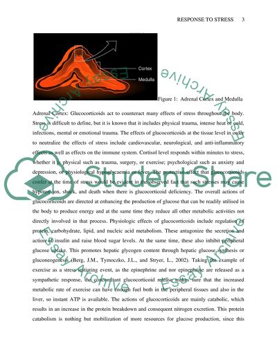
Compare the Roles of Adrenal Cortex and Adrenal Medulla in the Endocrine Response to Stress Introduction: Each adrenal gland is, in effect, structurally and functionally two endocrine glands in one in that it is comprised of an outer cortex surrounding an inner medulla. The cortex contains three histologically distinct zones from out to inward. These are the zona glomerulosa, zona fasciculata, and zona reticularis. In terms of secretion of hormones, the adrenal medulla and cortex behave in a different manner, both in terms of the hormones secreted and also in terms of stimulus for the hormone secretion. In response to the activation of its sympathetic nerve supply and factually with sympathetic stimulation, the neuroendocrine, chromaffin cells of the inner adrenal medulla secrete the catecholamines epinephrine and norepinephrine. These are synthesized in the same cells. However, once secreted they interact with four adrenergic receptors such as 1, 2, 1, 2, and 3 that mediate the cellular effects of the hormones. Stress the predominant stimulus that leads to secretion of these hormones. More accurately stimuli such as injury, anger, pain, cold, strenuous exercise, and hypoglycemia as a result of stress response generate sympathetic impulses in the cholinergic preganglionic fibers that provides the neural impetus to the chromaffin cells, and this ultimately culminates into the release of catecholamines. Biochemically, however, catecholamines stimulate glucose production in the liver, lipolysis in the adipose tissue, and promotes anaerobic metabolism in the muscles leading to lactate production. Whereas, the adrenal medulla responds to predominantly neural stimuli, the outer adrenal cortex that literally encapsulates the medulla and forms the bulk of the gland, responds only to endocrine stimulation and releases a number of hormones which have steroid structures chemically. These hormones are glucocorticoids, aldosterone, and adrenal androgens. The cells of the three zones secrete different steroid hormones: the cells of the zona glomerulosa secrete the mineralocorticoids; those of the zona fasciculata secrete glucocorticoids, while the cells of the zona reticularis secrete sex steroids or androgens (Brook, C., and Marshall, N. 2001). Figure 1: Adrenal Cortex and Medulla Adrenal Cortex: Glucocorticoids act to counteract many effects of stress throughout the body. Stress is difficult to define, but it is known that it includes physical trauma, intense heat or cold, infections, mental or emotional trauma. The effects of glucocorticoids at the tissue level in order to neutralize the effects of stress include cardiovascular, neurological, and anti-inflammatory effects as well as effects on the immune system. Cortisol level responds within minutes to stress, whether it is physical such as trauma, surgery, or exercise; psychological such as anxiety and depression, or physiological hypoglycaemia or fever. The protective effect that glucocorticoids confer at the time of stress would be evident in the observed fact that such stresses may cause hypotension, shock, and death when there is glucocorticoid deficiency. The overall actions of glucocorticoids are directed at enhancing the production of glucose that can be readily utilised in the body to produce energy and at the same time they reduce all other metabolic activities not directly involved in that process. Physiologic effects of glucocorticoids include regulation of protein, carbohydrate, lipid, and nucleic acid metabolism. These antagonize the secretion and action of insulin and raise blood sugar levels. At the same time, these also inhibit peripheral glucose uptake. This promotes hepatic glycogen content through hepatic glucose synthesis or gluconeogenesis (Berg, J.M., Tymoczko, J.L., and Stryer, L., 2002). Taking the example of exercise as a stress initiating event, as the epinephrine and nor epinephrine are released as a sympathetic response, the concomitant glucocorticoid release makes sure that the increased metabolic rate of exercise can have enough fuel both in the peripheral tissues and also in the liver, so instant ATP is available. The actions of glucocorticoids are mainly catabolic, which results in an increase in the protein breakdown and consequent nitrogen excretion. This protein catabolism is nothing but mobilization of more resources for glucose production, since this proteolytic action of glucocorticoids involves glycogenic amino acid precursors from peripheral supporting structures such as bone, skin, muscle, and connective tissue. Along with the amino acid mobilization, there is also inhibition of protein synthesis and peripheral amino acid uptake. This raises the amino acid concentration on blood plasma and in turn facilitates gluconeogenesis stimulated by glucagon the level of which is already up due to inhibition of insulin secretion and function by glucocorticoids in response to stress. By direct actions on liver, glucocorticoids stimulate synthesis of tyrosine aminotransferase and tryptophan pyrrolase which convert these amino acids to glucose. At the same time, glucocorticoids regulate fatty acid mobilization by enhancement of action of cellular lipase by lipid mobilizing hormones from the adrenal medulla, catecholamines. Inflammatory process in the body is a stress phenomenon, and glucocorticoids have anti-inflammatory properties. This works through the effects on the microvascular architecture and through suppression of proinflammatory cytokines such as IL-1a, IL-1b, IL-6, and TNF. This also directly inhibits mediators of inflammation such as eicosanoids, serotonin, PAF, and bradykinin. This is an example of loop by which stress, such as sepsis, increases adrenal hormone secretion resulting in elevated cortisol level that suppresses the immune response (Campbell, E.J.M., Dickinson, C.J., Slater, J.D.H., Edwards, C.R.W., and Sikora, E.K. (eds.), 1984). Figure 2: Immune Actions of Glucocorticoids (Constructed from Sapolsky RM, Romero LM, Munck AU., 2000) These maintain vascular responsiveness to circulating vasoconstrictors and oppose the increase in capillary permeability that is an innate response to acute inflammation. Leukocytes fight sepsis, and glucocorticoids produce a leukocytic response where leukocytes are prevented to leak through the vascular endothelium where they can kill bacteria in the blood stream. It reduces body core temperature that often accompanies stress response through inhibition of IL-1 which appears to be an endogenous pyrogen. This could be the mechanism of fever reduction of glucocorticoids and thus these act as an anti-stress hormone. Eosinophils which are involved in anaphylactic response, another stress event, are reduced by glucocorticoids through redistribution. The release of arachidonic acid from phospholipids is the key reaction of inflammatory response that gives rise to pain. Pain is a stress response. The inflammatory prokinetics prostaglandins and leukotrienes are inhibited by glucocorticoids through its inhibitory actions on phospholipase A2. This would block arachidonic acid release from phospholipids. Acid hydrolases that are involved in cytolytic actions as a result of inflammatory stress response are inhibited by glucocorticoids through reduction of antibody production and stabilization of lysosomal membranes. Glucocorticoid hormones are known to influence the CNS. Although the underlying mechanisms are unclear, cortisol acts on the CNS to produce euphoria, an effect that may also have a role in helping to mitigate the effects of stress (Griffin, J.E., and Ojeda, S.R., 2000). Figure 3: The Principal Physiologic Action of Glucocorticoids (Constructed from Sapolsky RM, Romero LM, Munck AU., 2000) The mineralocorticoid aldosterone is synthesized in the zona glomerulosa of the adrenal cortex. Angiotensin II stimulates aldosterone synthesis in the cells of the zona glomerulosa by increasing cytosolic calcium and activating protein kinase C. Angiotensin II is a potent pressor agent and exerts its action by a direct effect on arteriolar smooth muscle. Angiotensin II is the response to a reduction in circulating blood volume that leads to a corresponding reduction in renal perfusion pressure. Angiotensin II influences sodium homeostasis via changes in renal blood flow to maintain constant glomerular filtration rate and it stimulates the adrenal cortex to release aldosterone. Thus, this system regulates volume by modifying renal haemodynamics and tubular sodium transport. The sympathetic nervous system that is considered to be the major system responding to stress actually regulate the release of renin in response to assumption of the upright posture. Most glucocorticoids have some aldosterone like properties (Eaton DC et al., 2001). Adrenal Medulla: Alpha2-adrenergic receptors were initially characterized as presynaptic receptors that served in a negative feedback loop to regulate the release of norepinephrine. However, they are also involved in postsynaptic functions and play a role in blood pressure homeostasis. Alpha2-adrenergic receptors are implicated in diverse physiologic functions, particularly in the cardiovascular system and the central nervous system. The 1-adrenergic receptor plays an important role in regulating contraction and relaxation of cardiac myocytes. Several mechanisms are thought to be involved, including the phosphorylation of L-type Ca2+ channels in the sarcolemma, ryanodine-sensitive Ca2+ channels in the sarcoplasmic reticulum, troponin I, and phospholamban. The overall physiologic effect is an increase in contractility. The b2-adrenergic receptor mediates several physiologic responses, including vasodilatation, bronchial smooth muscle relaxation, and lipolysis, in various tissues. Abnormalities in the function of this adrenergic receptor may lead to hypertension (Laycock, J., and Wise, P., 1996). The 3-adrenergic receptor plays an important role in mediating catecholamine-stimulated thermogenesis and lipolysis. Catecholamines are released as part of the stress response to a physical or psychological insult such as severe blood loss, decrease in blood glucose concentration, traumatic injury, surgical intervention, or a fearful experience. Because catecholamines are part of the "fight or flight" response, their physiologic effects include arousal, alerting, papillary dilation, piloerection, sweating, bronchial dilation, tachycardia, inhibition of smooth muscle activity in the gastrointestinal tract, constriction of the sphincters, and relaxation of the uterine muscles (Ledingham, J.G.G., and Warrell, D.A. (eds.), 2000). Most of the events involved in coping with a stressful situation require the expenditure of energy. Catecholamines ensure substrate mobilization from the liver, muscle, and fat by stimulating the breakdown of glycogen by glycogenolysis and fat by lipolysis. This is in sharp contrast with the actions of glucocorticoids. Thus, an increase in circulating catecholamines is associated with elevations in plasma glucose and free fatty acid levels. Some of the most important effects of catecholamines are exerted in the cardiovascular system, where they cause tachycardia, produce peripheral vasoconstriction, and elevate vascular resistance. The physiological effects of adrenal medullary catecholamines must be considered as part of an overall sympathetic response since their release is always associated with an increase in the secretion of norepinephrine from sympathetic nerve terminals. While the adrenal medulla is not vital for survival, it does contribute to the response of the body to stress. Both epinephrine and norepinephrine raise the systolic blood pressure by stimulating heart rate and contractility, thereby increasing cardiac output. However, epinephrine reduces diastolic pressure as a result of causing vasodilatation of certain vessels, particularly those of skeletal muscle, while norepinephrine raises diastolic pressure by causing a more generalized vasoconstriction. Both catecholamines cause piloerection and dilatation of the pupils. Epinephrine also acts as a bronchodilator and reduces the motility of the gut (McEwen BS., 1998). Reference List Berg, J.M., Tymoczko, J.L., and Stryer, L. (2002). Biochemistry (5th edn), Chapters 15, 30, and 31. Freeman, New York. Brook, C., and Marshall, N. (2001). Essential endocrinology (4th edn). Blackwell Science, Oxford. Campbell, E.J.M., Dickinson, C.J., Slater, J.D.H., Edwards, C.R.W., and Sikora, E.K. (eds.) (1984). Clinical physiology (5th edn). Blackwell Scientific, Oxford. Eaton DC et al. (2001). Mechanisms of aldosterone's action on epithelial Na+ transport. J Membr Biol.;184:313. Griffin, J.E., and Ojeda, S.R. (2000). Textbook of endocrine physiology (4th edn). Oxford University Press, Oxford. Laycock, J., and Wise, P. (1996). Essential endocrinology. Oxford Medical Publications, Oxford. Ledingham, J.G.G., and Warrell, D.A. (eds.) (2000). Concise Oxford textbook of medicine, Chapters 7.1.7.16. Oxford University Press, Oxford. McEwen BS., (1998). Protective and damaging effects of stress mediators. N Engl J Med.;338:171. Sapolsky RM, Romero LM, Munck AU., (2000). How do glucocorticoids influence stress responses Integrating permissive, suppressive, stimulatory, and preparative actions. Endocr Rev;21:55-89. Read More
Cite this document
  • APA
  • MLA
  • CHICAGO
(“Endocrinology Essay Example | Topics and Well Written Essays - 1000 words”, n.d.)
Endocrinology Essay Example | Topics and Well Written Essays - 1000 words. Retrieved from https://studentshare.org/science/1507062-endocrinology
(Endocrinology Essay Example | Topics and Well Written Essays - 1000 Words)
Endocrinology Essay Example | Topics and Well Written Essays - 1000 Words. https://studentshare.org/science/1507062-endocrinology.
“Endocrinology Essay Example | Topics and Well Written Essays - 1000 Words”, n.d. https://studentshare.org/science/1507062-endocrinology.
  • Cited: 0 times
sponsored ads
We use cookies to create the best experience for you. Keep on browsing if you are OK with that, or find out how to manage cookies.
Contact Us