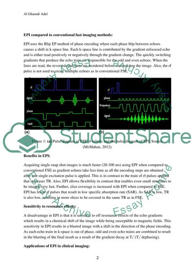
StudentShare
Contact Us
Sign In / Sign Up for FREE
Search
Go to advanced search...

BLIP Echo Planar Imaging Method - Essay Example

Cite this document
Summary
Q1. BLIP Echo Planar Imaging (EPI) method- Principles of EPI: EPI, like FSE (Fast Spin Echo), is an imaging method that reduces the imaging time as it acquires multiple phase encoding steps in each repetition time (TR). It is a fast and flexible method of imaging that provides with good contrast and resolution for the images…
Download full paper File format: .doc, available for editing
GRAB THE BEST PAPER93.5% of users find it useful
BLIP Echo Planar Imaging Method
Read Text Preview

Extract of sample "BLIP Echo Planar Imaging Method"

Download file to see previous pages

Sampling of k-space is different in EPI that enables high quality of images acquired. EPI uses quickly switching gradients to produce its echo train as against use of RF pulses in FSE. EPI frequency encoding gradient oscillates from positive to negative to create odd and even echoes. Each read gradient echo corresponds to one k-space with the positive and negative gradients changing the direction of the line that is read. The changing of direction of the line read results in a phase ‘blip’ causing a shift in Ky and this method of phase encoding is referred to as blip EPI.

Characteristics of EPI: EPI has three distinct characteristics in terms of speed, the variety of contrast and the flexibility in terms of available resolution for imaging small structures like the pituitary gland. EPI offers higher speeds (6 to 10 times) of imaging of short temporal events under motion. It provides with high image quality by collecting more averages with increased slice frequency per TR. The signal to noise ratio of the images is high and the resolution is also high. Its usefulness depends on the MR system used and the efficiency is determined by the ability to perform 1282 or 2562 in a very short time to avoid large off resonance artifacts.

It means that high quality images of moving patients can be obtained by using the speed of EPI. A variety of tissue contrasts (T1, T2 and T2*) are allowed by EPI for image acquisition and small structures are imaged using the flexibility in resolution where the field view can be reduced, the echo train length can be increased or geometric distortions can be reduced to increase the resolution. Also, just half of the data of the image can be gathered that can be further synthesized through conjugation to obtain the remaining data.

Flexibility in contrast can be used to produce an image similar in standards to that of an SE image with the same TE and TR by using an excitation pulse in the front part followed by a 1800 pulse (T2* dephasing) to create the first echo. EPI compared to conventional fast imaging methods: EPI uses the Blip EP method of phase encoding where each phase blip between echoes causes a shift in k-space line. Each k-space line is contributed by the gradient refocused echo and is either read positively or negatively through the gradient change.

The quickly switching gradients that produce the echo train are responsible for the odd and even echoes. When the lines are read, the reversed read lines are reordered before constructing the image. Also, the rf pulse is not used to create multiple echoes as in conventional FSE. Figure 1: (a) Pulse diagram of FSE, 4 echoes and (b) pulse diagram of EPI. Source: (McMahan, 2012). Benefits in EPI: Acquiring single snap shot images is much faster (20-100 ms) using EPI when compared to conventional FSE as gradient echoes take less time as all the encoding steps are obtained after one single excitation pulse is applied.

This is in contrast to the train of rf pulses applied that increases TR. Also, EPI allows flexibility in contrast that enables even small structures to be imaged very fast. Further, slice coverage is increased with EPI when compared to FSE. EPI has fewer rf pulses that result in low specific absorption rate (SAR). As SAR is low, TR is also low, resulting in more slices to be covered in the same TR as in FSE. Sensitivity to resonance effects: A disadvantage in EPI is that it is sensitive to off resonance effects of the echo gradients

...Download file to see next pages Read More
Cite this document
  • APA
  • MLA
  • CHICAGO
(“BLIP Echo Planar Imaging Method Essay Example | Topics and Well Written Essays - 2500 words”, n.d.)
Retrieved de https://studentshare.org/physics/1445011-mres
(BLIP Echo Planar Imaging Method Essay Example | Topics and Well Written Essays - 2500 Words)
https://studentshare.org/physics/1445011-mres.
“BLIP Echo Planar Imaging Method Essay Example | Topics and Well Written Essays - 2500 Words”, n.d. https://studentshare.org/physics/1445011-mres.
  • Cited: 0 times

CHECK THESE SAMPLES OF BLIP Echo Planar Imaging Method

Magnetic Susceptibility Technology

Reducing the time of echo train length: the longer the length of the echo train, the more time is required by the spin to gather phase error and the greater the distortion witnessed.... One of the ways of minimizing the total time of train length is by minimizing the echo spacing.... In FSE, the less the ESP or the echo spacing is, the less the blurring of the image gets.... In EPI, minimizing the echo spacing also results in a reduction in the blurring and geometric distortion....
22 Pages (5500 words) Essay

Magnetic Resonance Imaging of Bone Marrow: A Systematic Diagnostic Approach

hellip; Single or combined sequences of MR pulses such as (a) Spin echo T1 weighted, (b) T2 weighted fat saturated fast spin echo and (c) Short Time Inverse Recovery sequence (STIR) to determine the bone marrow compositions and the abnormalities.... These defects can be distinguished from usual chondral defects by comparing the centralization of the final lesion that are widely spread and lacks surroundings (Loren, 2008) Full thickness chondral defect is appeared with high signal joint surface lesion represented in black arrow is covered by normal low intense hyaline cartilage on this spin echo T2 weighted image....
8 Pages (2000 words) Essay

Echo-Planar Imaging in Magnetic Resonance Imaging

echo planar imaging, or EPI, is a method that can form an absolute image from a single data sample.... Echo-planar imaging is the fastest and most supple means of MR imaging nowadays.... BLIP EPI Method Echo-planar imaging (EPI) is skilled to considerably cut down the times of magnetic resonance (MR) imaging.... While studying the working of echo-planar imaging, it is helpful to put the usual spin-echo (SE) imaging side by side....
9 Pages (2250 words) Essay

Diffusion MRI Stimulation theory

This method is employs a chronological sequence to establish the microscopic random movement of water protons (Bammer 2003) and has thus been employed in the examination of numerous diseases due the various benefits that can be obtained as a consequence of its initiation (Kono et al.... 19) diffusion-weighted imaging has been one of the most pivotal advancements with regards to the progress of neurological imaging procedures in the past 10 years....
16 Pages (4000 words) Admission/Application Essay

To answer the past exam for sample exam,2002and 2003

?   Spiral scanning methods (square and circular):  Points in k space are also not collected uniformly in time (that is, in the line by line method we are familiar with).... ? The square method is, however, evenly spaced in k-space, therefore just needs reordering.... ? As you would have gathered by constructing the table in question 3, speed is of course the main advantage, and opens up the area of functional rather than anatomical imaging....
6 Pages (1500 words) Coursework

Gadolinium Enhanced Images in Obtaining Magnetic Resonance Venography

It also aims to justify the use of contrast medium in some cases, which is expensive, may have side effects, and is not the method of choice for patients with abnormal kidney function.... It also aims to justify the use of contrast medium in some cases, which is expensive, may have side effects, and is not the method of choice for patients with abnormal kidney function.... It constitutes, Non- imaging Procedures which involves the diagnosis of CVT based on the findings such as total blood count, sedimentation rate, chemistry panel, and prothrombin time and Also, D-Dimer measures, a fibrin split product, which can be seen as a fragment of a small protein (D-shaped) flowing in the blood....
44 Pages (11000 words) Research Paper

Echo Planar Image Sequences Data

PI is an imaging method that is fats and flexible.... (7 marks)echo planar image (EPI) sequences data collection has different ways of comparing with sequences in standard pulse.... (7 marks)echo planar image (EPI) sequences data collection has different ways of comparing with sequences in standard pulse.... echo planar is superior to conventional in investigations of the human physiological processes in the body.... Discuss the BLIP EPI method presented in Module 1 in detail....
9 Pages (2250 words) Assignment

Depth Pulse Sequences for Surface Coils

… April 28, 2012Qs 1.... he use of surface coil was introduced in 1980.... Surface coil consist of either one or two copper loops of wire.... They are positioned under the patient's organ close to the location of interest.... Surface coils are placed in a way that April 28, 2012Qs 1.... he use of surface coil was introduced in 1980....
9 Pages (2250 words) Essay
sponsored ads
We use cookies to create the best experience for you. Keep on browsing if you are OK with that, or find out how to manage cookies.
Contact Us