StudentShare
Contact Us
Sign In / Sign Up for FREE
Search
Go to advanced search...
Free

Hemolytic Disorders: Hereditary Elliptocytosis - Essay Example

Cite this document
Summary
The author of the paper "Hemolytic Disorders: Hereditary Elliptocytosis" argues in a well-organized manner that hereditary elliptocytosis is caused by mutations that cause disruptions in the cytoskeletal framework of red blood cells which are responsible for the elasticity of the circulating RBCs…
Download full paper File format: .doc, available for editing
GRAB THE BEST PAPER91.6% of users find it useful
Hemolytic Disorders: Hereditary Elliptocytosis
Read Text Preview

Extract of sample "Hemolytic Disorders: Hereditary Elliptocytosis"

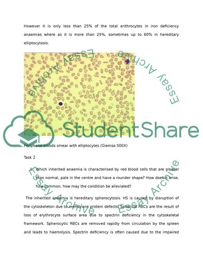
Task Haemolytic disorders A case of hereditary elliptocytosis Hereditary elliptocytosis is an inherited hemolytic disorder characterized by the presence of elongated red blood cells or poikilocytosis of irregular degrees. Hereditary elliptocytosis shows an autosomal dominant pattern of inheritance with at least 4 genetic loci implicated in the disorder pathogenesis. Hereditary elliptocytosis is caused by mutations that cause disruptions in the cytoskeletal framework of red blood cells which are responsible for the elasticity of the circulating RBCs. The formation of spectrin tetramers which form a major part of the cytoskeleton is disrupted by the mutations which results in less tolerant red blood cell membrane. The mechanical instability resulting from theses horizontal membrane defects is enough to cause haemolytic anaemia. Protein 4.1 or glycophorin C deficiencies are also attributed to the cause of hereditary elliptocytosis. Clinical symptoms range from an asymptomatic carrier status to various degrees of haemolytic anaemia. Chronic haemolysis in individuals also might accompany with complications such as splenomagaly, early gall bladder disease and jaundice. The disease has no reported sex predilection and is more common in individuals of Mediterranean and African descent. Diagnosis of the disease is based on the presence of elliptocytes in peripheral blood smear. The diagnosis can be confused with iron deficiency anaemias as the condition also shows the presence of elliptocytes in peripheral blood smear. However it is only less than 25% of the total erithrocytes in iron deficiency anaemias where as it is more than 25%, sometimes up to 60% in hereditary elliptocytosis. Peripheral bloods smear with elliptocytes (Giemsa 500X) Task 2 1. Which inherited anaemia is characterised by red blood cells that are smaller than normal, pale in the centre and have a rounder shape? How does it arise, how common, how may the condition be alleviated? The inherited anaemia is hereditary spherocytosis. HS is caused by disruption of the cytoskeleton due to membrane protein defects. Spherical RBCs are the result of loss of erythrocyte surface area due to spectrin deficiency in the cytoskeletal framework. Spherocytic RBCs are removed rapidly from circulation by the spleen and leads to haemolysis. Spectrin deficiency is often caused due to the impaired synthesis of the protein but sometimes also caused due to deficiency of binding proteins that integrate spectrin into the cell membrane. Hereditary spheroytosis is considered to be the most common haemolytic anaemia among Northern European descent (Perrotta S,2008). Other ethnic group incidence and prevalence are not clearly established. 2. Thalassaemias are disorders due to the reduced synthesis of either α or β globin hence defective Hb production. How do these disorders vary in severity and presentation? Both α and β Thalassemias results in inadequate amount of haemoglobin production due to the defective α and β globin chains available to construct haemoglobin dimers. Point mutations or partial deletions of the chromosome 11 cause defects in the synthesis of the β chain. The severity of β Thalassemia is dependent on the degree to which the β chain production is inadequate. β -Thalassemia major is a condition in which very liitle or no β chain is made. Peripheral blood smear of the condition shows poikilocytosis and anisocytosis, elliptocytes and teardrops. Clinical manifestations include severe transfusion dependent anaemia, splenomegaly and hepato megaly. Damage to organs such as heart, liver and endocrine organs might occur due to iron over load. Enlargement of the bones due to erythroid hyperplasia in the marrows also might occur. The patients mostly die before reaching adulthood. In β-Thalassemia minor the production of β chains are mildly reduced causing ineffective erythropoiesis in the marrow. The red blood cells in peripheral blood smear are microcytic and hypochromic. The anaemia seen in this condition is generally mild and might be mistaken for iron deficiency. In βThalassemia intermedia the β chain production is intermediate to the first two conditions. In the classical case of α Thalassemia entire α gene deletion from chromosome 16 is considered the cause. Point mutations in the gene are also attributed to the cause of the disorder. Clinical manifestations of the disorder vary with the number of genes deleted. Deletion of one α gene results in a silent carrier state without any haematological abnormalities. Deletion of two α genes results in hypochromia, microytosis and mild anaemia. Deletion of three α genes produces haemoglobin H which is made of four β chains which is unstable. Hgb H shows hypochromic and microcytic RBcs and precipitates in vivo causing haemolysis. Deletion of all four α genes results in the death of the foetus in the uterus a condition called hydrop foetalis. 3. Antibodies may be raised to red blood cells giving rise to autoimmune haemolytic anaemias. What is the difference between warm and cold haemagglutinin disease? Warm haemagglutinin disease is the most common autoimmune haemolytic anaemia mediated by auto antibodies IgG most active at 37˚ C against the surface antigens of red blood cells. Though most cases of warm haemagglutinin disease are idiopathic, diseases like Hodgkin’s disease, Non-Hodgkin’s lymphomas and autoimmune disorders like rheumatoid arthritis are also believed to cause this disease. Cold agglutinin disease mediated by IgM antibodies active at less than 30˚ C is another autoimmune haemolytic anaemia characterized by haemolysis and RBC agglutination in cold exposed areas of the body. This condition is associated with Mycoplasma pneumonia, lymphoma and rarely infectious mononucleosis. Chronic haemolytic anaemia along with haemoglobinuria and jaundice are clinical manifestations of the condition. 4. Haemolysis can occur due to physical intervention – describe the cause and consequences of March Haemoglobinuria. What other forms of physical intervention can cause haemolysis March haemoglobinuria is a condition that results from red blood cell trauma occurring in the feet due to marching running or jogging. Peripheral blood smear shows no RBC changes. Following are some of the different haemolysis caused by physical intervention: Cardiac Haemolytic anaemia: Haemolysis occurring due to red blood cells striking against artificial surfaces like prosthetic heart valves and arterial grafts or abnormal surfaces like atherosclerosis or aortic stenosis. Microangiopathic haemolytic anaemia: Damaged endothelial surfaces or fibrin strands on small vessels fragmenting red blood cells resulting malignant hypertension, disseminated intravascular coagulation and vascular neoplasms among many other damage. Thrombotic thrombocytopenia purpura: Red blood cell damage caused by platelet and fibrin deposition on small vessels. Adults with neurologic and mild renal problems are often victims of this condition. Haemolytic uremic syndrome: Fibrin and platelet deposition in small vessels seen in kidney cause fragmentation of RBCs. This condition is common in children with renal problems. References http://www.emedicine.com/med/topic2147.htm http://www.med-ed.virginia.edu/courses/path/innes/rcd/thalassemia.cfm http://www.aafp.org/afp/20011015/1379.html http://www.uq.edu.au/vdu/HDUThalassaemia.htm http://www.med-ed.virginia.edu/courses/path/innes/rcd/antibody.cfm http://www.med-ed.virginia.edu/courses/path/innes/rcd/trauma.cfm http://www.uq.edu.au/vdu/HDUAnaemiaTrauma.htm Perrotta S,2008 Gallagher PG, Mohandas N. Hereditary spherocytosis. Lancet. Oct 18 2008;372(9647):1411-26. Week 10 Blood Conservation Blood loss being an inevitable consequence of surgery, the importance of transfusion cannot be stressed enough. Transfusion of blood has become comparatively safer with rigorous donor selection, tighter quality control and screening. However the risk associated with transfusion still remains with the SHOT data showing the most frequent hazard remained to be incorrect blood component transfused. The risk associated with transfusion, shortage of blood, availability of transfusion alternatives and patient choices have increased interest in blood conservation strategies. Some of the blood conservation strategies include Autologous transfusion which involves giving back patients their own blood. Preoperative autologous donation uses blood taken from patients stored fresh or sometimes frozen prior to surgery. Acute normovolaemic haemodilution collects whole blood from patients in blood bags containing anticoagulants and replaced with crystalloid or colloid fluid before surgery and returned after the surgery. Cell salvage methods include processing collected blood lost during or after the surgery and replacing it. Postoperative cell salvage involves blood collection from surgical drains which is then re-infused following filtering and processing. Autologous transfusion reduces risks associated with blood transfusion but one of the most important blood conservation strategies is to avoid unnecessary transfusion, avoiding blood loss and optimizing preoperative Hb. Is autologous transfusion benefit to the patient or provider? Autologous transfusion peaked in its benefit when the HIV and other related infections caused a lot of panic around the world. However with advanced screening techniques used before transfusion and risks like bacterial infections still remaining in autologous infection the benefits has significantly declined in my opinion. However a psychological comfort provided to the patient on using their own blood is an advantage that cannot be disregarded. Week 11 Haemolytic Disease of the newborn portfolio task Haemolytic Disease of the new born is a condition in which the specific antibodies derived from the mother through placental transfer destroy the foetal red blood cells. The condition occur when the mother possess and IgG red cell antibody stimulated by previous transfusion or pregnancy and the foetus possess a corresponding antigen which in all probability is paternally derived. Clinical symptoms include anaemia, jaundice, hepatosplenomegaly, oedema and tachycardia while severe cases may result in congestive heart failure, kernicterus and even intrauterine death. Primary antenatal screening is done usually at 10-16 weeks of gestation and involves ABO and RhD typing, antibody screen at 37˚C and HIV, Hepatitis B and Syphilis tests. Secondary antenatal screening takes place in the 28th week of gestation and includes ABO and RhD typing and antibody screening at 37˚C. Following the secondary antenatal screening antibody levels should be monitored every two weeks. A titre of 32 or greater is likely to cause HDN other than Kell related antibodies which might affect the foetus irrespective of the titre. A greater antibody level requires an amniocentesis which is a source of foetal DNA which can be used to check the presence of the antigen in the foetus using foetal blood typing through the polymerase chain reaction. The amniotic fluid also will show an increased bilirubin in the case of excessive RBC breakdown which can be spectrophotometrically measured. Another test that can be done to confirm the foetal blood group is direct foetal blood sampling which can be carried out from 18 weeks of gestation. Postnatal testing involves taking cord blood from all RhD negative mothers and checking baby’s blood group including antigen typing if necessary. Maternal blood group and antibody screen is also done. Mothers of RhD positive babies are given 1500 iu prophylactic anti D. Size of the foetomaternal bleed is estimated by a Kleihauer test on maternal blood. Infants born to mothers who show clinically significant levels of antibodies should be kept under close watch for the first 48-72 hours of life. Why is routine ante natal Anti-D prophylaxis thought to be an important development in ante –natal care? Routine antenatal anti-D prophylaxis is a treatment option recommended to all pregnant women who are RhD negataive and are not sensitized to the RhD antigen. Sensitisation can occur in the third trimester of pregnancy or during childbirth. It is best to administer RAADP in such possibilities of sensitization occurring so that the foetal red cells can be destroyed before antibody production occurs in the mother. It can be routinely administered routinely in the third trimester against possible events of foetomaternal haemorrhage or abdominal trauma. RADDP can be administered as a dose of 250 iu up to 20 weeks of gestation and 1500 iu after 20 weeks of gestation. National Institute for Clinical Excellence provides full guidance on the administration of RAADP. Statistics show 17% of all births is to RhD negative women of which 59% of babies are RhD positive. HDN affected up to 1% of all births before the immunoprophylaxis. However the frequency of HDN has reduced significantly following prophylaxis. Semester 2 Week 4 A Brief encounter with Haematology Blood composition and structure Normal peripheral blood consists of red blood cells, white blood cells and platelets. Cells occupy 40% of the volume of the plasma in which it is suspended. White blood cells are the least numerous of the blood cells and includes the granulocytes neutrophils, eosinophils and basophils and the mononuclear lymphocytes and monocytes. Red blood cells or erithrocytes are the most numerous and is seen in circulation for 120 days before spleen removes it. The shape of erithrocytes is biconcave with approximately 7µm diameter. RBCs carry oxygen from the lungs to tissues and Haemoglobin the oxygen carrying pigment in RBCs impart the red colour to blood. Blood platelets are the second numerous cells which are seen in circulation only for 10-12 days. They take part in clotting blood to arrest blood loss. Platelets are generally anucleate with a granular cytoplasm and discoid in shape. Full blood count is a screening test which gives information regarding the cells present and their quantities. A number of stains are used to differentiate cells and some of them include the prototype Romanowsky stain using methylene blue and eosin, Wright’s Giemsa and Leishman stain . Stains are pH dependent. Freemethylene blue has a basic nature and stains acidic cellular components like DNA and RNA which is translated as a blue-violet colour to nucleic acids, granules of basophils and also to neutrophils granules. Free eosin stains basic components such as eosinophil granules and haemoglobin as it is acidic and gives it a pink or reddish colour. Improper staining such as prolonged staining or inadequate rinsing might result in poor appearance of the blood cells under the microscope. Flow cytometry is a technique that determines percentage of different cell populations passing through a flow cell. Immunophenotyping methods are used for HIV typing by determining CD4/CD8 counts on T-lymphocytes and Leukaemia typing which useor s cell markers for determining the lymphomas, acute and chronic differentiation and myeloid and lymphoid differentiation. In HIV typing with progression of the disease CD4 count falls and CD8 count rises. In leukaemia immunophenotyping fluorochrome conjugated antibodies label thw white blood cells and different cell populations are measured. Bone marrow biopsy include bone marrow aspirate which involves cytologically examining cells within the marrow for abnormalities and bone marrow trephine which studies the bone marrow architecture to assess the presence of fibrosis or distribution of an abnormal infiltrate. Cytogenetics studies the structure and inheritance of chromosome to understand chromosomal abnormalities and disorders. Read More
Cite this document
  • APA
  • MLA
  • CHICAGO
(“Pathology Science Essay Example | Topics and Well Written Essays - 1000 words”, n.d.)
Pathology Science Essay Example | Topics and Well Written Essays - 1000 words. Retrieved from https://studentshare.org/miscellaneous/1563269-pathology-science
(Pathology Science Essay Example | Topics and Well Written Essays - 1000 Words)
Pathology Science Essay Example | Topics and Well Written Essays - 1000 Words. https://studentshare.org/miscellaneous/1563269-pathology-science.
“Pathology Science Essay Example | Topics and Well Written Essays - 1000 Words”, n.d. https://studentshare.org/miscellaneous/1563269-pathology-science.
  • Cited: 0 times

CHECK THESE SAMPLES OF Hemolytic Disorders: Hereditary Elliptocytosis

The Symptoms and Diagnosis of a Patient Suffering from Haemolytic Anaemia

In congenital/hereditary type, the causes are “hereditary spherocytosis, Glucose -6-phosphate dehydrogenase (G6PD) deficiency, Pyruvate kinase deficiency, Sickle cell anaemia and Thalassaemia” (Davey, 2012, p.... Due to a hereditary biochemical lesion in individuals of mainly of Greek descent, Favism is common.... "The Symptoms and Diagnosis of a Patient Suffering from Haemolytic Anaemia" paper focuses on hemolytic anemia characterized by an increase in erythrocyte destruction caused by the trapping of cells in sinuses of the spleen and producing a decrease in the normal average life span of the erythrocyte....
4 Pages (1000 words) Essay

Hemolytic Disease of the Newborn

From the paper "hemolytic Disease of the Newborn" it is clear that non-responders may not produce anti-D even though during pregnancy, there were substantial fetal bleeds.... Running head: hemolytic Disease of the Newborn hemolytic Disease of the Newborn [The of the appears here] [The of institution appears here] ... hemolytic Disease of the Newborn is also called erythroblastosis fetalis.... hemolytic" means breaking down of red blood cells ...
7 Pages (1750 words) Essay

Hereditary Disorders in Children

Examples of hereditarily caused birth defects hereditary defects are one of the leading cause of death in children—“causing 1 in 5 deaths in babies through 12 months of age and as many as 40% of deaths in children through the age of 10 years.... hereditary disorders are also a significant cause of childhood morbidity and long-term disability.... Children with hereditary disorders often require care that is expensive and highly specialized....
4 Pages (1000 words) Essay

Clinicl Pathology

The heart pumps the oxygenated blood through the arteries.... The oxygenated blood goes to the organs to deliver oxygen and other chemical substances needed by the cells.... The blood goes back.... ... ... There are two main components of blood, the plasma and the cells.... The plasma is the liquid part of the blood....
12 Pages (3000 words) Coursework

Hemolytic Anemia in G6PD Patients

In the paper 'hemolytic Anemia in G6PD Patients,' the author discussed the ingestion of fava beans, which induced an oxidative stress to erythrocytes, causing acute hemolytic anemia.... Thus, some G6PD variants linked with rigorous enzyme deficiency, such as Union and Markham, cause no hemolytic problem, while some variants related to the less severe deficiency.... hemolytic Anemia in G6PD Patients ... In the case discussed below, the ingestion of fava beans induced an oxidative stress to erythrocytes, causing acute hemolytic anemia. ...
4 Pages (1000 words) Case Study

Types of Protein Defects That Can Be Associated with the Red Blood Cell

utations that obstruct the formation of spectrin tetramers result in hereditary elliptocytosis.... ereditary elliptocytosis ... ereditary elliptocytosis (HE) is a congenital hemolytic disorder in which erythrocytes get elongated into a cigar or oval shape.... G6PD deficiency is a widely seen hereditary disease among people of African, Southeast Asian and Mediterranean descent.... Yawata(2003: 21)suggests that “The abnormalities of ankyrin are the principle cause in the pathogenesis for hereditary spherocytosis”....
11 Pages (2750 words) Essay

What is Anemia

Hereditary Spherocytosis & hereditary elliptocytosis ... Any information regarding allergies, genetic blood disorders that run in the family, a change of urine/stool color must also be discussed in detail (Care of Patients with Hematologic disorders, Elsevier).... Care of Patients with Hematologic disorders.... Immune hemolytic Anemia ...
6 Pages (1500 words) Research Paper

Cat Dilated Cardiomyopathy and Immune Mediated Hemolytic Anemia in Daschunds

Genetic predisposition may be a factor; especially disorders affecting red blood cells, in the case of canine IMHA.... Other diseases, such as hormonal disorders may include autoimmune diseases as a complication.... In most mammals, the causes of auto-immune disorders are less understood than the means by which they can be addressed.... The paper "Cat Dilated Cardiomyopathy and Immune Mediated hemolytic Anemia in Daschunds" highlights that taurine is necessary for felines due to a variety of other health reasons; as deficiencies can lead to central retinal degeneration among other conditions....
10 Pages (2500 words) Research Paper
sponsored ads
We use cookies to create the best experience for you. Keep on browsing if you are OK with that, or find out how to manage cookies.
Contact Us