StudentShare
Contact Us
Sign In / Sign Up for FREE
Search
Go to advanced search...
Free

Computerized Tomography Portfolio - Report Example

Cite this document
Summary
The authoor of this paper "Computerized Tomography Portfolio" identifies the artifacts produced on the images during CT scans, describes the methods to reduce or remove these artifacts. The study highlights the physical-based, patient-based, helical, multi-slice, scanner-based artifacts…
Download full paper File format: .doc, available for editing
GRAB THE BEST PAPER91.5% of users find it useful
Computerized Tomography Portfolio
Read Text Preview

Extract of sample "Computerized Tomography Portfolio"

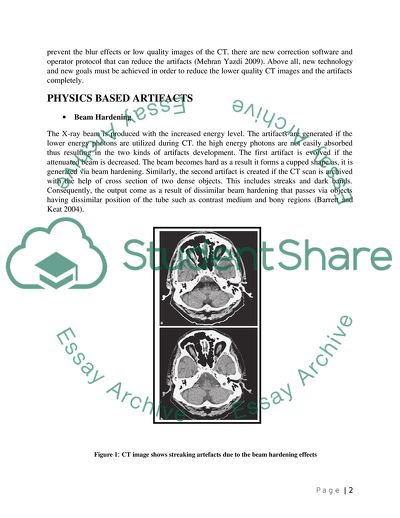
Identify the artifacts produced on the images during CT scans. Describe the methods to reduce or remove these artifacts. INTRODUCTION In the previousthree decades the diagnostic accuracy in the field of radiology has developed extremely especially after the invention of the Computed Tomography (CT). The non-invasive imaging technique is the excellent example of the Computed Tomography (CT). However; the loss of image quality is also marked via different kinds of CT artifacts (Watzke and Kalender 2004). The artifacts are divided into four main categories some of them are based on the principles of physics. The artifacts that rely on the physics are Beam hardening. Partial volume. Photon starvation. Under sampling. Some of the artifacts that are based upon the patients are mentioned below Metallic artifacts. Motion artifacts. Incomplete projections. The scanner based artifacts includes the most common artifact i.e. Ring artifacts The helical and the multi-slice CT artifacts stand last and can occur during the helical artifact in axial plane (Barrett and Keats 2004). The artifacts that are included into this category are mentioned below Helical artifacts in single-section scanning. Helical artifacts in multi-slice scanning. Cone beam effect. Multi-planner and three dimensional reformations. The studies revealed that the striping, shading and rings are responsible for the low image quality of the Computed Tomography (CT) therefore it is difficult to diagnose the disease if the CT images are smudge. Furthermore, in modern years new technology has been invented in order to prevent the blur effects or low quality images of the CT. there are new correction software and operator protocol that can reduce the artifacts (Mehran Yazdi 2009). Above all, new technology and new goals must be achieved in order to reduce the lower quality CT images and the artifacts completely. PHYSICS BASED ARTIFACTS Beam Hardening The X-ray beam is produced with the increased energy level. The artifacts are generated if the lower energy photons are utilized during CT. the high energy photons are not easily absorbed thus resulting in the two kinds of artifacts development. The first artifact is evolved if the attenuated beam is decreased. The beam becomes hard as a result it forms a cupped shape as, it is generated via beam hardening. Similarly, the second artifact is created if the CT scan is archived with the help of cross section of two dense objects. This includes streaks and dark bands. Consequently, the output come as a result of dissimilar beam hardening that passes via objects having dissimilar position of the tube such as contrast medium and bony regions (Barrett and Keat 2004). Figure 1: CT image shows streaking artefacts due to the beam hardening effects Image Source: (Brüning, Küttner, and Flohr ) Partial Volume The partial volume artifacts arise if images are scanned with the partial interference with the dense objects, as shown in Fig 2. The images that are not suitable for the diagnoses are the ones that include rigorous shading and streaking (Mahesh 2009). From Volume Averaging, artifact of motion is deleted and the images can be again formatted in various dimensions (Mitchell and Medzon 2005). Figure 2: Mechanism of partial volume artefacts, which occur when a dense object lying off centre protrudes part of the way into the x-ray beam Image Source: (Mahesh 2009) Photon Starvation The body part that is highly attenuated or consist high volume of tissues for example hips, thighs and shoulders provides high signs of streaks artifacts. Although, high noise is generated if the less amount of photons are getting through the detectors. The following artifacts are called as Photon Starvation artifacts (Mehran Yazdi 2009). Under Sampling The high quality CT images are reconstructed by producing a larger amount of projections that are captured correctly. Unfortunately, a large space is present that can cause loss of data and is known as under-sampling. This data lost is represented as sharp edges and small objects thus leading to an effect known as view aliasing (Barrett and Keat 2004). PATIENT-BASED ARTIFACTS Metallic Artifacts The severe artifacts are noticed if the CT images are contacted with the metallic objects. Due to the dense characteristic of metals the images that are archived from the CT are incomplete. For example dental fillings, surgical clips and hip prostheses can cause metallic artifacts (Mehran Yazdi 2009). The metallic artifacts caused by the scattered radiation thus resulting in the beam hardening and quantum noise. This can affect the quality of the images by vague details and reduced contrast (Watzke and Kalender 2004). Figure 3: metallic artefacts produced by scanning a patient with two hip prostheses Image Source: (Edward Boas) Motion Artifacts This type of artifacts occurred if the patient moved during the CT scan. The artifacts that are generated in the form of images are like streaks or blurring effects. The body movements are also classified into two kinds such as voluntary movements and involuntary movements. The movement of chest during the respiration is indicated as voluntary movement and the cardiac motion is indicated as involuntary movement. The motion artifacts are found common in the children and in patients related to injury or instability (Mehran Yazdi 2009). Incomplete Projections The chances of streaking or shading occurs because during the CT scanning the patients are required to lie with the examined part visible to the scan field. The artifacts are created because the insufficient information related to the examined body part is mentioned in the computer. For example, the presence of artifacts is visible by arms of the patient in the scanning area instead of moving them away from the scanning area (Barrett and Keat 2004). SCANNER-BASED ARTIFACTS Ring Artifacts In ring artifacts a ring is usually appears on the top of the CT images. This artifact is generated if the elements that are needed for the detector are not sufficient (Jan Sijbers 2004). Likewise, the ring artifact is formed by the result of one or many misaligned detectors that are positioned in the detector array of the rotating CT system (Jan Sijbers 2004). However, in many cases this kind of artefact is connected with the third generation or rotate-rotate CT systems (Nikolaou). In addition, the solid-state detectors that is generally sensitive to the ring artefacts as compared to the gas detectors (Barrett and Keat 2004). Figure 5 Ring Artifact Image Source: (Anthony R Mundy and others) HELICAL AND MULTI-SLICE CT ARTIFACTS Helical Artifacts In Single-Section Scanning The artifact is viewed in the helical scanner. The reconstruction process is responsible for the artifact. This can also be attributed to the fast change in the anatomic structures which occurs in the z direction for example at the top of the skull (Barrett and Keat 2004). Helical Artifacts In Multi-Slice Scanning There are many rows present in the multi-slice CT therefore the artifacts that are produced are in higher quantity than the single scanner and they appear like a windmill (Barrett and Keat 2004). Cone Beam Effect The two detectors that can help in dual source CT scanning is utilized to archived many sections as compared to the single slice CT per rotations. Therefore, it needs more collimation thus the shape of the X-Ray beam to be cone-shaped. These artifacts are more similar to the partial volume artifacts as the outer detectors more distinct (Barrett and Keat 2004). Multi-Planar and Three Dimensional Reformation The 3D imaging technology comes with different issues such as noise distribution and section thickness, thus resulting in the artifacts that appear like stair steps or zebra skinned shaped (Fleischmann et al. 2000). Figure 6: Maximum intensity projection image obtained with helical CT shows zebra artefacts Image Source: (Michael and others 2003) THE METHODS OF REDUCING ARTIFACTS Beam Hardening Artifacts At first this can be minimized by the technicians or operates that can place the metallic peace that can help to reduce beam hardening. Factually, beam hardening is minimized with the help of an operator technique, for instance, by positioning the patient or by making the gantry tilt for avoiding the bonny area (Barrett and Keat 2004). Photon Starvation Artifacts The photon starvation artefacts are been minimized by placing the adaptive filtration that can reduce the streak artefacts. However, multidimensional adaptive filtration are been utilized to reduce the noise in projections (Mehran Yazdi 2009). Moreover, techniques that are specialized with high resolutions can be incorporated in a bid to minimize ray aliasing. For instance, focal spot flying or quarter detector shift (Barrett and Keat 2004). Metal Artifacts In many cases patients are advised to remove all metal objects but for the non removable objects Metal Artifact Reduction method (MAR) is used. Moreover, Filtered Back Projection (FBP), maximum likelihood construction and beam hardening corrections are successfully used to remove metal artifacts (Oehler, Pfaffmann, and Buzug 2005). Motion Artifacts In motion artifacts proper positioning of patients is needed to reduce this artifact. In addition, sedations is used for pediatrics and children. Moreover, other applicable methods and techniques includes under scan and over scan modes, correction of software and cardiac gating (Barrett and Keat 2004). Conclusion The reflection from the discussion in this paper concludes that during the CT scan process, the artefacts created on the images are comprehensively explained along with providing various methods utilized for reducing or eliminating these artefacts. Likewise, the purpose of these artefacts is to reduce the quality of CT images along with displaying streak artefacts incorporating metal objects, starvation of photons, hardening of beam and motion full objects. Altogether, the presence of these artefacts is minimized by incorporating techniques to manufacturers of advance and modern scanners and scanner applications. Finally, the operators operating these CT scanning machines are expected to show some configurations including the selection of scan parameters along with correct physical position of the patient, as these factors will result in minimum artefacts. References Mahesh, Mahadevappa. 2009. MDCT Physics: The Basics--Technology, Image Quality and Radiation Dose . Philadelphia, PA: Lippincott Williams & Wilkins. Mitchell, Elizabeth J. and Ron Medzon. 2005. Introduction to Emergency Medicine . Philadelphia: Lippincott Williams & Willkins. Brüning, R., A. Küttner, and Thomas Flohr. Protocols for Multislice CT Springer. Nikolaou, Kyriakos. Multislice CT (Medical Radiology / Diagnostic Imaging) Springer. Anthony R Mundy, Holger Pettersson, John Clement Fitzpatrick, David B. Allison, and David Neal. The Encyclopaedia of Medical Imaging [S.l.] : NICER Institute ; [1998]-. Michael, MD Sabom, Michael Galanski, Md Prokop Mathias, Mathias Prokop, Aart J.,Md Van Der Molen, and Md Schaefer-Prokip Cornelia. 2003. Spiral and Multislice Computed Tomography of the Body . Germany: Thieme. Barrett, J. F., and N. Keat. 2004. Artefacts in CT: Recognition and Avoidance1. Radiographics 24 (6): 1679-1691. http://radiographics.rsnajnls.org/content/24/6/1679.abstract Fleischmann, D., G. D. Rubin, D. S. Paik, S. Y. Yen, P. R. Hilfiker, C. F. Beaulieu, and S. Napel. 2000. Stair-Step Artefacts with Single versus Multiple Detector-Row Helical CT1. Radiology 216 (1): 185-196. http://radiology.rsnajnls.org/content/216/1/185.abstract Jan Sijbers, A. P. 2004. Reduction of ring artefacts in high resolution micro-CT reconstructions. Physics in Medicine and Biology 49 (14): 1-7. http://webh01.ua.ac.be/visielab/papers/sijbers/pmb04.pdf Mehran Yazdi, L. B. 2009. Artefacts in Spiral X-ray CT Scanners: Problems and Solutions. International Journal of Biological and Medical Sciences 4 (3): 135-139. http://www.waset.org/journals/ijbms/v4/v4-3-24.pdf Oehler, M., L. Pfaffmann, and T. M. Buzug. 2005. Reduction of metal artefacts in computed tomography. International Congress Series 1281: 1310-1310. http://www.sciencedirect.com/science/article/B7581-4GFTP32-9V/2/ed1a172e4137cfc5f23539ce0c0085ff Sijbers, J., and A. Postnov. 2004. Reduction of ring artefacts in high resolution micro-CT reconstructions. Physics in Medicine and Biology 49 (14): N247-N253. Watzke, O., and W. Kalender. 2004b. A pragmatic approach to metal artefact reduction in CT: merging of metal artefact reduced images. European Radiology 14 (5): 849-856. http://dx.doi.org/10.1007/s00330-004-2263-y Read More
Cite this document
  • APA
  • MLA
  • CHICAGO
(Computerized Tomography Portfolio Report Example | Topics and Well Written Essays - 1750 words, n.d.)
Computerized Tomography Portfolio Report Example | Topics and Well Written Essays - 1750 words. https://studentshare.org/health-sciences-medicine/1779465-computerised-tomography-portfolio
(Computerized Tomography Portfolio Report Example | Topics and Well Written Essays - 1750 Words)
Computerized Tomography Portfolio Report Example | Topics and Well Written Essays - 1750 Words. https://studentshare.org/health-sciences-medicine/1779465-computerised-tomography-portfolio.
“Computerized Tomography Portfolio Report Example | Topics and Well Written Essays - 1750 Words”. https://studentshare.org/health-sciences-medicine/1779465-computerised-tomography-portfolio.
  • Cited: 0 times

CHECK THESE SAMPLES OF Computerized Tomography Portfolio

SPECT imaging

Single Photon Emission Computed Tomography Imaging Keiser Michelle Beth SPECT Imaging A Single photon emission computerized tomography (SPECT) allows an examiner to analyze the functions of internal organs of a patient's body.... A SPECT scan is similar to X-ray computed tomography or Magnetic Resonance Imaging, but different in a way that it utilizes a… SPECT scan gives a true image of the working of the organs, unlike X-rays that only depicts the state of the internal organ....
2 Pages (500 words) Assignment

Computerized Accounting

The essay "computerized Accounting" describes that the sales journal of a special journal is designed to record all the sales transactions.... The computerized accounting systems have fastened the process of preparing journal ledger entries automatically in a real time work environment....
2 Pages (500 words) Essay

EX 5-22 Computerized Accounting Systems

hellip; This essay discusses that most computerized accounting systems use electronic forms to record transaction information, such as an invoice.... This essay demonstrates and analyzes assuming the use of a two-column (all-purpose) general journal, a purchases journal, and a cash payment journal....
3 Pages (750 words) Essay

Computerized Working Memory Training

The paper "computerized Working Memory Training" highlights that better control of attention as well as better reasoning ability on the part of the individual.... hellip; The researchers determined that computerized working memory training was not only of great importance in the improvement of the subject's neuropsychological working memory demanding tests but also in improving the occupational performance and well as with the general degree of satisfaction that the subjects had as a result of their performance....
7 Pages (1750 words) Research Paper

Positron Emission Tomography

Emission tomography is a medical imaging branch that comprises of Position Emission tomography Techniques (PETT) and the Single Photon Emission Computed tomography (SPECT).... The Emission tomography images the characteristics of a physiological body by use of radioactive… For example, the metabolism of glucose, concentration of receptors and the flow of blood in the body can be spatially distributed by the Emission tomography....
1 Pages (250 words) Essay

Note making portfolio

c) It begun with the development of printers that gave 3D products and has since evolved to widen the application of 3D to other areas that adds value to life (Berman, 2012; Birtchnell and Urry, 2013). d) The exiting part of this technology is the potential of recycling the… ting objects and the powders that were not utilised giving the manufacturers the value in doing 3D printing related businesses (Berman, 2012; Birtchnell and Urry, 2013) The cost of 3D printing technology depends on the efficiency of the technology and the amount of units or Manufacturers would tie different price tags to their products depending on these indicators The potential for small market segments to order and acquire the applications using online approach makes it possible to run the technology on small productions hence applicable to many areas of economy....
5 Pages (1250 words) Essay

Computerized Tomography Technology

The "computerized tomography Technology" paper describes CAT scanning, which can be considered as a product of the major development in the field of medicine.... The initial computerized tomography technology was able to obtain the images with the help of 2 or 4 detectors, whereas, the technology has now developed to a level of 16 detectors....
7 Pages (1750 words) Assignment

Elderly Citizens Adapting to the Computerized World

The research “Elderly Citizens Adapting to the computerized World” is going to highlight the challenges when the elderly citizens are trying to acquire some computer knowledge and try to illustrate ways in which this situation can be corrected.... hellip; The author states that in some learning situations conducted, 56% of old people preferred books, while only 30% saw the essence of electronic learning materials....
9 Pages (2250 words) Assignment
sponsored ads
We use cookies to create the best experience for you. Keep on browsing if you are OK with that, or find out how to manage cookies.
Contact Us