StudentShare
Contact Us
Sign In / Sign Up for FREE
Search
Go to advanced search...
Free

Enterobacteriaceae - Laboratory Tests to Identify Differences - Research Paper Example

Cite this document
Summary
This paper "Enterobacteriaceae - Laboratory Tests to Identify Differences" focuses on the group of microorganisms that are causal agents in various diseases, notably intestinal ones. The isolation of strains is difficult because these bacteria share so many similar characteristics. …
Download full paper File format: .doc, available for editing
GRAB THE BEST PAPER93% of users find it useful
Enterobacteriaceae - Laboratory Tests to Identify Differences
Read Text Preview

Extract of sample "Enterobacteriaceae - Laboratory Tests to Identify Differences"

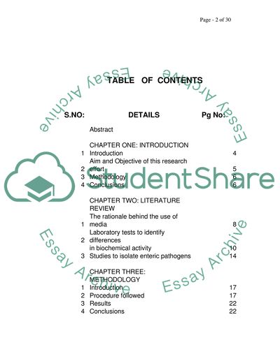
Enterobacteriaceae - Laboratory Tests to Identify Differences TABLE OF CONTENTS S.NO: DETAILS Pg No: Abstract CHAPTER ONE: INTRODUCTION 1 Introduction 4 2 Aim and Objective of this research effort 5 3 Methodology 6 4 Conclusions 6 CHAPTER TWO: LITERATURE REVIEW 1 The rationale behind the use of media 8 2 Laboratory tests to identify differences in biochemical activity 10 3 Studies to isolate enteric pathogens 14 CHAPTER THREE: METHODOLOGY 1 Introduction 17 2 Procedure followed 17 3 Results 22 4 Conclusions 22 BIBLIOGRAPHY 23 Abstract Enterobacteriaceae comprise that group of micro organisms that are causal agents in various diseases, notable intestinal diseases. The isolation and separation of strains is difficult, because these bacteria share so many similar characteristics. Experts have suggested different media in order to isolate specific strains of bacteria such as salmonella, among others. This research effort examines five different media and 12 strains of enterobactericeae and its finding is that MDCLS is the best medium of all five in effectively isolating the different strains, which is likely to be helpful in diagnosis when these bacteria are causal agents of disease. CHAPTER ONE INTRODUCTION 1.1: Introduction: Gram negative bacilli are found in the large intestines of human beings and they are (a) non sporing (b) non acid fast (c) sporing rods (d) motile through the use of flagella (e) aerobes and facultative anaerobes (f) ferment glucose with acid and gas and reduce nitrate to nitrite and most importantly function as opportunist pathogens in immunocompromised individuals. They are grouped together as Enterobacteriaceae because they exhibit similarity in morphological and biochemical properties similarities as well as DMA base compositions. (Bergeys manual, Kaufman, Edwards-Ewing) This family is subdivided into groups and tribes, genera, sub genera or species (Anant Paniker). Species may be further sub divided into biotypes, serotypes, bacteriophage types and colicintypes. However bacteria such as e.coli are pathogens that may be present in the enteric tract, urinary tracts, wounds appendices and the gall bladder. On the basis of their lactose action, they are divided into three groups: (i) lactose fomenters – examples: Escherichia, Klebseilla (ii) late lactose fomenters – examples: shingella sonnei and paracolons (iii) Non lactose fomenters – examples: Salmonella, Shigella and Proteus On a scientific basis, they may be classified as follows: Domain Bacteria Phylum Proteobacteria Class Gamma proteobacteria Order enterobacteriales Family enterobactaeriaceae Genera Escherichia Salmonella Shigella Yersinia 1.2: Aim and Objective of this research effort: Since enterobacteriaceae are primarily found in the intestines, they are important because they cause food to spoil and contaminate water through faeces, being pathogenic organisms that cause disease in the enteric tract, Others are ubiquitous and are found in soil, plants and water, causing trauma to mucus membranes. The coloform salmonella causes diarrhoea and enteric fevers, while shigella causes bacillary dysentery, Yersinia pestis causes bubonic and pneumonic plague, Yersinia enterocolitica may cause an appendicitis-like condition, vibrio cholera causes pandemics and vibrio parahaemolyticus causes gastroenteritis. Therefore, the aim of this research effort is to identify the different media that may be used to isolate enteric bacteria using the properties of the organism. The recognition of enteric bacteria in clinical and environmental samples leads to identification of strains and facilitates fast and effective disease diagnosis. This is also helpful in identifying the possible source of infection and in taking preventive measures where necessary. When the right causative organism is identified, it facilitates the administration of appropriate antibiotics. Through the use of selective and differential media, the pathogenic bacteria can be isolated from the other species of enterobacteriaceae. While there are several different pathogenic species, this research effort will focus upon the identification of salmonella and shigella since these are the causative strains responsible for most of the common diseases caused by infections in the intestinal tract. 1.3: Methodology: The methodology utilized in this research effort is an empirical, laboratory based plating of organisms belonging to the family enterobacteriaceae on different growth media by using Desoxycholate as the comparative measure. The four different media that were selected were (a) MacConkeys agar without salt (b) XLD agar-xylose lysine Desoxycholate agar (c) DCA agar- Desoxycholate citrate agar and (d) DCLS agar- Desoxycholate citrate lactose agar. One medium was independently prepared in the laboratory and this was MDCLS Agar. Twelve different strains of enterobacteriaceae were examined through this method. 1.4: Conclusions: This study enabled a comparison of five different media in order to assess which one was the best in providing a distinct identification of enterobacteriaceae. It was noted that the MDCLS agar medium that was used provided the clearest degree if isolation of the bacteria strain, by demonstrating clearly identifiable colonies, thus supporting the conclusion that this agar medium may be the most efficient when dealing with a sample where there may be multiple species present and clear isolation may be difficult. Since each of the strains was prepared as mixtures of three different species of bacteria, therefore the results support the use of the MCDLS medium as the best medium of the five used in this study. CHAPTER TWO LITERATURE REVIEW 2.1: The rationale behind use of media: Gram stained morphology is not successful in identifying strains of enterobacteriaceae. Endotoxins are heat-stable lipopolysaccharides produced by the outer membrane of all gram-negative bacteria and endotoxic shock as a result of gram-negative infection may be fatal and life-threatening, caused by enterobacteriaceae. The detection of endotoxin is by Limulus test, while new tests for detecting gram-negative sepsis are based on the development of monoclonal antibodies specific for lipid-A. On a solid growth media, an organism may grow as a colony which can provide some clues about size, shape, color and texture as well as reaction to the media used. However on blood agar plates, enterobacteriaceae grow in the form of as large, dull grey, dry or mucoid colonies. Therefore, performing haemolysis on the Blood agar plate may not be useful in distinguishing strains of Enterobacteriaceae, but the colonies appearing as waves provide an indication of the presence of the proteus species. However the presence of red colonies is an indication of the presence of lactose fermenting bacteria and therefore can differentiate the enterobactariaceae. The method of classification of enterobacteriaceae on the basis of their lactose action offers scope for strain identification. For example, salmonella and shigella are non lactose fermenting coliforms which are isolated from the feaces while lactose fermenting coliforms are isolated from specimens which are normally sterile like and are found in blood and urine. The enumeration of coliform bacteria is therefore dependent upon the lactose production with a simultaneous production of acid and gas. Through the addition of bile, Desoxycholate, Brilliant green and other substances, the growth of gram-positive bacteria can be restricted so that gram negative strains can be easily identified. E.Coli, Klebseilla and Enterobacter are true coliforms that ferment lactose vigorously and produce gas at 35-37 C within 24-48 hours. Those enterobaceriaceae which have minimal fermentation of lactose or are akin to non fermentors on media, such as Proteus, Morganella and Providencia may thus be used for the isolation of Shigella and Salmonella. These are unique in possessing enzyme Phenylalinine deaminase which can be easily tested.(Lindquist,1998). Enteric bacteria contain pyruvates and mixed acid fermentation converts the pyruvates by glycolytic pathways into acids, while the butanediol type of fermentation converts the pyruvate to neutral products. The net results of these reactions are evident on the biochemical media tested. Lactose is a disaccharide of glucose and galactose and its metabolism requires the presence of two enzymes.-b-galactoside permease and b-galactosidase.B-galactoside permease facilitates the transfer of lactose across the cell membrane whereas B-galactosidase hydrolyses the bond that holds the two sugars together. As a result, glucose is generally omitted from the primary isolation media that are used, such as MacConkeys Agar and EMB Agar. Late lactose fermenting organisms are thought to have normal B-galactosidase activity, but sluggish B-galactosidase permease activity. Therefore, the ONPG [o-nitro phenyl b-d galactopyrinoside] test is very useful to determine late lactose fermentors, such as Citrobacter and Salmonella Arizona (ONPG-+ve) from shigella and other salmonella species (ONPG –ve) 2.2: Laboratory tests to identify differences in biochemical activity: Escheria Coli is used as an index of faecal contamination. When evaluating the bacteria in the water supply, microbiologists look for E.coli and not for Salmonella or Shigella. (MMWR 1996). The characteristics of the.E.coli colony are such that on EMB agar, it grows with a typical metallic sheen. On Blood agar E.coli grows as Beta-hemolytic grey convex colonies. On MacConkeys agar it grows as flat lactose fermenting pink colonies, similar to the growth on DCA, SS, DCLS agar. The pink colour of the colony could be diffusing into the medium. On XLD agar E.coli grows as yellow convex colonies, while on Hektone enteric agar it grows as orange colonies. The tribe of Proteus are non coliforms, which ferment lactose very slowly or not at all. An analysis of Proteus in the laboratory reveals a UTI with alkaline urine. On Blood agar, the colony morphology is such that it appears as a swarming growth due to the motility of the organisms. On McConkey agar, it forms non lactose fermentor colonies. It produce H2S, seen as black precipitate on colonies on DCA agar, XLD, agar, DCLS agar. It is urease positive, with a distinct odour. It is PDA, LDA, TDA positive and LDC negative The tribe of shigella are gram-negative non-motile, non-lactose fermenting oxidase negative, non-capsulated bacilli. They are limited to human and primate enteric pathogens. (Russell 2004). They are almost always isolated from faeces and they do not cause urinary or generalized invasive infection (Bryan FL 1979). Classification of Shigella is based on biochemical and serological tests. They have somatic ’O’ polysaccharide antigen in their cell wall and some shigellas have ‘K’ sheath antigen. Blood agar, MacConkeys agar and other isolation media such as XLD, SS, HE, DCA, DCLS, in addition to enrichment broth like GN or selenite broth are used. On blood agar it grows as colorless colonies. On MacConkeys agar shigella grows as lactose negative colorless colonies, which is similar to the growth as seen on DCA, DCLS, and SS agar. On XLD agar it grows as colorless to light pink colonies. On HE agar it appears as green colonies. Klebseilla pneumoniae are gram-negative, lactose fermenting rods, living as saprophytes in human GI tract. Klebseilla are facultative anaerobes and oxidase negative. They utilize citrate and are positive for urease and lysine decarboxylase. They differ from E.coli in their IMViC reaction, since E.Coli is IMVic++--, while Klebseilla is IMViC --++. They are non motile rods. As far as their colony characteristics are concerned, on blood agar it gives a growth of large mucoid grey colonies, On MacConkeys agar large mucoid lactose fermenting red colour colonies, on DCA, SS, DCLS it shows pink LF colonies and on XLD and HEA it shows yellow mucoid colonies and yellowish green colonies. Enterobacter, Serratia and Citrobacter are not usually primary pathogens, but are characteristically involved as secondary opportunistic pathogens. The culture of Serratia may show red pigmentation, while Enterobacter sakazaki may be seen as yellow pigmented variant of Ent.cloacae. Citrobacter species may sometimes with other H2S producing colonies of Salmonella (which is NLF, H2S+) However Citrobacter freundi is ONPG +, LDC+ which are opposite reactions to salmonella and may serve to distinguish between the two. The salmonella organism is gram-negative motile rod, it does not ferment lactose, it is facultative and oxidase negative. Salmonella are fermentative with production of acid and gas. Its classical feature is production of H2S, which is seen on agars using desoxycholate and thiosuphate. Selective agars like Brilliant green agar or desoxycholate agars are used to isolate salmonella, since they inhibit coliforms but do not inhibit salmonella. Laboratory tests conducted to detect the presence of salmonella are generally based upon the kind of infection that the person is suffering from. For instance, stool and blood cultures are used for gastroenteritis, Widal test and blood cultures are used for enteric fever, while blood, stood, urine and clot cultures are used for speticemia. Samples from Blood, stool, and urine may be cultured repeatedly and isolated on to an enrichment media and differential media. In so far as salmonella colonies are concerned, On MacConkeys agar, it grows as non-lactose fermenting colorless colonies, on HEA, it develops blue-green colonies with a black center, on SS, DCA, DCLS, it forms colorless colonies with black center, on XLD agar it grows as red colonies with black center and on Bismuth sulphide agar it forms black colonies with metallic sheen. Enterobacter is a common human pathogen that causes upper respiratory tract infections and neonatal meningitis. Enterobacter grows as red to pink lactose fermenting colonies on MacConkeys agar, DCA, SS, XLD and DCLS agar. Organisms belonging to genus citrobacter are found in stool as normal flora. They cause UTI, wound infections, bacteraemia and neonatal meningitis. They are slow lactose fermentors on MacConkeys agar, ONPG positive, LDC negative and H2S positive. Colonies may be similar to Salmonella, but they are ONPG positive and LDC negative. They exhibit resistance to Ampicillin and first generation cephalosporins. Pseudomonas is considered among the medically important gram-negative environmental bacteria. are motile, obligate aerobic bacilli with limited fermentation activity and can survive over a wide range of temperature ( between 5C to 42 C).They have simple nutritional requirements which allow them to flourish in any moist environment. On solid media, Pseudomonas grows as large irregular colonies. It grows as pigmented colonies on Blood agar and other differential and routine media. The pigment is usually bluish green and is known as pyocyanin or yellowish green known as fluorescein. Pseudomonas grows with a fruity smell - an oxidase test, is strongly positive, and it shows an inert reaction to the API test. Pyocyanin typing is done in reference laboratories. Selective isolation is done on cetrimide containing agar medium, which inhibits many other organisms but stimulates pseudomonas to produce pyocyanin and fluorescein. 2.3: Studies to isolate enteric pathogens: Several studies have been performed in order to isolate two enteric pathogens – shingella and salmonella. The medium salmonella-shingella was used and showed a recovery rate of organisms of 60% (UK1). However other studies have contradicted the use of the SS medium because of the low isolation rate of Shingella as compared to Salmonella(4). However Hekteon Enteric Agar has been shown to have a higher level of selectivity for Shingella, as well as for other organisms; using a 1.5% bile salts concentration was also found to be completely inhibitive of e.coli and proteus mirabilis and of citrobacter to a considerable degree (19). The advantages of using Hekteon Enteric Agar are that it is highly selective and requires less effort, however its efficacy is not high in isolating salmonella from food products like Brilliant Green Agar(20). However by using a new plate medium, the salmonella species can be isolated with no false positives, through the phenotype characteristics, where acid is formed from propylene glycol in combination with beta-galactosidase - a chromogenic indicator – to clearly identify salmonella from proteus.(14). This method does not test lactose utilization, but rather is based upon the fact that all other species except for salmonella typhi produce bright red colors – while salmonella is colorless. Where primary plating of stool specimens is concerned, CHROM Agar Salmonella (CAS) has been found to be preferable to Hekteon Enteric Agar because (i) it is less time consuming (ii) displays the characteristic mauve colonies and (iii) generates fewer false positive results (iv) higher sensitivity and specificity as compared to SS Agar with an increased incubation period of 24-48 hours (6) (v) allows for the performance of rapid and inexpensive confirmatory tests, such as the disk oxidase test or direct microscopy. ABC has also been suggested as another new chromogenic medium which exploits the presence of alpha-galactosidase activity of Salmonella species and produces green colony of Salmonella species with high sensitivity and specificity – however this is limited in that the S. typhi cannot be distinguished from the other enteric organisms (23). However other studies have contraindicated the use of the ABC medium for direct isolation of Salmonella species, due to its susceptibility to competing flora and mannitol lysine crystal violet brilliant green (MLCB) or Xylose Lysine Deoxycholate (XLD) is preferable for use, either singly or in combination (when such use is justified), for direct isolation of salmonella with highest recovery rate (UK3). Another study corroborating this opinion, stated that some of the strains of Salmonella did not show any alpha galactosidase activity (22) and that put the limitation of the use of ABC medium even though it has the best diagnostic performance when compared with COMPASS Salmonella agar and CHROMagar Salmonella Agar (13). Similarly, Rambach Agar has also been suggested for the detection of the salmonella sub-species I to IV using the color colony, however the color criterion cannot be applied in the case of subspecies IIIa, IIIb and V, which produce Beta-galactosidase which makes them indistinguishable from e.coli and other lactose fermenter organisms.(UK2). On the other hand, Chromogenic Samonella esterase (CSE) Agar can distinguish Salmonella species with better specificity and sensitivity as compared to Rambach agar(UK3). Therefore, on an overall basis, it appears that COMPASS and CHROMagar Salmonella agar can be considered the best single-plate media for the detection and presumptive identification of Salmonella due to the close efficiencies of these two media. Where the salmonella species needs to be specifically isolated through the direct streaking of feaces and overnight enrichment with Selenite F broth. Brilliant Greeen Sulphite (BS) is recommended to be the best due to its 100% recovery and isolation rate of the salmonella typhi. The next best medium is Novobiocin-Brilliant Green-Glucose Agar, which has a distinct advantage over other enteric media in its differential colonial morphology. The second advantage is that direct isolation of Salmonella spp. from human stool samples can be obtained with the highest sensitivity and positive predictive values (PPV) (61%) using novobiocin-brilliant green-lactose agar (NBGL) medium (2).But the medium showed the worst sensitivity in direct plating for the isolation of non-typhoid Salmonella spp. from stool samples even after enrichment (1). CHAPTER THREE METHODOLOGY 3.1: Introduction: Since the aim and objective of this research study was to examine different media that may be utilized for the isolation of different strains of enterobacteriaceae, a laboratory method was deemed to be the most effective research method. Prior to embarking on the laboratory aspects, the researcher first undertook a literature review of the properties of various organisms. The literature review also examined other studies that have been done for the isolation of species such as salmonella and shingella wherein certain media have been identified as being more conducive to isolation of different strains on the basis of the properties of the organisms. 3.2: Procedure followed: The four different media that were obtained ready made and prepared contained the following compositions, and the directions for preparation as provided are also included: MacConkeys agar (without salt Formula gm/liter Peptone 20.0 Lactose 10.0 Bile salts 5.0 Neutral red 0.075 Agar 12.0 pH 7.4 + or –0.2 Directions;- Suspend 47 grams in 1 litre of distilled water. Bring to boil to dissolve completely. Sterilise by autoclaving at 121 c for 15 minutes. Mix well before pouring. Dry the surface of the gel before inoculating the plates. Appearance of the plates: Dark red coloured plates XLD Medium; - Xylose lysine desoxycholate agar medium. Formula gms/litre Yeast extract 3.0 L-lysine Hcl 5.0 Xylose 3.75 Lactose 7.5 Sucrose 7.5 Sodium desoxycholate 1.0 Sodium chloride 5.0 Sodium thiosulfate 6.8 Ferric ammoniumcitrate 0.8 Phenol red 0.08 Agar 12.5 PH 7.4 ,+ or –0.2 Directions Suspend 53 grams in 1 litre of distilled water. Heat with frequent agitation. As soon as the medium cools, pour onto plates. Dry the agar surface before use. Appearance of plates; Red coloured gel. DCA agar-Desoxycholate citrate agar (Hynes) Formula gms/litre Lab-lemco powder 5.0 Peptone 5.0 Lactose 10.0 Sodium citrate 8.5 Sodium thiosulphate 5.4 Ferric ammoniumcitrate 1.0 Sodium desoxycholate 5.0 Nuetral red 0.02 Agar 12.0 PH 7.3 + or –0.2 Directions Suspend 52.0 grams of agar in 1 liter of distilled water . Bring to boil, with frequent agitation.to dissolve completely. Do not autoclave. Pour the plates. Dry the agar surface before use. Appearance of plates; Pink coloured gel. DCLS agar; Desoxycholate citrate lactose sucrose agar Formula gm/litre Special peptone 10.0 Sodium citrate 10.5 Sodium thiosulphate 5.0 Lactose 5.0 Sucrose 5.0 Sodium desoxycholate 2.5 Neutral red 0.03 Agar 12.0 PH 7.2 +/- 0.02 Directions Suspend 50.0 grams in 1 litre of distilled water. Bring to boil to dissolve the medium completely. Cool to 50 C and pour plates. Dry the surface before use. Appearance of plates; - Red coloured gel. MDLS agar; Modified Desoxycholate citrate lactose Agar. Formula gms/litre Lactose 10.0 Peptone 10.0 Glucose 1.0 Sodium desoxycholate 0.5 Sodium thiosulphate 0.5 Ferric ammoniumcitrate 0.5 Sodium chloride 3.0 Neutral red 0.033 Agar 15.0 PH 7.5 +/- 0.1 Direction The above ingredients were dissolved in 1 litre of distilled water and the pH of the mixture was adjusted by adding 10% NaOH. The medium was then autoclaved at 121 C for 15 mins. When cooled to 70 C, 20 ml was poured into sterile petri plates and stored in refrigerator. Appearance of plates; - Red coloured gel. Twelve different test organisms were used in this study. The five lactose fermenting organisms that were used were 414- E.coli, 415- E.coli (mucoid), 169- Klebseilla aerogenes, 173- Enterobacter cloacae and 413- Citrobacter freundii, The non lactose fermenting organisms that were used were: 129- Proteus mirabilis, 131- Proteus vulgaris 233- Salmonella enteritis, 340- Salmonella typhi, 200- Shigella sonnei, 237- Pseudomonas aeruginosa,145- Pseudomonas aeruginosa. The MCDLS agar prepared in house was stored in the refrigerator and used within a period of 14 days after preparation. All the test organisms were stored at –80 C in lyophilised ampoules. These were among the culture collection library of Manchester University, Department of Medical Microbiology. After reconstution the cultures were grown on Columbia Blood agar and Muller hinton agar and the viability and sterility of the cultures were checked. Cultures were tested by API 20 E and the strain was verified. Using nutrient broth as the suspension medium, the test organisms were converted into suspensions and adjusted using standard McFarland turbidity controls. Ten separate mixtures were made with each comprising three separate types of organisms. For each media, a calibrated loop was used to perform inoculation. Before the process of inoculation, the surface of each plate was dried in an incubator at 370C and each mixture was inoculated on the five media at the same time. The plates were incubated for 24 hours and 48 hours and the description of the colony appearance as well as the nature of the precipitate produced was recorded at the end of 24, and 48 hours. The colony characters were then compared with the literature as described by Oxoid. For the MDCLS agar medium however, the appearance of the colony was noted at the end of 24 and 48 hours, and then compared with the characteristics as described by Arikan et al, since the MDCLS Agar media was a modification of the DCLS agar proposed by Arikan et al. The growth characteristics of the different organisms were evaluated using a polymicrobial inoculam. The focus was to note which media were able to highlight the characteristics of a particular organism in the mixtures. The type of agar that was able to produce identifiable colonies was also noted, and in particular the production of precipitates or diffusion around the colony on a particular medium. Pink color diffusion was noted among lactose fermentors, while it was also observed that non lactose fermentors producing H2S produced black colonies due to the production of sulphide from thiosulphate. At the end of the study, all the 122 strains were suspended in 1 ml of 15% glycerol in nutrient broth and stored at -150C for future reference. 3.3: Results: Based upon the extend to which colony characteristics were clearly identifiable through the use of different media, the efficacy of all the various media were compared. The home prepared MDCLS media was shown to be superior to the other media, because it was able to produce identifiable colonies, which helped to positively identify the enterobacteriaeae strains. 3.4: Conclusions: This study has carried out a laboratory process using five different media and it has been clearly established that not all media are the same. The MDCLS media has been shown to have the best isolation rate in identifying enterobactariaceae such that the strains can be separated. This serves to establish the importance of examining more media in a laboratory setting in order to discover which media may best promote the isolation and identification of enterobacteriaceae. The identification of such media would considerably facilitate the process of disease diagnosis through the isolation of different strains. BIBLOGRAPHY [1]. Dusch, Herbert and Altwegg, Martin, 1995. “Evaluation of five new plating media for isolation of Salmonella species” Journal of Clinical Microbiology, 33(4): 802-804 [2]. Ruiz, Joaquin, Nunez, Maria-Luz, Diaz, Julian, Lorente, Isabel, Perez, Jeronimo and Gomez, Joaquin, 1996. “Comparison of five plating media for isolation of Salmonella species from human stools.” Journal of Clinical Microbiology, 34(3): 686-688 [3]. Novicki, Thomas J, Daly, Judy A, Mottice, Susan L and Carroll, Karen C, 2000. “Comparison of Sorbitol MacConkey Agar and a two step method which utilizes enzyme linked immunosorbent Assay toxin testing and a chromogenic Agar to detect and isolate enterohemorrhagic Escherichia Coli”, Journal of Clinical Microbiology, 38(2): 547-551 [4]. Pollock, Helen, M and Dahlgren, Bonnie J, 1974. “Clinical Evaluation of enteric media in the primary isolation of Salmonella and Shigella.” Applied Microbiology, 27(1): 197-201 [5] D’Mello, Anthony and Yotis, William W, 1987. “The action of Sodium Deoxycholate on Escherichia Coli” Applied and environmental Microbiology, 53(8):1944-1946 [6] Maddocks, Susan, Olma, Tom and Chen, Sharon, 2002. “Comparison of CHROMAgar Salmonella medium and Xylose-Lysine-Desoxycholate and Salmonella-Shigella agars for isolation of salmonella strains from stool samples.” Journal of Clinical Microbiology, 40(8): 2999-3003 [7] Hansen, D.S., Jensen, A.G, Nerskov-Lauritsen, N. Skov, R and Bruun, B,. “Direct identification and susceptibility testing of enteric bacilli from positive blood cultures using VITEK (GNI+/GNS-GA) [8] Closs, O, 1971. “Two tube test for the rapid identification of prompt lactose fermenting genera within the family Enterobacteriaceae” Applied Microbiology, 22(3): 325-328 [9] “Evaluation of BBL CHROMagar Orientation Medium for detection and presumptive identification of urinary tract pathogens.” Journal of Clinical Microbiology, 35(11): 2773-2777 [10] Thaller, Maria Christina, Berlutti, Francesca, Pantanella, Fabrizio, Pompei, Raffaello and Satta, Guiseppe, 1992. “Modified MacConkey Medium which allows simple and reliable identification of Providentia Stuartii.” Journal of Clinical Microbiology, 30(8): 2054-2057 [11] Filius, P.M.G., Netten, D. Van, Roovers, P.J.E., Vutto, A.G. and Gyssens, I.C., 2003. “Comparative evaluation of three chromogenic agars for detection and rapid identification of aerobic gram negative bacteria in the normal intestinal microflora.” Clinical Microbiology and Infection, 9(9): 912 [12] Cassar, Robert and Cuschieri, Paul, 2003. “Comparison of Salmonella chromogenic medium with DCLS agar for isolation of salmonella species from stool specimens.” Journal of Clinical Microbiology, 41(7): 3229-3232. [13] Perez, J.M., Cavalli, P, Roure, C, Renac R, Gille, Y and Fredieyere, A.M., 2003. “Comparison of four chromogenic media and Hekteon Agar for detection and presumptive identification of Salmonella strains in Human Stools.” Journal of Clinical Microbiology, 41(3): 1130-1134 [14] “New Plate medium for facilitated differentiation of salmonella spp from Proteus spp and other enteric bacteria.” Applied and environmental microbiology, 56(2): 301-303 [15] O’Hara, Caroline Mohr, Brenner, Frances W and Miller, Michael, J, 2000. “Classification, identification and clinical significance of proetus, providencia and Morganella” Clinical Microbiology Reviews, 13(4): 534-546 [16] Iversen, Carol, Druggan, Patrick, and Forsythe, Stephen, 2004. “A selective differential medium for Enterobacter sakazkii: a preliminary study” International Journal of Food Microbiology, 96(2): 133-139 [17] Pao, Stephen, Patel, Dhartika, Kalantari, Aref, Tritschler, Joseph P and Wildeus, Stephan, 2005. “Detection of salmonella strains and escherichia coli: O157:H57 in feces of small ruminants and their isolation with various media.” Applied and Environmental Microbiology, 71(4): 2158-2161 [18] Devenish, J.A., Cierin, B.W. and Brodsky, M.M., 1986. “Novocin brilliant green glucose agar –new medium for isolation of Salmonellae.” Applied and environmental microbiology, 52(3): 539-545 [20] Goo, Velma Y L, Ching, George Q.L., and Gooch, John M, 1973. “Comparison of Brilliant green agar and Hekteon enteric agar media in the isolation of salmonellae from food products.” Applied Microbiology, 26(3): 288-292 [21] Wilson, G, 2004. “Rapid and economical method for biochemical screening of stool isolates for Salmonella and Shigella species.” Journal of Clinical Microbiology, 42(10): 4821-4823 [22] Perry, J.D., Riley, G. Gould, F.K., Perez, J.M. Boissier, E, Ouedraogo, R.T. and Freydiere, A.M., 2002. “Alafosfalin as a selective agent for isolation of salmonella from clinical samples.” Journal of clinical microbiology, 40(11): 3913-3916 [23] Perry, John D, Ford, Michael, Tayloe, Jeffrey, Jones, Amanda L, Freeman, Roger, and Gould, Frances K, 1999. “ABC Medium: Anew chromogenic agar for selective isolation of salmonella spp” Journal of clinical Microbiology, 37(3): 766-768 [24] Gaillot, Oliver, Di Camillio, Patrick, Berche, Patrick, Courcol, Rene and Savage, Collette, 1999. “Comparison of CHROMagar Salmonella medium and Hekteon enteric agar for isolation of salmonella from stool samples.” Journal of Clinical Microbiology, 37(3): 762-765 [25] Cassar, Robert and Cuschieri, Paul, 2003. “Comparison of Salmonella chromogenic medium with DCLS agar for isolation of salmonella species from stool specimens.” Journal of Clinical Microbiology, 41(7): 3229-3232. [26] Perez, J.M., Cavalli, P, Roure, C, Renac R, Gille, Y and Fredieyere, A.M., 2003. “Comparison of four chromogenic media and Hekteon Agar for detection and presumptive identification of Salmonella strains in Human Stools.” Journal of Clinical Microbiology, 41(3): 1130-1134 [27] Dunne, Corwin and Martin, William, L, 1971. Comparison of media for isolation of Salmonellae and Shigella from Fecal specimens.” Applied Microbiology, 23(1): 17-22 [28] Kuhn, H, Wonde, Barbel, Rabsch, W and Reissbrodt, R, 1994. “Evolution of Rambach agar for detection of salmonella subspecies I to VI” Applied and environmental Microbiology, 60(2): 749-51 [29] Cooke, Venitia M, Miles, R.J., Price, R.G., and Richardson, A.C., 1999. “A new chromogenic ester agar medium for detection of salmonellae.” Applied and environmental microbiology, 65(1): 807-812 [30] Nye, K.J., Falton,D, Forsham, D, Gee. B, Graham, C, Hoew, S, Messer, S, Turner T and Warren, R.E., 2002. Journal of Clinical Pathology, 55: 286-288 [31] Dusch, Herbert and Altwegg, Martin, 1993. “Comparison of Rambach Agar, SM-ID Medium, and Hekteon Enteric agar for primary isolation of non typhi salmonellae from stool samples.” Journal of Clinical Microbiology, 31(2): 410-412 [32] Ohkusu, Kiyofumi, 2000.”Cost effective and rapid presumptive identification of gram negative bacilli in routine urine, pus and stool cultures: Evaluation of the use of CHROMagar orientation medium in conjunction with simple biochemical tests.” Journal of Clinical Microbiology, 38(1): 4586-4592 [33] Kocagor, Sevin, Badak, Fatma and Gur, Denir, 2006. “Evaluation of Chromogenic medium for rapid detection of extended spectrum Beta lactamase producing salmonella spp” Indian Journal of Medical Research, October: 443-446 Read More
Cite this document
  • APA
  • MLA
  • CHICAGO
(Enterobacteriaceae - Laboratory Tests to Identify Differences Research Paper, n.d.)
Enterobacteriaceae - Laboratory Tests to Identify Differences Research Paper. Retrieved from https://studentshare.org/health-sciences-medicine/1708224-new-selective-media-for-the-identification-of-enterobacteriaceae
(Enterobacteriaceae - Laboratory Tests to Identify Differences Research Paper)
Enterobacteriaceae - Laboratory Tests to Identify Differences Research Paper. https://studentshare.org/health-sciences-medicine/1708224-new-selective-media-for-the-identification-of-enterobacteriaceae.
“Enterobacteriaceae - Laboratory Tests to Identify Differences Research Paper”, n.d. https://studentshare.org/health-sciences-medicine/1708224-new-selective-media-for-the-identification-of-enterobacteriaceae.
  • Cited: 0 times

CHECK THESE SAMPLES OF Enterobacteriaceae - Laboratory Tests to Identify Differences

Laboratory Testing and Evaluation Report

laboratory Testing and Evaluation Contents Number Topic Pages 1 Introduction 1-2 2 Structure of Materials 3 3 Expected Theoretical Results 4-5 4 Actual Results 6-7 5 Conclusion 8 Bibliography 9 Introduction The laboratory testing and evaluation study is focused on two material grades specified in JIS G3141 (2011).... Mechanical properties are to be assessed in this laboratory testing and material evaluation case study....
6 Pages (1500 words) Essay

Clinical and Laboratory Diagnosis of Salmonellosis

iochemical tests:KIAGasH2SMRVPIndCitUreMotPadLysArgOrnONPGK/A+++--+-+-++/-+-Serological tests of Salmonellosis: Widal test (H and O agglutination for typhoid and paratyphoid patients), CIEP, Haemagglutination, ELISA, Bactericidal Antibody test, Adherence test for detection of IgM antibodies, RIA, Co-agglutination test, Latex agglutination test, PCR, Diazo test of Urine, bacteriophage typing (Josephine A.... erological tests of Shigellosis: Slide agglutination with antisera for serogroup and serotype, PCR, ELISA, Monoclonal Antibodies test....
4 Pages (1000 words) Essay

Causality and Inference: Tests of Difference and Relationship

The presence of a weapon word (such as “dagger” or “bullet”) should increase the accessibility of an aggressive word (such as… This study by Anderson, Benjamin, and Bartholow (1998) tested this hypothesis. In this project, data is used from the case, Weapons and Aggression.... The study consisted of 32 subjects (17 male and 15 female) with average age about 24 years ranging Each subject named both aggressive and non-aggressive words following both weapon and non-weapon “primes”....
5 Pages (1250 words) Assignment

Pathogenic Microorganisms

The differential staining of the bacteria helps to identify the unknown bacteria present in the sample.... lot of tests were done to determine the presence of the bacterial load in the microorganisms.... Some of them include Gram staining, Catalase test, Oxidase test, coagulase test, API Strip test, the effect of disinfectant hypochlorite, cleaning agents, and morphological tests.... he presence of the pathogens is found using the biochemical tests and the API strip test....
6 Pages (1500 words) Term Paper

Laboratory Scenarios

Safety precautions in the laboratory are an essence that has to be adhered to by anyone who is concerned with chemicals and other reagents especially in the chemistry laboratory.... The chemicals and other reactants found in these laboratories pose a great danger to the personnel… These risks may range from inhaling poisonous and irritating gases, burns from chemicals that are highly reactive and other far reaching implication that can be caused by the According to the National Institute for Occupational safety and Health (2006), anyone entering or using the lab has to have detailed knowledge of the dangers and risks involved, and the risks presented by each of the reagents in the laboratory....
5 Pages (1250 words) Essay

Scientific Lab

The purpose of the experiment was to identify unknown bacteria from mixed culture #18 using biochemical and morphological test: The tests done include: Culturing and Gram staining for Morphology.... With regards to the aim of the experiment, which is to identify unknown bacteria from mixed culture #18 using biochemical and morphological test, the two organisms identified had distinct characteristics which were looked for following laboratory tests.... The biochemical tests were: Lactose fermentation, Tryptophan digestion (Indole),… Basing on the results the most likely bacteria were: for the unknown A, was Klebsiella pneumoniae which is a typical rod shaped bacterium, a Gram-negative, lactose fermenting, facultative anaerobic, encapsulated and non-motile....
4 Pages (1000 words) Research Paper

Enterotube case study

In order to reach a proper diagnostic conclusion several tests needs to be conducted to accurately identify the exact species of the pathogenic bacteria responsible for causing the symptoms in the patient.... Even though the procedure is less reliable than conventional molecular methods yet the advantage of using this system lies in the fact that all the tests can be done simultaneously using just one single colony (Grunberg et al, 1969, p....
4 Pages (1000 words) Lab Report

The Laboratory Technique to Grade Soil

This paper "The Laboratory Technique to Grade Soil" focuses on the fact that in geotechnical engineering different laboratory techniques/tests are used to determine the properties of the soil.... The laboratory techniques that mostly used are Compression, Atterberg limit, Cone penetrometer test....
6 Pages (1500 words) Lab Report
sponsored ads
We use cookies to create the best experience for you. Keep on browsing if you are OK with that, or find out how to manage cookies.
Contact Us