StudentShare
Contact Us
Sign In / Sign Up for FREE
Search
Go to advanced search...
Free

IRAK-1 Bypasses Priming and Directly Links TLRS to Rapid - Research Paper Example

Cite this document
Summary
According to the research findings "IRAK-1 Bypasses Priming and Directly Links TLRS to Rapid ", the differential requirement for IRAK-1 in the early and late pathways allowed the researchers to investigate the importance of rapid NLRP3 inflammasome pathway in vivo using IRAK-1–deficient animals…
Download full paper File format: .doc, available for editing
GRAB THE BEST PAPER93% of users find it useful
IRAK-1 Bypasses Priming and Directly Links TLRS to Rapid
Read Text Preview

Extract of sample "IRAK-1 Bypasses Priming and Directly Links TLRS to Rapid"

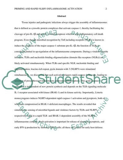
IRAK Bypasses Priming and Directly Links TLRS to Rapid NLRP3 Inflammasome Activation Tissue injuries and pathogenic infections always trigger the assembly of inflammasomes that is defined as cytosolic protein complexes that activate caspase-1, thereby facilitating the cleavage of pro-IL-1β and pro-IL -18 and pyroptosis which is a pro inflammatory cell death program. Even though microbial recognition by Toll including receptors (TLRs) is known to induce the synthesis of the major caspase-1 substrate pro-IL-1β, the function of TLRs is considered limited in up-regulation of the inflammasome components. During a virulent microbe infection, TLRs and nucleotide-binding oligomerization (domain-like receptors (NLRs)) are likely activated simultaneously. When TLRs and specific NLR; nucleotide binding and oligomerization, leucine-rich repeat, pyrin domain-with 3 (NLRP3) were stimulated simultaneously, we discovered that such activation triggers rapid caspase-1 cleavage, leading to secretion of presynthesized inflammatory molecules and pyroptosis. This acute caspase-1 activation is independent of new protein synthesis and depends on the TLR-signaling molecule IL-1 receptor-associated with kinase (IRAK-1) and its kinase activity. Importantly, Listeria monocytogenes induces NLRP3-dependent rapid caspase-1 activation and pyroptosis, both of which are compromised in IRAK-1-deficient macrophages. The results revealed that simultaneous sensing of microbial ligands and virulence factors by TLRs and NLRP3, respectively, leads to a rapid TLR- and IRAK-1-dependent assembly of the NLRP3 inflammasome complex. Such activation is important for release of alarmins, pyroptosis, and early IFN-γ production by memory CD8 T cells; all these are critical for early host defense. 1. Simultaneous Stimulation of TLRs and NLRP3 Leads to Acute Inflammasome Activation. Bone marrow was extracted from the experimental bones, erythrocytes were lysed, and the white cells were seeded in tissue culture dishes in media containing 10% fetal bovine serum and conditioned media containing mouse M-CSF. The remaining suspension cells were harvested the following day, and plated on fresh plates to faciliated selection by growth and adhesion. After 6 days, more than 99% of the surviving cells were macrophages and ready for stimulation. They were then divided into samples labeled A, B, C, D, E, F, G, and H. (A) BMDMs were stimulated with LPS, CpG, Poly I: C (pIC), R837, or Pam3CSK4 (Pam3) together with ATP for 30min, after which cell lysates were evaluated for caspase-1 cleavage by Western blot analysis. (B and C) BMDMs of indicated genotypes stimulated with LPS and ATP for 30 min were evaluated for caspase-1 cleavage by Western blot analysis. (D and E) BMDMs were treated with 50 ng/mL cycloheximide (CHX) (D) or10μM Bay11-7082 (E) for 60 min before stimulation with LPS or R837 together with ATP for 30 min, followed by evaluation for caspase-1 activation by Western blot analysis. (F) BMDMs from WT, IRAK-1 KO, and IRAK-2KO mice were stimulated with LPS together with ATP for 30 min and evaluated for caspase-1 activation by Western blot analysis. (G–I) BMDMs from the indicated mouse strains were left unprimed or were primed with LPS for 4 hours, followed by stimulation with ATP for 30 minutes. Lysates were probed for caspase-1 activation by Western blot analysis. Data are representative of three to five independent experiments. DKO, IRAK-1, and IRAK-2 double KO. Results The responses of bone marrow-derived macrophages (BMDMs) were tested to simultaneous stimulation of TLRs and NLRP3. WT BMDMs stimulated simultaneously with a TLR ligand and ATP for 30 minutes activated caspase-1. Ligands for TLR4, TLR9, TLR7, TLR2-LPS, CpG, R837, and Pam3CSK4—triggered rapid caspase-1 cleavage in BMDMs co-stimulated with ATP, but poly I: C, a TLR3 ligand, did not. Kinetically, stimulation of BMDMs with ATP and LPS for as little as 15 or 20 min led to rapid caspase-1 activation (SI Appendix, Fig.S1). Rapid inflammasome activation was abolished in both TLR-and NLRP3-deficient BMDMs, suggesting a necessary role for both TLRs and NLRP3 (SI Appendix, Fig. S2A–C). Interestingly, TLR4-driven rapid caspase-1 activation occurred only in Toll/IL-1 receptor domain-containing adaptor inducing IFN-beta (TRIF)KO BMDMs, and was absent in both myeloid differentiation primary response gene 88(MyD88) KO andMyD88/TRIF double-KO BMDMs (Mean et al., 2014). As noted previously, TLR3 signaling did not trigger rapid caspase-1 activation, suggesting that TRIF and its downstream components do not directly activate the NLRP3 inflammasome. Thus, rapid caspase-1 activation downstream of all TLRs depends entirely on the adapter MyD88. Previous studies have shown that TLR signals in both MyD88- and TRIF- dependent pathways (10) leads to NF-κB–dependent up-regulation of inflammasome components, particularly NLRP3, suggesting the need for inflammasome “priming” before activation. Combined stimulation of BMDMs with LPS and ATP, pre-treated with cycloheximide or an NF-κB inhibitor in (E), led to caspase-1 cleavage comparable to that seen in untreated cells, suggesting that rapid NLRP3 inflammasome activation is independent of “priming,” given that both NF-κ B activation and new protein synthesis are not necessary . Collectively, these data suggest that constitutive expression of NLRP3 (10) is sufficient to activate caspase-1 when cells receive signals from both TLR and NLRP3 ligands simultaneously. 2. Dependence of Rapid NLRP3 Inflammasome Activation on IRAK-1. In examining the mechanism of priming-independent caspase-1 activation, the study focused on the role of signaling components directly downstream of MyD88, in particular the IRAK family of molecules. This was also prompted by the finding that IL-1β, and not TNF-α, induced rapid caspase-1 activation. IL-1βbut not TNF-α can induce rapid NLRP3 inflammasome activation. BMDMs from WT mice were stimulated with LPS, TNF-α and IL-1βwith and without ATP for 30 minutes. Results Only LPS or IL-1β were able to induce caspase-1 cleavage. Macrophages express very low levels of IL-1R and this could explain the lower level of caspase-1 cleavage induced by IL-1 when compared to LPS. This suggests the existence of a TLR- and IRAK-1 dependent pathway that leads to rapid NLRP3 inflammasome assembly and caspase-1 activation. Interestingly, priming of macrophages for 4 hours with LPS abolished the requirement of IRAK-1 for caspase-1 cleavage. 3. IRAK-1 Associates with Inflammasome Components and Regulates NLRP3 Inflammasome Assembly Further tests were done to determine whether IRAK-1 interacts with inflammasome components. (A) IRAK-1 KO BMDMs were stimulated with LPS, ATP, or LPS plus ATP for the indicated times, and co-immunoprecipitation was performed by precipitating with anti-ASC, followed by Westernblot analysis for IRAK-1 and ASC. Light chain of the antibody. (B) Whole-cell lysates from WT BMDMs stimulated as described in A were blotted for the indicated proteins. (C) Whole-cell lysates from WTBMDMs stimulated with LPS alone or LPS together with ATP for the indicated times were immunoblotted for the indicated proteins. Data are representative of three independent experiments. (D and E) Immunostaining of endogenous ASC, IRAK-1 and NLRP3 in WT (D) and IRAK-1 KO (E) BMDMs stimulated with LPS and ATP for 15 min. (F) cells containing ASC or IRAK-1 specks in WT and IRAK-1 KOBMDMs. (G) Immuno staining of endogenous IRAK-1 and NLRP3 in IRAK-1 KD knock-in mouse BMDMs stimulated with LPS and ATP for 15 min. Results The result depicted an association of IRAK-1 with ASC when cells were stimulated with either ATP or a combination of LPS and ATP. It was found out that stimulation of WT BMDMs with LPS led to lack of IRAK-1 detection within15 minutes of TLR4 activation, but simultaneous exposure of BMDMs to LPS and ATP prevented polyubiquitination and degradation of IRAK-1 allowing it to associate with ASC. Interestingly, failure of IRAK-1 detection coincided with the absence of IκBα degradation at 15 min (Fig. 2B). Further kinetic analysis of IRAK-1 detection and IκBα phosphorylation degradation revealed that these events were delayed by simultaneous stimulation with LPS and ATP (Fig. 2C). These results suggest that combined activation of TLR and NLRP3 favors IRAK-1–ASC interaction and inflammasome activation, thereby delaying in NF-κB activation. 4. Is Rapid IRAK-1–Dependent NLRP3 Inflammasome Activation Important for Secretion of Presynthesized IL-18? The next investigation was on the physiological relevance of the rapid NLRP3 inflammasome activation pathway. Although they do not express pro-IL-1β, BMDMs are known to express pro-IL-18 without any TLR stimulation. In addition, caspase-1 activation triggers pyroptosis, a proinflammatory cell death program that could play a major role in host defense by eliminating infected cells and releasing inflammatory cellular contents. Results WT BMDMs secreted IL-18 but IRAK-1 KO BMDMs did not, despite similar pro-IL-18 expression levels. WT BMDMs failed to secrete IL-18 when stimulated only with LPS or ATP, suggesting the importance of simultaneous activation of TLR4 and NLRP3. IRAK-1 deficiency did not hinder the ability of TLR-primed BMDMs to secrete either IL-1β or IL-18, consistent with their ability to cleave caspase-1 when primed by TLR ligands. 5. L. Monocytogenes Induces Rapid IRAK-1–Dependent NLRP3 Inflammasome Activation and Pyroptosis. Given that many pathogens express both TLR and inflammasome activators, the experiment hypothesized that infection with a live pathogen would activate TLRs and NLRs simultaneously or in rapid sequence and induce acute inflammasome activation. The pathogen L. monocytogenes has been implicated in the activation of several inflammasome complexes including the NLRP3 inflammasome (Mean et al., 2014). Listeria monocytogenes infection induces NLRP3-dependent inflammasome activation and pyroptosis. WT or NLRP3 deficient BMDMs were infected with L. monocytogenes for 1 hour and analyzed for pyroptosis by (A) PI incorporation, (B) LDH release and (C) cleaved caspase-1 release and HMGB-1 release. Results When WT BMDMs were exposed to L. monocytogenes for 1 hour (without previous priming by TLR ligands), they underwent pyroptosis, as proved by PI uptake, caspase-1 cleavage, HMGB-1 release, and LDH release, all of which were absent in NLRP3 KO BMDMs. Although rapid activation of caspase-1 by L. monocytogenes is induced in an NLRP3-dependent manner, prolonged infection of BMDMs using L. monocytogenes led to the absence in melanoma 2 (AIM2)-dependent cleavage of caspase-1. This could be attributed to the induction of AIM2 protein by type I IFNs induced by L. monocytogenes. 6. IRAK-1 Promotes Host Response to L. Monocytogenes. (A) WT, IRAK-1 KO, and caspase-1KO mice were injected i.p. with L. monocytogenes (1×10⁷CFU/mouse). 20 minutes later, the cells in the peritoneal cavity were stained with anti-CD45, anti-F4/80, and PI. (Upper) Proportions of CD45+ cells staining positive for F4/80. The Lower Proportions of CD45+F4/80+ cells stained positive PI that suggested production of (B and C) IFN-γ that could be facilitated by polyclonal memory CD8 T cells from mice infected i.v. with 1×10⁶CFU of L. monocytogenes for 12 hours. Results From the experiment’s literature review, it is known that the early inflammasome activation pathway promotes host defense against infections. Therefore, injection of L. monocytogenes into the peritoneal cavity resulted in rapid disappearance of resident BMDMs in WT mice but no change in both caspase-1 and IRAK-1 KO mice. A higher proportion of WT BMDMs incorporated PI, suggesting that the peritoneal BMDMs from WT mice were depleted because of pyroptotic cell death. IFN-γ has been shown to play important roles in host defense against L. monocytogenes infection. The innate source of IFN-γ is an important part of host defense during the early stage of infection. Discussion The findings of these studies reveal a critical early cellular response pathway in BMDMs induced by simultaneous engagement of TLRs and NLRP3. This is distinct from the earlier studies priming-dependent NLRP3 inflammasome activation pathway, in which TLR and NLRP3 are engaged in a sequential manner. Traditionally, TLRs have only been known for sensitization of NLRP3 inflammasome activation through a priming phase .TLR signaling was considered to be directly involved in delivering signals that trigger assembly of the inflammasome complex. The study expounds on the role of TLR signaling beyond that of inflammatory gene induction through NF-κB. It has shown that during early NLRP3 inflammasome activation, MyD88 dependent TLRs play a direct role through the MyD88-IRAK1 signaling axis, and that dual signals from TLRs and NLRP3 provoke inflammasome activation. The early inflammasome activation pathway appears to operate mechanistically differently from the late priming- dependent pathway (Mean et al., 2014). The experiment revealed that the TLR signaling molecule IRAK-1 plays a unique role in rapid inflammasome activation and seems to regulate NLRP3 inflammasome assembly and activation at several different levels. On the other hand, abrogation of kinase activity of IRAK-1 does not completely prevent rapid inflammasome activation, suggesting that IRAK-1 might have additional kinase-independent functions that regulate NLRP3inflammasome activation. Cellular sensing of either a TLR ligand or a NLRP3 ligand causes relocalization of ASC from the nucleus into the cytosol, but these signals are insufficient to induce inflammasome complex formation. TLR-induced ASC relocalization is IRAK1–dependent, but ATP-induced ASC relocalization is IRAK-1–independent. Inflammasome complex formation as measured by ASC speck formation is IRAK-1dependent; however, it occurs only when cells are stimulated by LPS and ATP simultaneously. Nonetheless, further work is needed to tease out the exact biochemical nature of this inflammasome assembly. Interestingly, when both TLRs and NLRP3 were activated, compared with TLR activation alone, IRAK-1’s disappearance was delayed, suggesting redistribution of IRAK-1 between the NF-κB and inflammasome pathways. Similarly, IκBα phosphorylation and degradation was also delayed and reduced in magnitude, indicating that the use of IRAK-1 by the inflammasome pathway reduced the availability of IRAK-1 for activating NF-κB. Thus, when encountering TLR and inflammasome activators at the same time, these may occur in case of the presence of virulent pathogen, IRAK-1 serves as a controlling node for the cells to choose between the prosurvival NF-κB pathway, which will lead to new gene synthesis that takes time, and the pro death inflammasome pathway, which immediately eliminates the niche for survival and replication of pathogens and secretes proinflammatory cell contents. The study also revealed that although rapid NLRP3 inflammasome activation is defective in IRAK-1–deficient BMDMs, priming of BMDMs with LPS abrogates the requirement for IRAK-1 for inducing inflammasome activation. It is possible that elevated NLRP3 protein levels bypass additional regulators of inflammasome activation. It should be noted that IRAK-1 and IRAK-2 combined are still necessary for late inflammasome activation. Whether this is related to a failure of NLRP3 up regulation or to a lack of other signals transduced by these two IRAKs remains to be investigated. The rapid NLRP3 inflammasome pathway could enable the host to mount a true innate response immediately on pathogen invasion, before the transcriptional induction of inflammatory cytokines and chemokines. The rapidity of caspase-1 cleavage and pyroptosis suggests that this pathway contributes to detection and limitation of early infection by depriving pathogens such as Listeria of an intracellular sanctuary for survival and replication, and by initiating local inflammation through the release of presynthesized IL-18 and other pro inflammatory mediators, such as HMGB-1. These rapid events are likely critical for decreasing the early pathogen burden and do not depend on new protein synthesis that could be targeted by virulence factors. It is possible that the late inflammasome activation pathway induced in IRAK-1–deficient mice might compensate for the lack of early activation. Future work with lower doses of Listera using natural routes of infection should be done to provide insight into the importance of early-phase and late-phase inflammasome activation in protecting against Listeria. The differential requirement for IRAK-1 in the early and late pathways allowed the research to investigate the importance of rapid NLRP3 inflammasome pathway in vivo using IRAK-1–deficient animals. In particular, the research determined that IL-18 secretion resulting from early inflammasome activation plays an important role in inducing IFN-γ production by memory CD8 T cells, which occurs early in the course of infection (12 h) (Mean et al., 2014). In contrast, the late inflammasome pathway induced after priming of cells by TLR ligands leads to de novo synthesis of IL-1 family members, such as pro-IL-1β, and subsequent processing and secretion, which could be important for a powerful systemic inflammatory response. References Mean, L. K, Hu.W, Dale.T, Jennings.M, Brewer .T, Li.X, Nanda.S , Cohen.P,Thomas.J and Pasare.C.(2014). IRAK-1 bypasses priming and directly links TLRs to rapid NLRP3 Inflammasome Activation. PNAS Journal vol 111 pg 3-8. Read More
Cite this document
  • APA
  • MLA
  • CHICAGO
(“43.Keng-Mean Lin, Wei Hu, Ty Dale Troutman, Michelle Jennings, Travis Research Paper”, n.d.)
43.Keng-Mean Lin, Wei Hu, Ty Dale Troutman, Michelle Jennings, Travis Research Paper. Retrieved from https://studentshare.org/health-sciences-medicine/1641532-43keng-mean-lin-wei-hu-ty-dale-troutman-michelle-jennings-travis-brewer-xiaoxia-li-sambit-nanda-philip-cohen-james-a-thomas-and-chandrashekhar-pasare-irak-1-bypasses-priming-and-directly-links-tlrs-to-rapid-nlrp3-inflammasome-activation-201
(43.Keng-Mean Lin, Wei Hu, Ty Dale Troutman, Michelle Jennings, Travis Research Paper)
43.Keng-Mean Lin, Wei Hu, Ty Dale Troutman, Michelle Jennings, Travis Research Paper. https://studentshare.org/health-sciences-medicine/1641532-43keng-mean-lin-wei-hu-ty-dale-troutman-michelle-jennings-travis-brewer-xiaoxia-li-sambit-nanda-philip-cohen-james-a-thomas-and-chandrashekhar-pasare-irak-1-bypasses-priming-and-directly-links-tlrs-to-rapid-nlrp3-inflammasome-activation-201.
“43.Keng-Mean Lin, Wei Hu, Ty Dale Troutman, Michelle Jennings, Travis Research Paper”, n.d. https://studentshare.org/health-sciences-medicine/1641532-43keng-mean-lin-wei-hu-ty-dale-troutman-michelle-jennings-travis-brewer-xiaoxia-li-sambit-nanda-philip-cohen-james-a-thomas-and-chandrashekhar-pasare-irak-1-bypasses-priming-and-directly-links-tlrs-to-rapid-nlrp3-inflammasome-activation-201.
  • Cited: 0 times

CHECK THESE SAMPLES OF IRAK-1 Bypasses Priming and Directly Links TLRS to Rapid

EXPRESSION ON B CELLS IN HEALTHY INDIVIDUALS AND IN CHRONIC LYMPHOCYTIC LEUKIMIA

This review considers the role that Toll-like receptors (tlrs) have on immune system functioning, with a specific focus on CD180 (also known as RP105) and its role on CLL.... One class of molecules that have been shown to have an important role in the effectiveness of the immune system is Toll-like receptors (tlrs).... Thus, tlrs play a significant role in the immune system and related diseases....
10 Pages (2500 words) Literature review

Gastric Bypass

This procedure is now effectively carried out by laparoscopy, which is a less invasive procedure involving smaller incisions and the recovery time is also rapid (Goldstone).... The extensive bypass procedure is a more complicated surgery in which the lower portion of the stomach is removed completely and the small pouch region of the stomach is directly connected to the last segment of the small intestine thereby bypassing both the duodenum and the jejunum regions....
5 Pages (1250 words) Term Paper

Activation of inflammasome

PRRs are classified into trans-membrane receptors: Toll like receptors (tlrs) and C-type lectin receptors (CLRs), the cytoplamic families of PRRs: Nucleotide-binding oligomerization (NOD)-like receptor (NLRs), RIG-I like receptors (RLRs) and cytoplasmic DNA receptors (CDRs)6.... tlrs are type-I trans-membrane proteins composed of an ectodomain domain rich in leucine-rich repeats that play a role in the recognition of PAMPs, trans-membrane region and cytosolic Toll-IL-1 receptor (TIR) domain which are involved in the activation of downstream signaling pathways....
14 Pages (3500 words) Dissertation

Factory Gate Pricing (FGP) Definition

Business thought of this concept in order to look for economies in moving goods between layers and links of the logistics chain, but it has also become a basis of negotiations.... This essay Factory Gate Pricing talks that FGP is a price without the transport cost element.... ... ...
9 Pages (2250 words) Essay

The Immune System

The Toll-like receptors (tlrs) have an intracellular domain linking to the Disinclining pathway.... This paper ''The Immune System'' tells us that infection of a host cell with bacteria can result in a fatal condition known as septic shock or blood poisoning.... When certain bacterial types invade an organism, they bind to the host's cell membranes stimulating a cascade of events eventually resulting in NFB activation....
15 Pages (3750 words) Lab Report

Toll-Like Receptor and Signaling Pathway

Fig 2 shows different ligand molecules recognized by mammalian tlrs.... Experiments reported in last few years reveled conserved, wide spread distribution of TLR among living system starting from plants to mammals.... It was also shown that there are significant diversity found among different group of organisms but still they have similar function....
7 Pages (1750 words) Essay

Innate immunity in tuberculosis

Tuberculosis, one of the most important infectious diseases that kill about 3 millions of people per year, caused by aerobic, non – spore – forming, non – motile bacillus, Mycobacterium tuberculosis, contains a coat that is waxy and stains red using an acid – fast stain.... ... ... This disease that commonly affects the lung was first isolated by a German physician, Robert Koch, in 1882 who subsequently received a Nobel Prize following its discovery (Schiffman, 2008)....
12 Pages (3000 words) Essay

Links Simulation: Service Science, Management, and Engineering

This paper ''links Simulation'' tells us that a firm named links Services Management Simulation is one of the most refined and outperforming organizations.... Owing to the above elucidation, links Services Management has its own goals that it works upon to achieve there are the goals of Link Simulation: to create the best services in the market by this the firm invests its energy and time to produce better services that fit the consumer through its availability and affordability....
7 Pages (1750 words) Essay
sponsored ads
We use cookies to create the best experience for you. Keep on browsing if you are OK with that, or find out how to manage cookies.
Contact Us