StudentShare
Contact Us
Sign In / Sign Up for FREE
Search
Go to advanced search...
Free

Risks associated with ultrasound procedure - Essay Example

Cite this document
Summary
Tjie essay "Risks associated with ultrasound procedure" concerns the phenomenon of ultrasound and its impact. Reportedly, ultrasound is the mechanical transmission of energy all the way through a medium at a frequency above the upper limit of human hearing…
Download full paper File format: .doc, available for editing
GRAB THE BEST PAPER94.4% of users find it useful
Risks associated with ultrasound procedure
Read Text Preview

Extract of sample "Risks associated with ultrasound procedure"

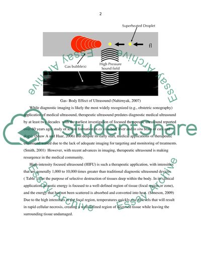
Risks Associated with Ultrasound Procedure Introduction Ultrasound is the mechanical transmission of energy all the way through a medium at a frequency above the upper limit of human hearing (i.e., above roughly 20 kHz). (White, 2006) At frequencies well above this limit, medical ultrasound applies high frequency (i.e., typically 0.5 to 20 MHz) acoustic energy to a region of interest for the purpose of diagnostic imaging or therapeutic treatment. (Vaezy, 2007) Ultrasonic imaging involves mechanical energy, in which pressure waves pass across tissue. Image is formed by the reflection and dispersing back to the transducer. Risks Associated with Ultrasound Procedures There are several physical effects of ultrasound, which are classified as follows: 1. Thermal effects It involves heating of tissues due to the absorption of ultrasonic waves by the tissues. Some heat is also generated at the surface of transducer; 2. Cavitation Cavitation involves the production of gas bubbles at extreme negative pressures; 3. Gas- Body Effect Exposure to Ultrasonic waves stimulates bioeffects within mammalian tissue through gas body activation which is non-thermal mechanism. (Vella, 2003) 4. Other mechanical effects Radiation forces may get access to body fluids and create pressure at tissue interfaces. These physical effects of ultrasound are discussed below in detail. Gas- Body Effect of Ultrasound (Nahirnyak, 2007) While diagnostic imaging is likely the most widely recognized (e.g., obstetric sonography) application of medical ultrasound, therapeutic ultrasound predates diagnostic medical ultrasound by at least two decades with the earliest investigation of focused therapeutic ultrasound reported over 60 years ago; study of lesion formation in ex vivo beef liver and in situ brain of cats and dogs). (Shaw A and Haar, 2006) But despite its early start, medical applications of therapeutic ultrasound waned due to the lack of adequate imaging for targeting and monitoring of treatments. (Smith, 2001) However, with recent advances in imaging, therapeutic ultrasound is making resurgence in the medical community. High-intensity focused ultrasound (HIFU) is such a therapeutic application, with intensities that are generally 1,000 to 10,000 times greater than traditional diagnostic ultrasound devices ( Table 1) for the purpose of selective destruction of tissues deep within the body. In its clinical application, acoustic energy is focused to a well-defined region of tissue (focal region or zone), and the energy that has not been scattered is absorbed and converted into heat. (Soneson, 2009) Due to the high intensities in the focal region, temperatures quickly rise to levels that will result in rapid cellular necrosis, creating a well-defined region of necrosed tissue while leaving the surrounding tissue undamaged. Penetration of Ultrasonic Waves in the Skin (Shaw, 2006) This lesion may be on the order of one to a few centimeters in length and one to a few millimeters in diameter. Electron microscopy has revealed that a narrow boundary of about six to ten cells in width exists between live cells and dead cells at the edges of the lesion. Such localization of the damaged region is clinically advantageous by permitting targeting in close proximity to sensitive structures (e.g., nerves). (Haar, 2001) Studies have demonstrated instantaneous thermal necrosis when temperatures in excess of 56oC are maintained for 1 second or longer. As such, high-intensity therapies seek to rapidly (i.e., within just a few seconds) achieve focal temperature rises in excess of 56oC. While these high-intensity ultrasound technologies offer many clinical advantages, they also present several limitations and challenges. (Haar, 2007) Limitations include reduced effectiveness through air-filled regions (e.g., lungs, bowels) and bone (e.g., brain, ribcage), costly and limited real-time monitoring of treatment, and the potential for incomplete ablation of targeted tissues (a concern for treatment of cancerous lesions). One of the primary challenges of high-intensity ultrasound is the prevention of unintended thermal ablation/damage to surrounding pre-focal and post-focal tissues. Of particular concern is the potential for energy absorption in bone, leading to thermal conduction to nearby tissues. For example, bone is a highly attenuating medium that has the potential to generate unintended temperature rises when exposed to high-intensity ultrasound and to then conduct its thermal energy to nearby tissues, presenting potentially significant safety concerns for diagnostic and therapeutic uses of high-intensity ultrasound systems. Sufficiently high temperatures can damage surrounding tissues such as nerves, resulting in potential acute and/or chronic discomfort, pain, and/or impaired function. (Haar, 2008) Table 1. Typical parameters of HIFU versus diagnostic ultrasound Little work has been done to quantify the effect of the presence of bone on the extent and magnitude of heating and of cellular necrosis within and around a region of tissue exposed to high-intensity ultrasound. (Protopappas, 2007) And, while there are FDA guidance documents and/or international consensus standards for lower-intensity applications of diagnostic ultrasound and physiotherapy (i.e., FDA guidance for diagnostic US, 1997; IEC 60601-2-5, 2000; IEC 1689, 1996), currently there are no available references for high-intensity systems (there are efforts in progress). Furthermore, the only consensus standards that address heating due to the presence of bone (i.e., AIUM/NEMA UD 3-2004 and IEC 60601-2-37, referred to as the “Output Display Standards”) are limited in the following ways: they predict steady-state temperature rise, only (not transient); they do not account for nonlinear propagation in tissue (as would occur in high-intensity applications); and, they do not directly account for thermal heating from shear waves in bone. Kourtis (2007) conducted an analytical study of longitudinal and shear wave propagation in tissue. The study found negligible heating from shear waves in tissue, compared with longitudinal waves, despite higher attenuation coefficients (67x103 dB/cm and 1.33 dB/cm at 5 MHz, respectively). Consequently, analytical and computational efforts described within this dissertation modeled the tissue as a viscous fluid (also as homogeneous). (Protopappas, 2007) Generation of an Ultrasound (Vaezy, 2007) Ren and Zhou (2009) conducted an experimental study of the heating in an in vivo dog thigh (muscle and femur) from 20 minutes of focused hyperthermia. (Ren X-L, Zhou X-D, Yan R-L, Liu D, Zhang, 2009) The study used transducers that ranged from 6 to 8 Watts of power, operated from 1 to 3.58 MHz. The temperatures were measured with 0.7 mm manganin-constantin thermocouples (this type is presently undefined) placed in an array in the dog thigh muscle; one thermocouple was placed in a hole drilled in the thigh bone. The study found that the maximum temperature occurred at the surface of the bone – this finding is not in agreement with Nahimyak (2007) which reported the maximum temperature to be ~ 7 to 8 mm within the bone in the cancellous region. (Nahirnyak, 2007) Myers (2005) conducted a computational study of the propagation and heating in a muscle-bone system from focused (Gaussian) hyperthermia. The study was limited to normal incidence, only; it did not model shear waves in the bone; it presumed the thermal conductivity and specific heat of the bone were equal to that of the tissue; and, it evaluated steady-state temperature rises, not transient temperatures. The study evaluated the heating from a 0.057 Watt, Gaussian-shaded transducer (I0 = 0.1 W/cm2) operating at 2 MHz. The article concluded that the temperature rise due to reflected wave (back into tissue) is significant and should be considered. It furthermore concluded that the location of the focus (be it at various points in the tissue or inside the bone) has essentially no effect on the location at which maximal heating occurs, and the maximum temperature increase was found to be ~ 13 degrees Celsius, just inside the bone (perfusion was not modeled), when the focus was at the interface. This conclusion is consistent with Fujii et al. (1999), except that Fujii found the maximum to be at interface (for normal incidence) – the Wu & Du finding that the maximum heating was just inside the bone surface is consistent with Myers (2005) and also with the findings of this research which found the peak temperature rise to occur at roughly ½ mm distal to the surface of the bone. (Myers, 2004) The lack of standardized methods to characterize the acoustic output and spatial intensity distributions of high-intensity transducers and to test and evaluate the thermal effects of bone has hampered preclinical research studies and subsequently has slowed the FDA regulatory review process for such devices. Most pregnant women in the UK are now offered at least one ultrasound examination as a routine part of their antenatal care. There is, however, considerable debate, in both the lay and medical press, over whether ultrasound should be used to examine all pregnant women rather than just those with clinical indications. Clement's (2004) review of routine ultrasound reported that `no clear benefit in terms of a substantive outcome measure like perinatal mortality can yet be discerned to result from the routine use of ultrasound'. (P. 1357) Exposure to Ultrasonic waves stimulates bioeffects within mammalian tissue through gas body activation which is non-thermal mechanism. Already present bodies of gas can be stimulated due to pressure amplitudes. For greater-pressure amplitudes, aggressive cavitation action with inertial disintegration of microbubbles can be initiated from hidden nucleation areas or from the deterioration of gas bodies. This reflex perturbation at the site of stimulation results in the development of biological effects on neighbouring cells and organs. Cavitational bioeffects were first observed during Shockwave lithotripsy within mammalian tissues, like injury in the kidney and hemorrhage etc. The thermal index is defined as the amount of power needed to generate a highest increase in temperature of 1°C. If the thermal index is 1, it means power generating a rise of temperature up to 1°C. If the thermal index is 2, it shows the double amount of power but would not essentially show a maximum increase of temperature up to 2°C. Since increase of temperature depends upon the type of tissue and is predominantly reliant on the existence of bone, hence the thermal index is further categorised into the following indices: 1. TIS i.e. thermal index used for soft tissue; 2. TIB i.e. thermal index with bone closed to the focus; 3. TIC i.e. thermal index for bone at the exterior. When scanning fetus, the maximum temperature rise would probably take place at the bone and thermal index with bone closed to the focus may cause the ‘worst case’ to happen. Both the thermal and mechanical index should be exhibited if the ultrasound system is able to go beyond an index of 1. Conclusion Due to the ability of ultrasound to transmit through tissue and take effect at a location within the body, remote from the transducer, medical ultrasound offers minimally invasive (i.e., can be used extra corporeally) diagnostic and therapeutic treatments that are faster than open surgery, reduce the need for general anesthesia, reduce complications and pain, reduce recovery time, permit access to remote/inaccessible locations, and permit localized treatment with minimal damage to surrounding tissues. Certain physical hazards have found to be associated with high intensity ultrasound procedure i.e. thermal effect, caviatation, gas body effect and mechanical effects like radiation pressure etc. References Clement, G. T, White PJ, and Kynynen K. 2004. “Enhanced ultrasound transmission through the human skull using shear mode conversion.” J. Acoust. Soc. Am., 115 (3), 1356-1364. Haar, G. 2001. “Acoustic surgery.” Physics Today, 29-34. Haar, G. 2007. “Therapeutic applications of ultrasound.” Progress in Biophysics and Molecular Biology, 93, 111-129. Haar, G. 2008. “The resurgence of therapeutic ultrasound – a 21st century phenomenon.” Ultrasonics, 48, 233. Myers, M. R 2004. “Transient temperature rise due to ultrasound absorption at a bone-tissue interface.” Journal of the Acoustical Society of America, 115 (6), 2887-2891. Myers, M. R. 2005. “Effect of pulse characteristics on temperature rise due to ultrasound absorption at a bone-tissue interface.” Journal of the Acoustical Society of America, 117 (5), 3281-3287. Nahirnyak, V, Mast, T. D, and Holland, C. K. 2007. “Ultrasound-induced thermal elevation in clotted blood and cranial bone.” Ultrasound in Med. & Biol., 33 (8), 1285-1295. Protopappas VC, Baga DA, Fotiadis DI, Likas AC, Papachristos AA, and Malizos KN. 2005, “An ultrasound wearable system for the monitoring and acceleration of fracture healing in long bones.” IEEE Transactions on Biomedical Engineering, 52 (9), 1597-1608. Protopappas, V. C, Kourtis IC, Kourtis LC, Malizos KN, Massalas CV, and Fotiadis DI 2007, “Three dimensional finite element modeling of guided ultrasound wave propagation in intact and healing long bones.” J. Acoust. Soc. Am., 121 (6), 3907-3921. Ren, X-L, Zhou X-D, Yan R-L, Liu D, Zhang J, He G-B, Han Z-H, Zheng M-J, and Yu M. 2009, “Sonographically guided extracorporeal ablation of uterine fibroids with high-intensity focused ultrasound: midterm results.” J. Ultrasound Med.- Letters to the Editor, 28, 95-103. Shaw, A and Haar G. 2006, “Requirements for measurement standards in high-intensity focused ultrasound (HIFU) fields.”, NPL Report DQL AC 015, Middlesex, UK. Smith, NB, Temkin JM, Shapiro F, and Hynynen K. 2001, “Thermal effects of focused ultrasound energy on bone tissue.” Ultrasound in Med. 7 Biol., 27 (10), 1427-1433. Soneson, J 2009, “A user-friendly software package for HIFU simulation.” Proceedings of 8th International Symposium on Therapeutic Ultrasound, Minneapolis, MN, ,September 2008, AIP Press, Melville, NY. Vaezy, S. and Zderic V. 2007, “Hemorrhage Control Using High-intensity Focused Ultrasound.” Int. J. Hyperthermia, 23, 203-211. Vella, G. J, Humphrey, V. F, Duck, F.A, and Barnett SB 2003, “Ultrasound-induced heating in a foetal skull bone phantom and its dependence on beam width and perfusion.” Ultrasound in Medicine and Biology, 29 (6), 779-788. White, P. J, Clement GT, and Hynynen K 2006, “Longitudinal and shear mode ultrasound propagation in human skull bone.” Ultrasound in Med. & Biol., 32 (7), 1085-1096. Read More
Cite this document
  • APA
  • MLA
  • CHICAGO
(“Risks associated with ultrasound procedure Essay”, n.d.)
Risks associated with ultrasound procedure Essay. Retrieved from https://studentshare.org/health-sciences-medicine/1571472-risks-associated-with-ultrasound-procedure
(Risks Associated With Ultrasound Procedure Essay)
Risks Associated With Ultrasound Procedure Essay. https://studentshare.org/health-sciences-medicine/1571472-risks-associated-with-ultrasound-procedure.
“Risks Associated With Ultrasound Procedure Essay”, n.d. https://studentshare.org/health-sciences-medicine/1571472-risks-associated-with-ultrasound-procedure.
  • Cited: 0 times

CHECK THESE SAMPLES OF Risks associated with ultrasound procedure

In Vitro Fertilization

Certain risks are associated with IVF which can affect both the mother and the fetus.... In Vitro Fertilization Name University Abstract In vitro fertilization (IVF) first performed by two doctors, Patrick Steptoe and Robert Edwards, is a scientifically advanced procedure that provides an effectual solution for those couples who are suffering from infertility caused by any reason.... hellip; Since the successful attempt in 1978, many couples in various parts all over the world have opted for this procedure and have enjoyed rewarding outcomes....
9 Pages (2250 words) Research Paper

Imaging Modality for Acute Cholecystitis due to Gallstones

To get a better understanding on which modality to use, several factors such as the method of generating the image, strengths and weaknesses, costs, and the associated risks of the practice need emphasis.... Defined as collection, analysis, modification, duplication, and visualization of images, imagining has remained an interesting filed of study due to the present technological advancement....
6 Pages (1500 words) Essay

Central Venous Catheter Technique in Small Children

There are certain risk factors associated with this procedure like the risk of infection or thrombosis, which may lead to various complications... The success of this procedure depends upon a number of factors including the general condition of the child as described by Grebenik (Grebenik 2004).... These include an experienced hand performing the procedure, the site of insertion of cannula, the presence of vascular anomaly, clotting problems or any previous cannulation procedure performed in the past....
10 Pages (2500 words) Research Paper

Major Effects of Abortion

However, even as modern medicine has paved the way for  safer reproduction processes it has also allowed people greater options for controlling… Access to abortions and the legality of the procedure are controversial issues in the United States as they bring into question the balance of rights of potential parents versus their unborn children....
9 Pages (2250 words) Essay

Pregnancy Risks & Scanning Methods - Computed Tomography, Magnetic Resonance Imaging

These methods include Computed Tomography (CT), Magnetic Resonance Imaging (MRI) and Ultrasound (US) and they are all utilised to examine and evaluate… The purpose of this paper is to examine the possible biological risks and hazards that are associated with these three methods when imaging a pregnant patient.... This will be done by the examination of the scientific In the process recommendations of how to reduce and ameliorate these risks are critiqued and examined A computed tomography (CT) scan utilises x-rays to provide detailed pictures of structured inside the body of pregnant women (WebMD, 2013)....
8 Pages (2000 words) Essay

Special Features of Ultrasound in Medical Physics

This coursework describes special features of ultrasound in medical physics.... This paper outlines the treatment of diseases, medical imaging techniques, features of an ultrasound device, advantages and disadvantages of using it.... Moreover, one of the technologies of medical physics, ultrasound will be discussed in detail, and different related aspects of ultrasound will be analyzed in the paper....
9 Pages (2250 words) Coursework

The Risks Associated with Ionizing Radiation in Medical Imaging Practice

The ultrasound uses the acoustic pulses for echo-ranging imaging or in case of magnetic resonance imaging (MRI) radio-waves are combined with high-field magnets to produce images.... Both ultrasound and MRI make use of non-ionizing radiations.... This paper highlights the risks linked with the use of ionizing radiation in medical imaging practice and the necessary precautions that needs to be taken while handling it....
10 Pages (2500 words) Assignment

Advantages of Fetal Marker Scanning

? As in all screening tests, there are false-positives and false-negatives with ultrasound.... Most of those markers indicating pathology are associated with an abnormal karyotype and because first trimester scans are performed more often, the frequency of marker observation has risen in the same way (Whittle, 1997).... Most, but not all soft markers are associated with an increased risk for Trisomy 21 (Down syndrome) (Ibrahim& Newman, 2005)....
11 Pages (2750 words) Essay
sponsored ads
We use cookies to create the best experience for you. Keep on browsing if you are OK with that, or find out how to manage cookies.
Contact Us