StudentShare
Contact Us
Sign In / Sign Up for FREE
Search
Go to advanced search...
Free

Human Circulatory System: Anatomy and Physiology - Research Paper Example

Cite this document
Summary
The paper "Human Circulatory System: Anatomy and Physiology" states that the abnormal physiology leading to diseases of the circulatory system are numerous and a complete description is beyond the scope of this paper. However, certain disease types can be mentioned here…
Download full paper File format: .doc, available for editing
GRAB THE BEST PAPER95.2% of users find it useful
Human Circulatory System: Anatomy and Physiology
Read Text Preview

Extract of sample "Human Circulatory System: Anatomy and Physiology"

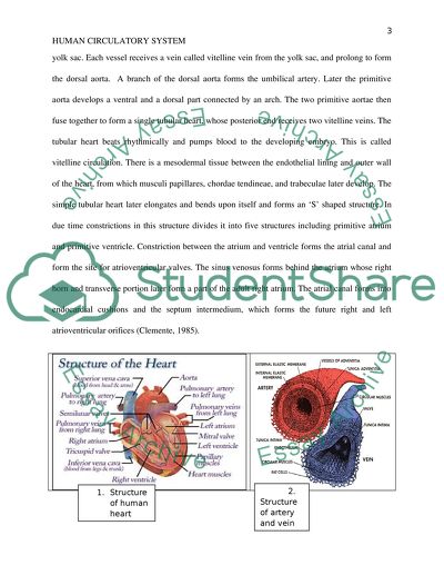
? Human Circulatory system: Anatomy and Physiology First, middle initial, last) (Your college Human Circulatory system: Anatomy and Physiology The human circulatory system is made up of two systems, the cardiovascular system and the lymphatic system. The two systems work in cohort. The cardiovascular system consists mainly of heart, arteries, arterioles, capillaries, venules, and veins. The lymphatic system consists of the lymph, capillaries, the lymphatic vessels and the lymph nodes. It is estimated that if all the blood vessels were connected end to end, they would measure more than 60,000 miles. The amazing function of the heart is to pump blood though these 60,000 miles; blood which carries essential nutrients, hormones and oxygen to some 3 trillion cells of the body. The exchange of nutrients and wastes between blood and the cells occur at the capillaries. The human cardiovascular system is a closed system, which gets narrower as the vessels reach the cells and finally widens as the vessels reach the heart. Such a system allows a large surface area for exchange of nutrients and gases at the cell-capillary interface. Blood circulation is so important for the body that if the heart stops beating even for a few minutes, it can be fatal. The heart beats for an average 100,000 times a day pumping about 2000 gallons of blood per day throughout the body. It pumps the entire blood volume of an adult body of 5.6 liters for three times in a minute. Anatomically, heart is made up of cardiac muscles and consists of four chambers, separated by valves. The blood vessels are made up elastin membranes and smooth muscle cells (Clemente, 1985). The lymph node is the main functional unit of the lymphatic system. Here the immune system is primed and most of the immune cells are activated to fight the foreign substances in the body. The rudimentary heart is a pair of tubular vessels, the primitive aortae, and develops in the space of the pericardial area. These then develop a direct connection with the vessels of the yolk sac. Each vessel receives a vein called vitelline vein from the yolk sac, and prolong to form the dorsal aorta. A branch of the dorsal aorta forms the umbilical artery. Later the primitive aorta develops a ventral and a dorsal part connected by an arch. The two primitive aortae then fuse together to form a single tubular heart, whose posterior end receives two vitelline veins. The tubular heart beats rhythmically and pumps blood to the developing embryo. This is called vitelline circulation. There is a mesodermal tissue between the endothelial lining and outer wall of the heart, from which musculi papillares, chordae tendineae, and trabeculae later develop. The simple tubular heart later elongates and bends upon itself and forms an ‘S’ shaped structure. In due time constrictions in this structure divides it into five structures including primitive atrium and primitive ventricle. Constriction between the atrium and ventricle forms the atrial canal and form the site for atrioventricular valves. The sinus venosus forms behind the atrium whose right horn and transverse portion later form a part of the adult right atrium. The atrial canal forms into endocardial cushions and the septum intermedium, which forms the future right and left atrioventricular orifices (Clemente, 1985). Figure 1. Structure of human heart, and blood vessels. Sources: 1. http://kardiol.com/?p=4 2. http://www.accessexcellence.org/AE/AEC/CC/heart_anatomy.php The heart and the great vessels are contained in a conical fibro-serous sac, between the lungs. The heart is a hollow and muscular organ. It measures 12 cm in length and 8-9 cm in width, and weighs around 280 grams. It is divided into four chambers, the upper two are called left and right atrium, while the bottom two are called left and right ventricle. The atria are separated from ventricles by the coronary sulcus and the interatrial groove separates the two atria. Anterior longitudinal sulcus and the posterior longitudinal sulcus are two grooves separating the two ventricles. Right atrium can hold about 57cc of blood. It has a lager cavity called the sinus venarum and a smaller portion called auricula. The superior- and inferior- vena cava drain blood into the right atrium from upper and lower half of the body respectively. The coronary sinus, guarded by a valve, also opens into this atrium draining blood from the heart’s body itself. There is an atrioventricular opening guarded by the tricuspid valve between the right atrium and the right ventricle. The right ventricle has an opening for the pulmonary artery, guarded by the semilunar valves. The left atrium is smaller than the right atrium and consists of a larger principal cavity and a smaller auricula. Four pulmonary veins, without any valves, drain blood into the left atrium. The left atrioventricular opening is found between the left atrium and the left ventricle, guarded by the bicuspid or the mitral valve. The left ventricle, at the apex of the heart, is larger than the right ventricle. The aortic opening guarded by the aortic semilunar valves is also found in this chamber. Structurally, the heart is made up of muscle fibers and fibrous rings. Its walls are made up of outer epicardium, middle muscular myocardium and inner endocardium. The fibrous rings surround the artrioventricular openings and the arterial orifices. Interspersed between the muscle fibers of the heart are the purkinje’s fibers. The artrioventricular bundle of His is the only connection between the atrium and the ventricles forming the atrioventricular node and the sinoatrial node. These nodes send systolic contractions from atria to ventricles (Clemente, 1985). Now a days image based models are available, which can give detailed anatomical structure and fiber orientation of the heart (Deng, Jiao, Ye, & Xia, 2012). Functionally, the heart pumps blood to all parts of the body by the arteries. This pumping action occurs by regular contractions, about 70/min; each contraction followed by a rest period constitutes the cardiac cycle. Each cardiac cycle, lasting about 0.8 second, has three phases: (1) atrial systole, a short simultaneous contraction of the atria, (2) ventricular systole, a more prolonged contraction of both ventricles, and (3) a period of rest for the whole heart. The venous openings followed immediately by the atrial contraction forces the blood into the ventricles through the atrioventricular openings. Passage of blood back into the atria is prevented by the closing of the tricuspid and the bicuspid valves when the ventricles contract. When the pressure in the ventricles get higher than that in the aorta and the pulmonary artery, the valves of these blood vessels open and the blood from the right ventricle flow into the pulmonary artery from the right ventricle and into the aorta from the left ventricle. When the ventricles relax, the pressure in the respective vessels closes the semilunar valves and prevents blood form reentering into the ventricles. During the rest period, the tricuspid and the bicuspid valves also relax and blood flows from veins into the atria due to negative intrathoracic pressure (Clemente, 1985). Anatomically, all blood vessels are tubular structures. They are made up of 3 main layers: (1) tunica adventitia, the outermost strong covering consisting of connective tissue, collagen and elastin; it helps the vessel to anchor to the adjacent tissue, (2) tunica media, made up of smooth muscles and elastic fibers. It is thicker in arteries than veins. When these muscles contract, the lumen becomes narrow (vasoconstriction) and gets wider (vasodilation) upon relaxation, and (3) tunica intima, made up of smooth endothelial cells and elastic membrane. Elastic lamina lies between each layer. The walls of the larger vessels have their own blood supply too! They have their own arteries and veins called vasa vasorum (vessels of vessels). The veins, but not the arteries, contain valves in them to prevent backflow of the blood. These valves are bicuspid and made up of elastic tissue. Physiologically, high upstream blood pressure causes the blood in arteries to move forward and also press outward on the arterial wall. This is called hydrostatic pressure. The bigger arteries stretch when the pressure increases, but recoils back to prevent further drop in pressure. Blood flows due to pressure gradient, from high pressure to low pressure, so this drop in pressure is important for proper blood flow in the body by the arteries. Veins, on the other hand can expand to hold large blood volume. Veins carry about 64% of the body’s blood. Blood flow in the veins is more complex, compared to the arteries. This is because, the blood pressure is low in them, flow rate depends on muscle contraction and relaxation of the legs, blood has to move against the gravity, veins collapse in absence of blood, and they have to carry large volume of blood. Since most of the veins are embedded in muscles, muscle contraction helps the blood to move forward to the heart (Clemente, 1985). Like nerve cells, heart muscle cells are able to generate action potential (AP), but they last a bit longer in the heart. Heart muscle cells at different region possess different AP. AP in the heart has five stages, 0 to 4. Phase 0 is the immediate depolarization, which makes the voltage positive. Here the permeability to sodium ions increase and that to potassium ions decrease. Little repolarization begins as soon as the permeability to sodium ions decrease, which constitute phase 1. The membrane potential then becomes and remains steady at 0 millivolts. This is phase 2 and is termed as the plateau of the AP. At this point the rate of inward flow of calcium ion equals that of outbound potassium ions. Since the permeability to the calcium ions begin to decrease and that to potassium ions begin to increase, the voltage does not remain zero. Repolarization begins again and this marks the phase 3. Until the next AP initiation, the voltage decreases to its original value and remains there. This is stage 4, where the muscle cell is unstimulated. These APs initiate contractions in the heart, making it a pump. Cardiac muscles communicate to each other though gap junctions. Cardiomyocyte or cardiac muscle cells are made up of hundreds of myofibrils. These fibrils contain action and myosin proteins in them. Myosin contains a myosin head. Each AP causes the cardiomyocytes to release calcium ions from the reserve. These calcium ions allow myosin heads to bind to the actin molecules and pulls them causing the cell to contract (Oracle ThinkQuest). The lymphatic system consists of the lymph, capillary network present in various tissues and organs, the vessels which carry lymph from capillaries to the large veins in the neck, and the lymph nodes or glands present in the pathways of the vessels where the lymph is filtered and lymphocytes are added to the lymph. Lymph has the composition similar to blood plasma and is transparent, slightly yellowish in color, watery fluid with specific gravity of 1.015. Apart from constituents of plasma, lymph has larger proteins, lymphocytes, foreign particles, and frequently RBCs. The capillaries consist of single layer of endothelial cells and are bathed in intercellular tissue fluids. Lymph is made by physical process like filtration, diffusion, osmosis, and active secretion by the endothelial cells of the capillaries. They are present in almost all tissues, like skin, subcutaneous tissue, joint capsules, pericardial cavity, salivary gland, liver, spleen, nasal cavity, bronchia, heart, kidneys, ovary, prostate, testes, the alimentary canal etc. The lymphatic vessels collect lymph from the capillaries. They are made up of three layers; the outer coat is elastic and transparent, inner coat has smooth muscles, and the external coat has connective tissue. These vessels have semilunar valves like those present in veins, but are more in number and their number increases as the vessels approach the lymph nodes, and areas of neck. The lymph glands are small bean shaped bodies, interspersed in the path of lymphatic vessels, such that the lymph passes through them on their way to blood. A lymph node is made up of several lymphoid lobules, which are the structural and functional units of the lymph nodes. The capsule of the lymph node is collagenous and has a subcapsular sinus. Lymph carrying macrophages, dendritic cells, and lymphocytes from the sinus drains into the node cortex and the medulla. Activated B cells mature into plasma in the cortex, which have nodules with germinal centers. T cells are processed in the medulla of the lymph nodes. Afferent lymphatic vessels bring lymph into the node and efferent vessels exit the node (Clemente, 1985). Recent evidence shows that lymphatic endothelial cells play an important role in the trafficking of lymphocytes and aid in the regulation of tolerance and immunity. They do this by the presence of cell adhesion molecules on their surface (Tewalt, Cohen, Rouhani, & Engelhard, 2012). Figure 2. The Structure of a lymph node and the circulatory system Source: 1. http://www.acm.uiuc.edu/sigbio/project/updated-lymphatic/lymph7.html 2. http://www.healingdaily.com/exercise/effects-of-rebounding-on-the-lymphatic- system.htm Functions of the lymphatic system includes following: Collection of wastes from the intercellular fluid Filtering lymph at the nodes Filtering blood of bacteria, viruses, and foreign particles, by the spleen Fight infection by raising an immune reaction The abnormal physiology leading to diseases of the circulatory system are numerous and a complete description is beyond the scope of this paper. However, certain disease types can be mentioned here. As far as cardiovascular system is concerned, there are six types of diseases. They are 1) Ischemic heart diseases, which is due to poor or absence of blood circulation in the body of the heart itself. 2) Stroke, abnormal circulation of blood in the brain. 3) Peripheral vascular diseases, usually the lower extremities.4) Heart failure, when the pump cannot keep up with body’s need of blood. 5) Rheumatic heart disease where a bacterial infection in the childhood affects the valves of the heart and have to be replaced, and 6) Congenital heart disease, where a birth defect leads to abnormal structure of heart (PHAC, 2010). Lymphedema is the most common disease of the lymphatic system where the organs of the system get inflamed. Obesity, with higher body mass index has been attributed to the lymphedema of the lower extremity (Greene, Grant, & Slavin, 2012). Other diseases of the system include lymphangioma, protein-losing enteropathy, intestinal lymphangiectasia, complex vascular malformations, infectious diseases, and lymphangioleiomyomatosis (Lee, Bergan, & Rockson, 2001). The human circulatory system encompasses the cardiovascular system and the lymphatic system. The cardiovascular system provides nutrition and oxygen to the tissues and organs of the body, while the lymphatic system gathers all the wastes and foreign substance from the interstitial spaces and processes them for their disposal. The two systems work in close collaboration with each other. The cardiovascular system is a closed system while the lymphatic system is an open system. Both the systems can have acquired and inherent anatomical as well as physiological problems, some of them being fatal. Reference List Clemente, C. (1985). Gray's Anatomy of the Human Body (30th Ed.). Philadelphia: Lea & Febiger. Deng, D., Jiao, P., Ye, X., & Xia, L. (2012). An image-based model of the whole human heart with detailed anatomical structure and fiber orientation. Comput Math Methods Med. 2012:891070. doi: 10.1155/2012/891070 Greene, A.K., Grant, F.D., & Slavin, S.A. (2012). Lower-extremity lymphedema and elevated body- mass index. N Engl J Med. 366(22):2136-7. Lee, B., Bergan, J., & Rockson, S.G. (Eds.). (2001). Lymphedema, London: Springer-Verlag. Oracle ThinkQuest. (n/a). The cardiac Action Potential. Retrieved from http://library.thinkquest.org/C003758/Function/The%20Cardiac%20Action%20Potential.htm. PHAC. (2010). Six Types of Cardiovascular Disease. Retrieved from http://www.phac-aspc.gc.ca/cd-mc/cvd-mcv/cvd-mcv-eng.php Tewalt, E.F., Cohen, J.N., Rouhani, S.J., & Engelhard, V.H. (2012). Lymphatic endothelial cells- key players in regulation of tolerance and immunity. Front Immunol. 3:305.doi: 10.3389/fimmu.2012.00305. Read More
Cite this document
  • APA
  • MLA
  • CHICAGO
(“Anatomy and phisiology of the circulatory system Research Paper”, n.d.)
Retrieved from https://studentshare.org/health-sciences-medicine/1459678-anatomy-and-phisiology-of-the-circulatory-system
(Anatomy and Phisiology of the Circulatory System Research Paper)
https://studentshare.org/health-sciences-medicine/1459678-anatomy-and-phisiology-of-the-circulatory-system.
“Anatomy and Phisiology of the Circulatory System Research Paper”, n.d. https://studentshare.org/health-sciences-medicine/1459678-anatomy-and-phisiology-of-the-circulatory-system.
  • Cited: 0 times

CHECK THESE SAMPLES OF Human Circulatory System: Anatomy and Physiology

The Effects of Postural Changes, Exercise and Cognitive Stress on Human Arterial Pressure and Pulse Rate

The circulatory system, which plays a major part in coronary health, functions mainly in the transportation of blood carrying nutrients and oxygen to all parts of the body.... However, the circulatory system is subjected to various pathological conditions.... Pathological conditions may result from the abnormality of any component or processes in the circulatory system.... The primary importance of studying the components and the processes of the circulatory system is to understand the relationship between the functions and the structures, and for proper diagnosis of the diseases affecting the circulatory system....
3 Pages (750 words) Essay

Anatomy and physiology

Although my textbook ‘Essentials of Human anatomy and physiology' explains the mechanism of muscular movements in great detail, one has to see these muscles displayed the way they are in this exhibition to appreciate what “actually” goes on under our skin.... he Exhibition eased me into the experience by starting with the skeletal system.... he muscular system was more graphic and I had to pause in the gallery for a moment to take in the red and white figures displayed to highlight the movements of the muscles....
5 Pages (1250 words) Essay

Urinary system

These organs are ultimately responsible for the important job of interacting with the circulatory system to remove harmful wastes from the body and to maintain appropriate water and salt levels in the blood.... In fact, these systems are so closely related that a specific type of physician, called a urologist, is responsible for treating aspect of all three systems The urinary system is composed of many subunits, which may be better understood by studying the gross anatomy of the urinary system, the overall function and metabolic importance of the urinary system, and the complex unit formations and regulatory mechanisms that allow the kidney to function....
5 Pages (1250 words) Term Paper

HUMAN PHYSIOLOGY AND NUTRITION

Thin walls of the alveoli enhance quick diffusion of The human circulatory system can generally be described as an organ system concerned with transfer of amino acids, the lymph and electrolytes on one hand and gases, blood cells and hormones on the other.... he circulatory system comprises basically of two principle mechanisms that is; cardiovascular and lymphatic systems, the former being concerned with blood distribution and the latter being involved in return of excess filtered blood plasma from between cells and refers to as lymph....
8 Pages (2000 words) Case Study

The Human Anatomy and Possible Threats

The circulatory system is responsible for the circulation of blood and lymph around the body (Swearingen, 2012).... The main systems of the human body are the cardiovascular system, the digestive system, the endocrine system, the urinary system, the immune system, the muscular system, the nervous system, the reproductive system, and the respiratory system (Schlossberg and Zuidema, 2007)....
12 Pages (3000 words) Essay

Exploring Endocrine and Respiratory Systems

While many tissues are capable of utilizing other energy sources such as fat and protein, brain and the red blood cells can only use glucose.... Glucose is normally stored in form of glycogen in the liver when there is high.... ... ... Glycogenesis is the process through which glycogen stored in the body tissues is converted into glucose when the blood glucose goes down....
2 Pages (500 words) Essay

Musculoskeletal, nervous, respiratory, and circulatory systems in ergonomics

General anatomy and Musculoskeletal System.... Circulatory systemThe circulatory system is an organ system that allows blood to circulate and transport gases (O2 and CO2), nutrients (electrolytes and amino acids), hormones, waste, and blood cells to and from cells.... Mismatch in work demands and abilities of the circulatory system causes muscle fatigue and increases risk of injury.... Human physiology: From cells to systems, New York: Cengage Learning....
2 Pages (500 words) Essay

Coronary and Pulmonary Circulations

The essay 'Coronary and Pulmonary Circulations' is a vivid example of a visual and accessible presentation of material, gives a clear understanding on the topic of anatomy and physiology of blood circulation in the heart and lungs.... The major blood vessels which constitute this circulation are the left main coronary artery and the right main coronary artery, both of which originate at the base of the aorta from openings called the coronary Ostia located behind the aortic valve leaflets (Klabunde, 'Cardiovascular physiology Concepts')....
6 Pages (1500 words) Essay
sponsored ads
We use cookies to create the best experience for you. Keep on browsing if you are OK with that, or find out how to manage cookies.
Contact Us